

 PDF
PDF ePub
ePub Citation
 Print
Print


Abstract
Purpose
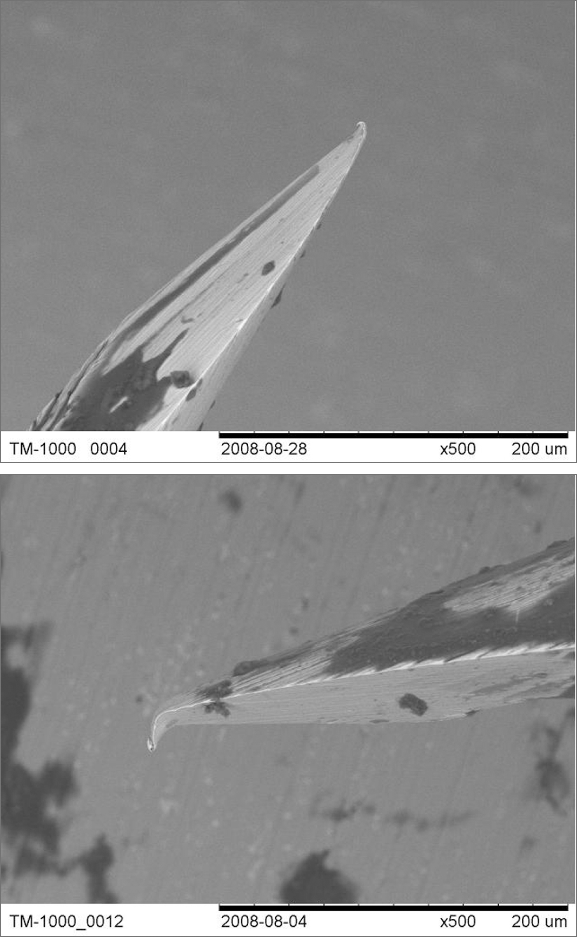
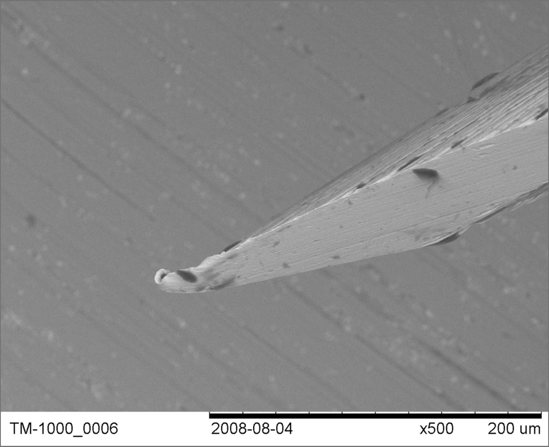
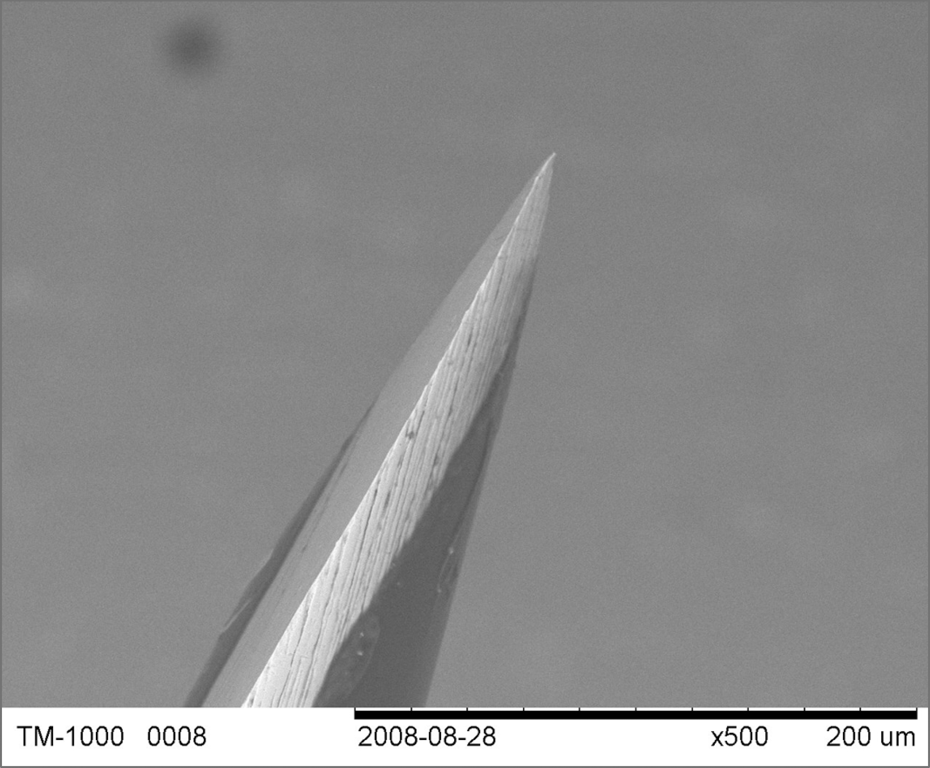
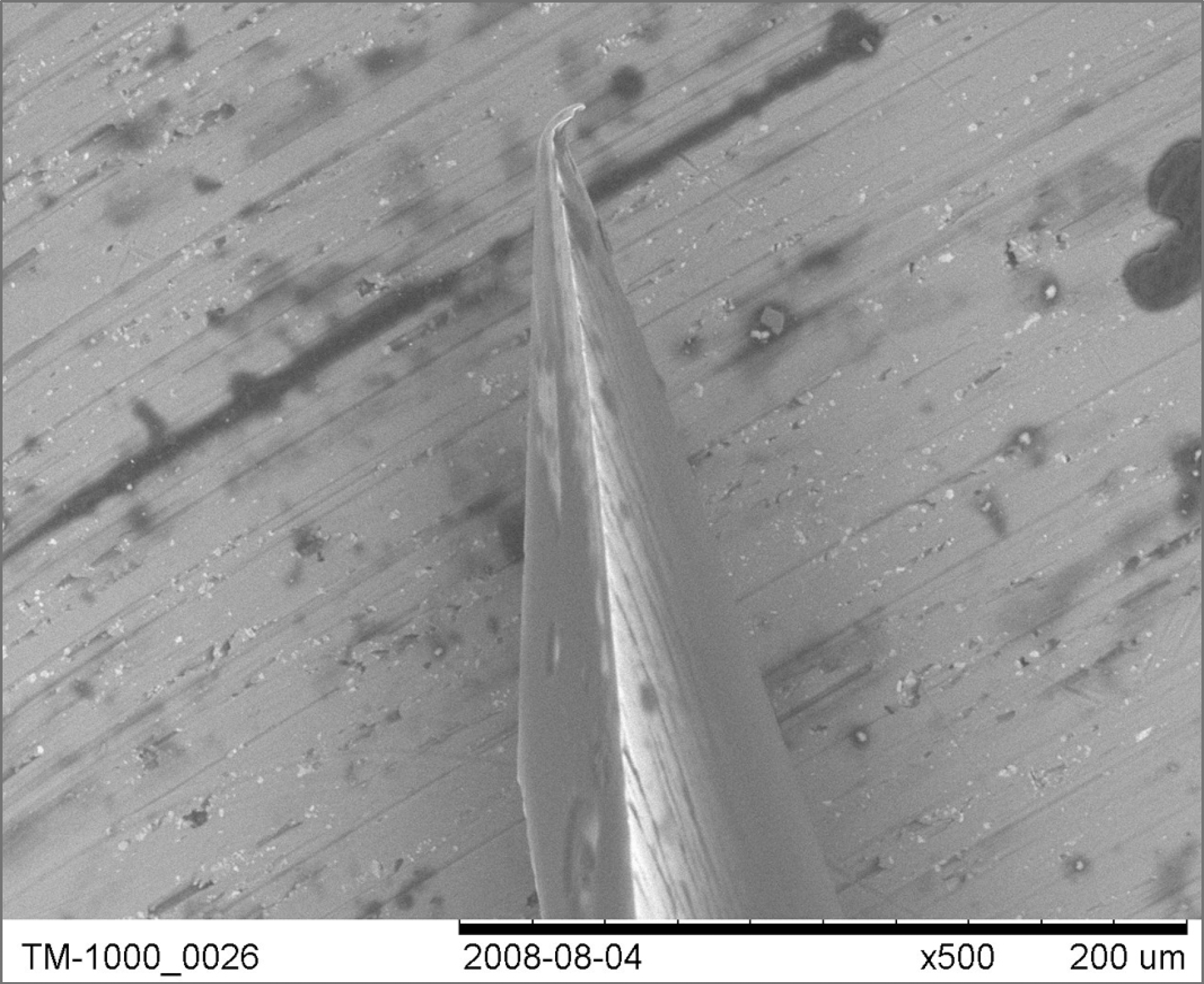
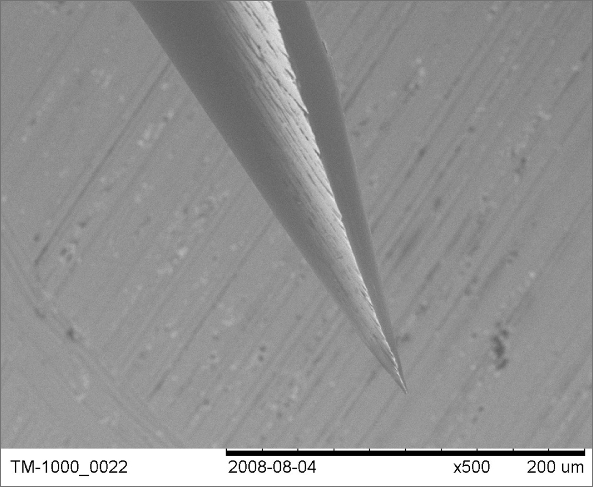
To observe the degree of damage in a 30-gauge injection needle by observing the changes in needle tip following an intravitreal injection with the use of a scanning electron microscope.
Methods
The present study evaluated 11 injection needles collected following the use of an intravitreal injection of bevacizumab. Ten unused injection needles were selected as the control group. Needle examination was performed using a scanning electron microscope.
Results
Following 11 intravitreal injections, seven bent needle tips, two stubbed needle tips and two almost normal needle tips were observed following intravitreal injections. In the control group, a single damaged needle tip was observed.
Conclusions
Significant damage to the needle tip was observed following intravitreal injection using a 30-gauge injection needle. The results indicate that needles should be manipulated carefully during an intravitreal injection. Additionally, in the control group where no procedures were performed, a single injection needle with damaged status was found. These results indicate that needles should be replaced in cases in which resistance is perceived during the procedure.
References
1. Jager RD, Aiello LP, Patel SC, Cunningham ET Jr. Risks of intra-vitreous injection: a comprehensive review. Retina. 2004; 24:676–98.

2. Gragoudas ES, Adamis AP, Cunningham ET Jr, et al. For the VEGF Inhibition Study in Ocular Neovascularization Clinical Trial Group, Pegaptanib for neovascular age-related macular degeneration. N Engl J Med. 2004; 351:2805–16.
3. Jeganathan VS, Verma N. Safety and efficacy of intravitreal anti-VEGF injections for age-related macular degeneration. Curr Opin Ophthalmol. 2009; 20:223–5.
4. Bakri SJ, Beer PM. The effect of intravitreal triamcinolone acetonide on intraocular pressure. Ophthalmic Surg Lasers Imaging. 2003; 34:386–90.
5. Jonas JB, Kreissig I, Söfker A, Degenring RF. Intravitreal injection of triamcinolone for diffuse diabetic macular edema. Arch Ophthalmol. 2003; 121:57–61.
6. Aiello LP, Brucker AJ, Chang S, et al. Evolving guidelines for intra-vitreous injections. Retina. 2004; 24:S3–19.
7. Rodrigues EB, Meyer CH, Schmidt JC, et al. Unsealed sclerotomy after intravitreal injection with a 30-gauge needle. Retina. 2004; 24:810–2.
8. Hayhoe S, McCrossan M, Smith A, et al. Single-use acupuncture needles: scanning electron-microscopy of needle-tips. Acupunct Med. 2002; 20:11–8.
9. Friedenwald JS. Contribution to the theory and practice of tonometer. Am J Ophthalmol. 1937; 20:985–1024.
10. Goodside V. Ocular rigidity. A clinical study. Arch Ophthalmol. 1959; 62:839–41.
11. Mehra KS. Ocular rigidity in emmetropes. Acta Ophthalmol. 1965; 43:105–8.
12. Pallikaris IG, Kymionis GD, Ginis HS, et al. Ocular rigidity in living human eyes. Invest Ophthalmol Vis Sci. 2005; 45:409–14.
13. Lee IS, Youn DH. Ocular rigidity in Korean. J Korean Ophthalmol Soc. 1979; 20:461–7.
 XML Download