Abstract
Purpose
To report the case of a patient with ciliochoroidal detachment after brief exposure to patterned scanning laser photocoagulation.
Case summary
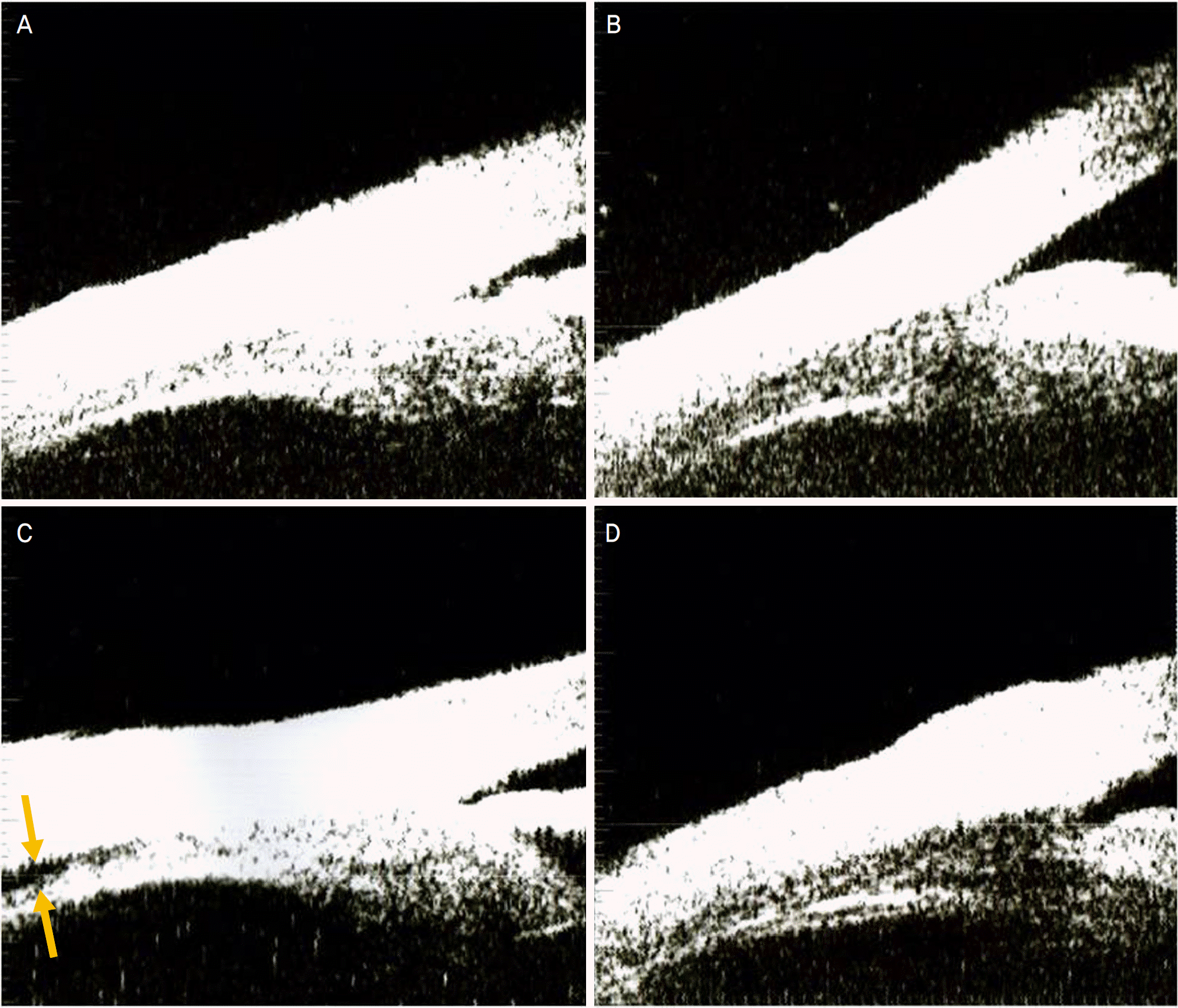
We examined a 62-year-old woman with early proliferative diabetic retinopathy and observed neovascularization and macular edema upon fundus examination. The patient underwent patterned scanning laser photocoagulation with an exposure time of 0.03 sec over the entire retina in a single pass. In vivo, the ciliary body and choroid were examined using ultrasound biomicroscopy (UBM), before, immediately after, 3 and 7 days after panretinal photocoagulation. Ciliochoroidal detachment was observed 3 days after panretinal photocoagulation and spontaneously disappeared by 7 days after photocoagulation. The change in IOP coincident with ciliochoroidal detachment were not significant.
Conclusions
Ciliochoroidal detachment after panretinal photocoagulation may lead to complications such as angle-closure glaucoma. Patterned scanning laser photocoagulation with short exposure time should be practiced only with careful attention to the possible development of cilochoroidal detachment.
Go to : 
References
1. Photocoagulation treatment of proliferative diabetic retinopathy: clinical application of Diabetic Retinopathy Study (DRS) findings, DRS Report Number 8. The Diabetic Retinopathy Study Research Group. Ophthalmology. 1981; 88:583–600.
2. Neubauer AS, Ulbig MW. Laser Treatment in Diabetic Retinopathy. Ophthalmologica. 2007; 221:95–102.

3. Yuki T, Kimura Y, Nanbu S, et al. Ciliary Body and Choroidal Detachment after Laser Photocoagulation for diabetic Retinopathy. Ophthalomology. 1997; 104:1259–64.

4. Kawahara S, Nagai Y, Kayakami E, et al. Ciliochoroidal detachment following sclera buckling surgery for rhegmatogenous retinal detachment. Nippon Ganka Gakkai Zasshi. 2000; 104:344–8.
5. Fourman S. Angle-closure glaucoma complicating ciliochoroidal detachment. Ophthalmology. 1989; 96:646–53.

6. Stockl FA, Saheb NE. Angle-closure glaucoma secondary to ciliochoroidal detachment. Can J Ophthalmol. 1998; 33:280–2.
7. Gentile RC, Berinstein DM, Leibmann J, et al. High-resolution ultra-soud biomicroscopy of the pars plana and peripheral retina. Ophthalmology. 1998; 105:478–84.
8. Liemann JM, Weinreb RN, Rtich R. Angle-closure glaucoma associated with occult annular ciliary body detachment. Arch Ophtalmol. 1998; 116:731–35.
9. Framme C, Alt C, Schnell S, et al. Selective targeting of the retinal pigment epithelium in rabbit eyes with a scanning laser beam. Invest Ophthalmol Vis Sci. 2007; 48:1782–92.

10. Jain AT, Blumenkraz MS, Paulus Y, et al. Effect of pulse duration on size and character of the lesion in retinal photocoagulation. Arch Ophthalmol. 2008; 126:78–85.

11. Sanghvi C, McLauchlan R, Delgado C, et al. Initial experience with the Pascal photocoagulator: a pilot study of 75 procedures. Br J Ophthalmol. 2008; 92:1061–4.

12. Huamonte FU, Peyman GA, Goldberg MF, Locketz A. Immediate fundus complications aftrer retinal scatter photocoagulation I. Clinical picture and pathogenesis. Ophthalmic Surg. 1976; 7:88–99.
13. Blondeau P, Pavan PR, Phelps CD. Acute Pressure Elevation Following Panretinal Photocoagulation. Arch Ophthalmol. 1981; 99:1239–41.

Go to : 
 | Figure 1.Ultrasound biomicroscopy (UBM) images of the 62-year-old woman with proliferative diabetic retinopathy. (A) UBM before treatment. Ciliochoroidal area was intact. (B) UBM right after panretinal photocoagulation using patterned scanning laser. No specific change was seen compared to UBM before treatment. (C) 3 days after panretinal photocoagulation. Slit-like ciliochoroidal detachment appeared at pars plana (arrows). (D) 7 days after panretinal coagulation. Cilochoroidal detachment disappeared without any specific treatment for ciliochoroidal detachment. |


 PDF
PDF ePub
ePub Citation
 Print
Print


 XML Download