

 PDF
PDF ePub
ePub Citation
 Print
Print


Abstract
Purpose
To report a case of intraocular cilium revealed by diagnostic vitrectomy in a case of stubborn uveitis that was un-responsive to steroid therapy.
Case summary
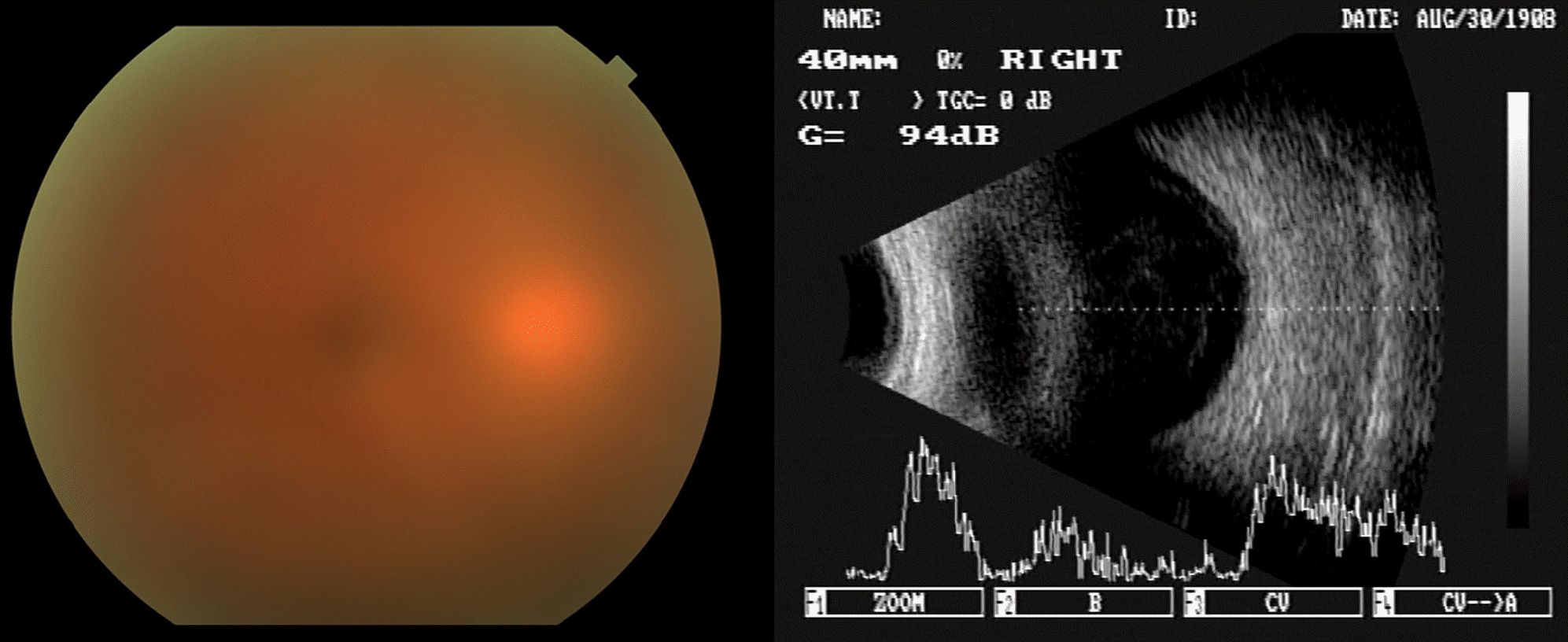
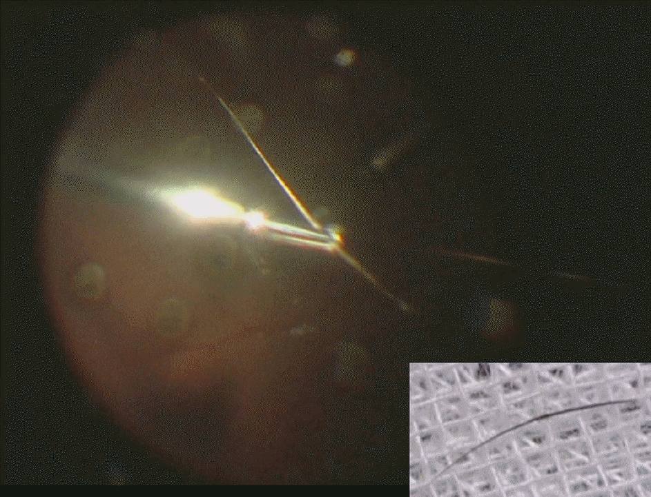
A 39-year-old man was referred to our hospital due to decreased vision in his right eye that started two months prior to presentation. He had previously been treated for a diagnosis of iridocyclitis. The patient’s history revealed a blunt trauma to the right eye while wearing glasses after which he developed a microhyphema and was treated for traumatic iritis at another clinic 3 months ago. He was treated with topical and oral steroids after being diagnosed with iridocyclitis and had recently been prescribed additional oral cyclosporine because his condition had not improved. Ocular examination revealed inflammatory cells in the anterior chamber and vitreous cavity with hand motion vision. Ultrasonography revealed a hazy vitreous cavity but the retina was flat. Diagnostic vitrectomy with intravitreal antibiotic injection was performed and an intraocular foreign body presumed as a cilium was detected without an entrance wound on the exterior or interior surface of the eye. After removal of the foreign body, the patient’s vision was completely recovered.
References
1. Fortuin M, Blanksma L. An unusual complication of perforating wounds of the eye. Doc Ophthalmol. 1986; 61:197–203.

2. Gottlieb F, Finestone J, Akerman JL. Intravitreal cilia and retinal detachment. Ann Ophthalmol. 1982; 14:541–4.
3. Gopal L, Banker AS, Sharma T, et al. Intraocular cilia associated with perforating injury. Indian J Ophthalmol. 2000; 48:33–6.
4. Seawright AA, Bourke RD, Gray PJ, Cooling R. Intravitreal cilia in phakic penetrating eye injury. Aust N Z J Ophthalmol. 1997; 25:133–5.
5. Islam N, Dabbagh A. Inert intraocular eyelash foreign body following phacoemulsification cataract surgery. Acta Ophthalmol Scand. 2006; 84:432.
6. Oh KT-(Kean), Oh KT-(Kong), Singerman LJ. An eyelash in the vitreous cavity without apparent etiology. Ophthalmic Surg Lasers. 1996; 27:243–5.
7. Kertes PJ, Al-Ghamdi AA, Brownstein S, et al. An intraocular cilium of uncertain origin. Can J Ophthalmol. 2004; 39:279–81.
8. Wirth MG, Helbig H. Can eyelashes migrate? Klin Monatsbl Augenheilkd. 2005; 222:238–40.
9. Kozart DM, Yanoff M, Katowitz JA. Tolerated eyelash embedded in the retina. Arch Ophthalmol. 1974; 91:235–6.
10. Humanyun M, de la Cruz Z, Maguire A, et al. Intraocular cilia: report of six cases of 6 weeks' to 32 years' duration. Arch Ophthalmol. 1993; 111:1396–401.
11. Snir M, Kremer I. Eyelash complications in the anterior chamber. Ann Ophthalmol. 1992; 24:9–11.
12. Ahn M. Noninfectious endophthalmitis caused by an intraocular foreign body retained for 16 years. J Korean Ophthalmol Soc. 2001; 42:793–6.
13. Margolis R. Diagnostic vitrectomy for the diagnosis and management of posterior uveitis of unknown etiology. Curr Opin Ophthalmol. 2008; 19:218–24.
14. Verbraeken H. Diagnostic vitrectomy and chronic uveitis. Graefes Arch Clin Exp Ophthalmol. 1996; 234:S2–7.
15. Margolis R, Brasil OF, Lowder CY, et al. Vitrectomy for the diagnosis and management of uveitis of unknown cause. Ophthalmology. 2007; 114:1893–7.
16. Tommaso R, Hermann D, Paolo M, et al. Intraocular cilium masquerading as a parasite. Retinal Cases and Brief Reports. 2008; 2:70–2.
 XML Download