Abstract
Purpose
To compare 23-gauge transconjunctival sutureless vitrectomy (TSV) and sutured vitrectomy in the aspect of intraocular pressure (IOP) changes and complications.
Methods
Through a retrospective chart review, 45 sutureless patients (48 eyes) and 48 sutured patients (52 eyes) who had undergone 23-gauge transconjunctival vitrectomy by one surgeon (J.H. Sohn) were compared. IOP was measured routinely pre-operativley, at 1 day, 1 week, and 1 month post-operatively. Postoperative IOP, hypotony (IOP<7 mmHg) rates and complications associated with hypotony were assessed respectively. In addition, the cases involving intraocular air or gas tamponade or cataract extraction were compared.
Results
One day after the surgery, 3 eyes of the sutureless group developed hypotony, which persisted in 2 eyes through post-operative 1 week. Two eyes of the sutureless group developed hypotony 1 week after the surgery. Most of the hypotony cases were transient, but choroidal detachment occurred in 2 cases, and retinal detachment occurred in 1 case. In contrast, none of the sutured group developed hypotony. Intraocular air or gas tamponade significantly raised IOP 1 day after the surgery. When the operation was combined with cataract extraction, IOP was reduced postoperative 1 week and 1 month.
Conclusions
The well-known risk factor of 23-gauge sutureless vitrectomy is postoperative hypotony. The present study showed postoperative hypotony can result in retinal detachment as a complication in contrast to previous studies. The authors conclude that suturing the wound for the prevention of hypotony is better, especially in cases with high risk of hypotony or defi-nite wound leakage.
Go to : 
References
1. Machemer R, Buettner H, Norton EW, Parel JM. Vitrectomy: a pars plana approach. Trans Am Acad Ophthalmol Otolaryngol. 1971; 75:813–20.
2. Fujii GY, De Juan E Jr, Humayun MS, et al. Initial experience using the transconjunctival sutureless vitrectomy system for vitreoretinal surgery. Ophthalmology. 2002; 109:1814–20.
3. Fujii GY, De Juan E Jr, Humayun MS, et al. A new 25-gauge instrument system for transconjunctival sutureless vitrectomy surgery. Ophthalmology. 2002; 109:1807–12.
5. Fine HF, Iranmanesh R, Iturralde D, Spaide RF. Outcomes of 77 consecutive cases of 23-gauge transconjunctival vitrectomy surgery for posterior segment disease. Ophthalmology. 2007; 114:1197–200.

6. Tewari A, Shah GK, Fang A. Visual outcomes with 23-gauge transconjunctival sutureless vitrectomy. Retina. 2008; 28:256–62.

8. Yanyali A, Celik E, Horozoglu F, Nohutcu AF. Corneal topographic changes after transconjunctival(25-gauge) sutureless vitrectomy. Am J Ophthalmol. 2005; 140:939–41.
9. Woo SJ, Park KH, Hwang JM, et al. Risk factors associated with scle-rotomy leakage and postoperative hypotony after 23-gauge transconjunctival sutureless Vitrectomy. Retina. 2009; 29:456–63.

10. Schweitzer C, Delyfer MN, Colin J, Korobelnik JF. 23-Gauge transconjunctival sutureless pars plana vitrectomy: results of a prospective study. Eye (Lond). 2009; 23:2206–14.

11. Kim MJ, Park KH, Hwang JM, et al. The safety and efficacy of transconjunctival sutureless 23-gauge vitrectomy. Korean J Ophthalmol. 2007; 21:201–7.

Go to : 
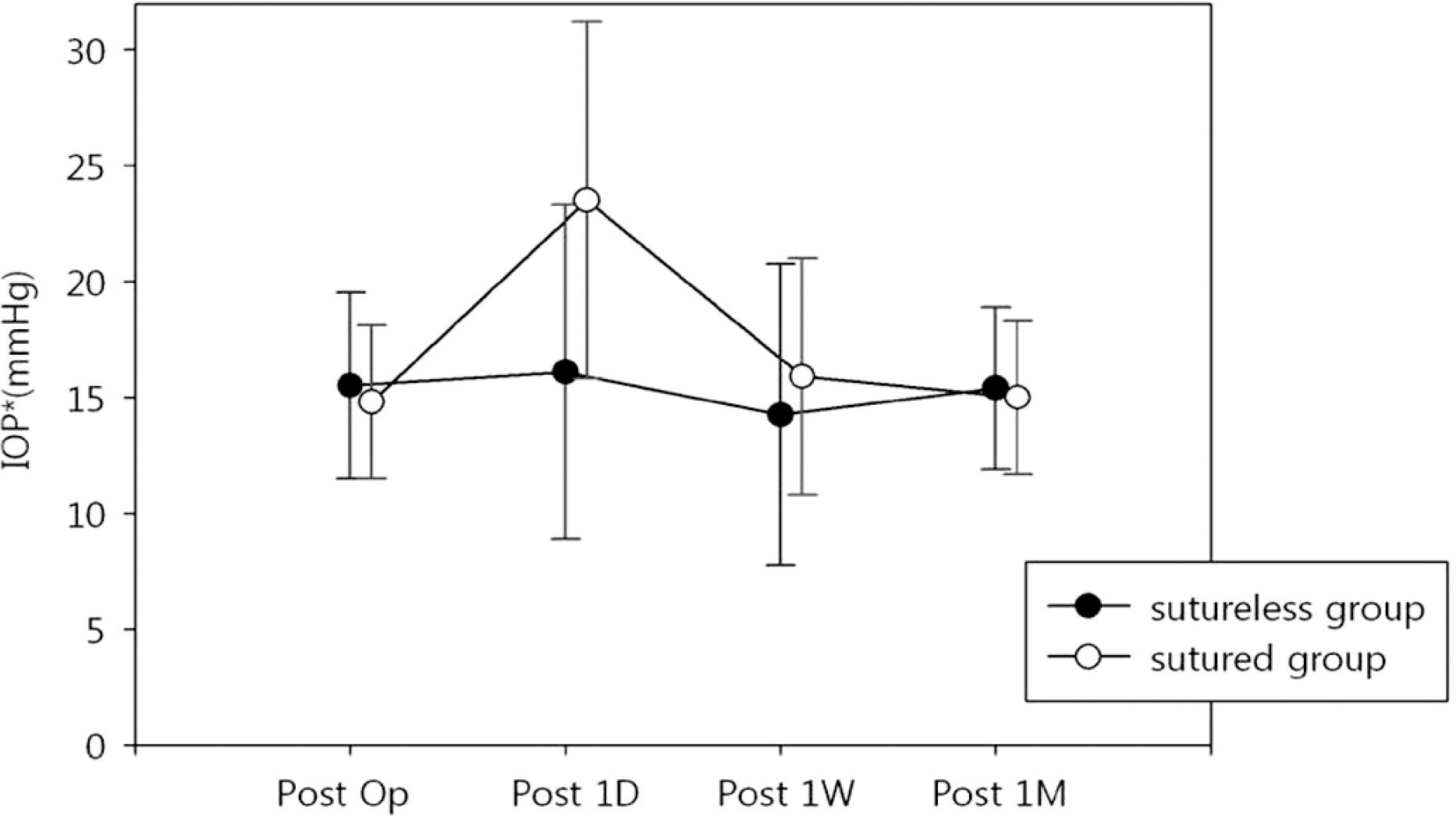
 | Figure 1.Preoperative and postoperative IOP changes in 23-gauge vitrectomy. Post operative 1 day IOP is higher in sutured group than sutureless group (IOP: intraocular pressure, Op: operation, D: day, W: week, M: month). |
Table 1.
Preoperative diagnosis
|
No. of eyes (%) |
||
|---|---|---|
| Sutureless (%) | Sutured (%) | |
| Vitreous hemorrhage due to PDR*, tractional retinal detachment | 26 (54.2) | 27 (51.9) |
| Diabetic macular edema | 2 (4.2) | 0 (0) |
| Vitreous hemorrhage due to RVO† | 3 (6.3) | 4 (7.7) |
| Macular edema due to RVO† | 5 (10.4) | 1 (1.9) |
| Macular hole | 2 (4.2) | 4 (7.7) |
| Epiretinal membrane | 2 (4.2) | 4 (7.7) |
| Vitreous opacity due to uveitis | 1 (2.1) | 3 (5.8) |
| IOL‡ dislocation | 4 (8.3) | 3 (5.8) |
| Lens dislocation | 3 (6.3) | 1 (1.9) |
| Macular hole, Retinal detachment | 0 (0) | 2 (3.8) |
| Retinal detachment | 0 (0) | 2 (3.8) |
| Central RVO† (RON§) | 0 (0) | 1 (1.9) |
Table 2.
Comparison of intraocular pressure changes between sutured and sutureless group associated with cataract extraction and AFX or GFX
| Pre-operation | Postoperative 1 day | Postoperative 1 week | k Postoperative 1 month | |
|---|---|---|---|---|
| All group | ||||
| With cataract extraction | 14.5 | 21.3 | 12.6 | 13.84 |
| Without cataract extraction | 15.7 | 18.9 | 16.9 | 16.17 |
| P-value* | 0.106 | 0.152 | <0.001 | <0.001 |
| With AFX† or GFX‡ | 14.9 | 21.9 | 15.5 | 15.2 |
| Without AFX† or GFX‡ | 15.7 | 14.9 | 13.7 | 14.9 |
| P-value* | 0.657 | <0.001 | 0.319 | 0.746 |
| Sutured group | ||||
| With cataract extraction | 14.3 | 24.1 | 13.6 | 14.1 |
| Without cataract extraction | 15.2 | 23.0 | 17.8 | 15.7 |
| P-value* | 0.37 | 0.604 | 0.012 | 0.046 |
| With AFX† or GFX‡ | 14.7 | 24.9 | 16.4 | 15.0 |
| Without AFX† or GFX‡ | 14.9 | 19.1 | 13.8 | 14.8 |
| P-value* | 0.861 | 0.027 | 0.248 | 0.818 |
| Sutureless group | ||||
| With cataract extraction | 16.1 | 17.6 | 11.3 | 13.5 |
| Without cataract extraction | 14.6 | 15.1 | 16.1 | 16.6 |
| P-value* | 0.189 | 0.205 | 0.006 | 0.002 |
| With AFX† or GFX‡ | 15.1 | 18.2 | 14.4 | 15.4 |
| Without AFX† or GFX‡ | 16.5 | 11.5 | 13.5 | 15.1 |
| P-value* | 0.734 | 0.001 | 0.88 | 0.835 |
Table 3.
Comparison of intraocular pressure changes between sutured and sutureless group
|
Intraocular pressure change (mmHg) |
||||
|---|---|---|---|---|
| Pre-operation | Postoperative 1 day | Postoperative 1 week | Postoperative 1 month | |
| Without cataract extraction | ||||
| Sutured group | 15.2 | 23.0 | 17.8 | 15.7 |
| Sutureless group | 16.1 | 15.1 | 16.1 | 16.5 |
| P-value* | 0.376 | <0.001 | 0.329 | 0.405 |
| With cataract extraction | ||||
| Sutured group | 14.3 | 24.1 | 13.7 | 14.1 |
| Sutureless group | 14.6 | 17.8 | 11.0 | 13.5 |
| P-value* | 0.794 | 0.007 | 0.053 | 0.44 |
| With AFX† or GFX‡ | ||||
| Sutured group | 14.7 | 24.9 | 16.4 | 15.0 |
| Sutureless group | 15.1 | 18.2 | 14.4 | 15.4 |
| P-value* | 0.595 | <0.001 | 0.199 | 0.626 |
| Without AFX† or GFX‡ | ||||
| Sutured group | 14.9 | 19.1 | 13.8 | 14.8 |
| Sutureless group | 16.5 | 11.5 | 13.5 | 15.1 |
| P-value* | 0.366 | 0.004 | 0.862 | 0.808 |


 PDF
PDF ePub
ePub Citation
 Print
Print


 XML Download