Abstract
Purpose
To report the case of a patient with amaurosis fugax that occurred following a Valsalva maneuver.
Case summary
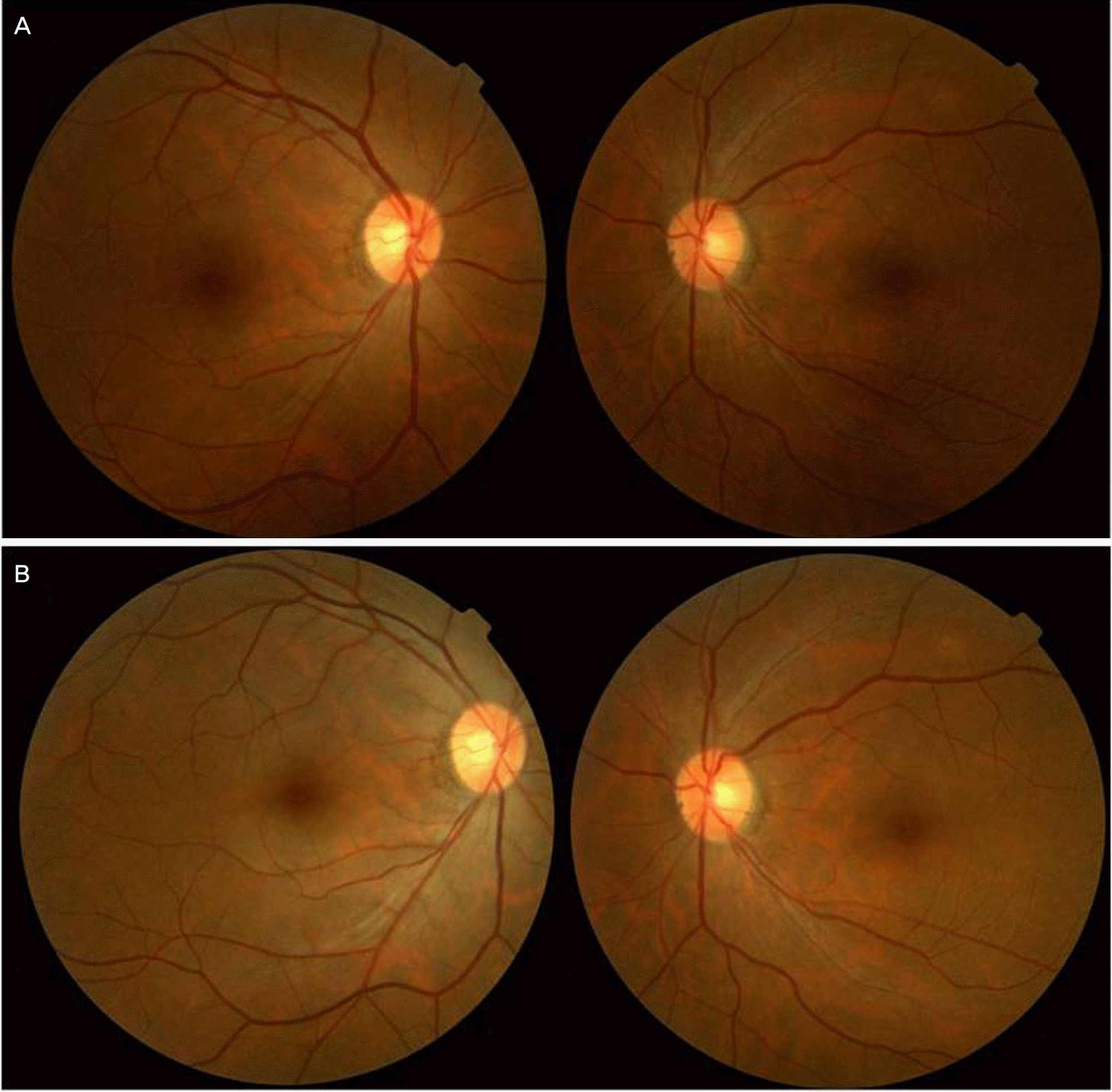
A 40-year-old man presented with amaurosis fugax of the right eye, which had occurred several times during the previous month. After coughing, the visual acuity of the right eye decreased temporarily during the first episode. Subsequently, any time a Valsalva maneuver, such as coughing, occurred, this symptom reappeared. Initially, this symptom persisted for five to ten minutes and occurred once or twice a day, but it gradually increased in frequency. The physical examination was normal, and his best corrected visual acuity was 20/20 bilaterally. Neither specific findings in the slit lamp examination nor abnormal findings in the fundus examination were detected. On fluorescein fundus angiography, no abnormal finding was observed before the symptom was triggered by a Valsalva maneuver, but after the symptom was triggered by coughing, the choroidal and retinal arterial phases were delayed. Hematological and neurological examinations, including magnetic resonance imaging, magnetic resonance angiography, and cerebral angiography, were all normal. Therefore, he was diagnosed with amaurosis fugax generated by a Valsalva maneuver.
Go to : 
References
2. Miller FW, Santoro TJ. Nifedipine in the treatment of migraine abdominal and amaurosis fugax in patients with systemic lupus erythematosus. N Engl J Med. 1984; 311:921.
3. Shaw He Jr, Osher RH, Sith JL. Amaurosis fugax associated with SC hemoglobinopathy and lupus erythematosus. Am J Ophthalmol. 1979; 87:281–5.

4. Aasen J, Kerty E, Russell D, et al. Amaurosis fugax: clinical, Doppler, and angiographic findings. Acta Neurol Scand. 1988; 77:450–5.

5. Burger SK, Saul RF, Selhorst JB, Thurston SE. Transient monocular blindness caused by vasospasm. N Engl J Med. 1991; 325:870–3.

6. Jehn A, Dettwiler BF, Fleischhauer J, et al. Exercise-induced abdominal amaurosis fugax. Arch Ophthalmol. 2002; 120:220–2.
7. Hsu HY, Chao AC, Chen YY, et al. Reflux of jugular and retrobulbar venous flow in transient monocular blindness. Ann Neurol. 2008; 63:247–53.

8. Amaurosis Fugax Study Group. Current management of amaurosis fugax. Stroke. 1990; 21:201–8.
9. North American Symptomatic Carotid Endarterectomy Trial Collaborators. Beneficial effect of carotid endarterectomy in abdominal patients with high-grade carotid stenosis. N Engl J Med. 1991; 325:870–3.
10. Winterkorn JM, Kupersmith MJ, Wirtschafter JD, Forman S. Brief abdominal: treatment of vasospastic amaurosis fugax with calcium-channel blockers. N Engl J Med. 1993; 329:396–8.
Go to : 
 | Figure 1.(A) Fundus photographs of the patient. During an asymptomatic period. (B) During acute attack of symptoms by Valsalva maneuver. There is no difference in the fundus finding observed before and after symptom. |
 | Figure 2.Fluorescein fundus angiography of the right eye. (A) During an asymptomatic period, filling of the retinal artery was completed at 20 seconds after injection. (B) filling of the retinal vein was completed at 32 seconds after injection. (C) During an attack of symptom by valsalva maneuver, filling of the retinal artery was started at 28 seconds after injection. (D) filling of the retinal artery was still incomplete at 40 seconds after injection. |


 PDF
PDF ePub
ePub Citation
 Print
Print


 XML Download