Abstract
Purpose
To report a case of macular hole following intravitreal ranibizumab injection in choroidal neovascularization associated with age-related macular degeneration.
Case summary
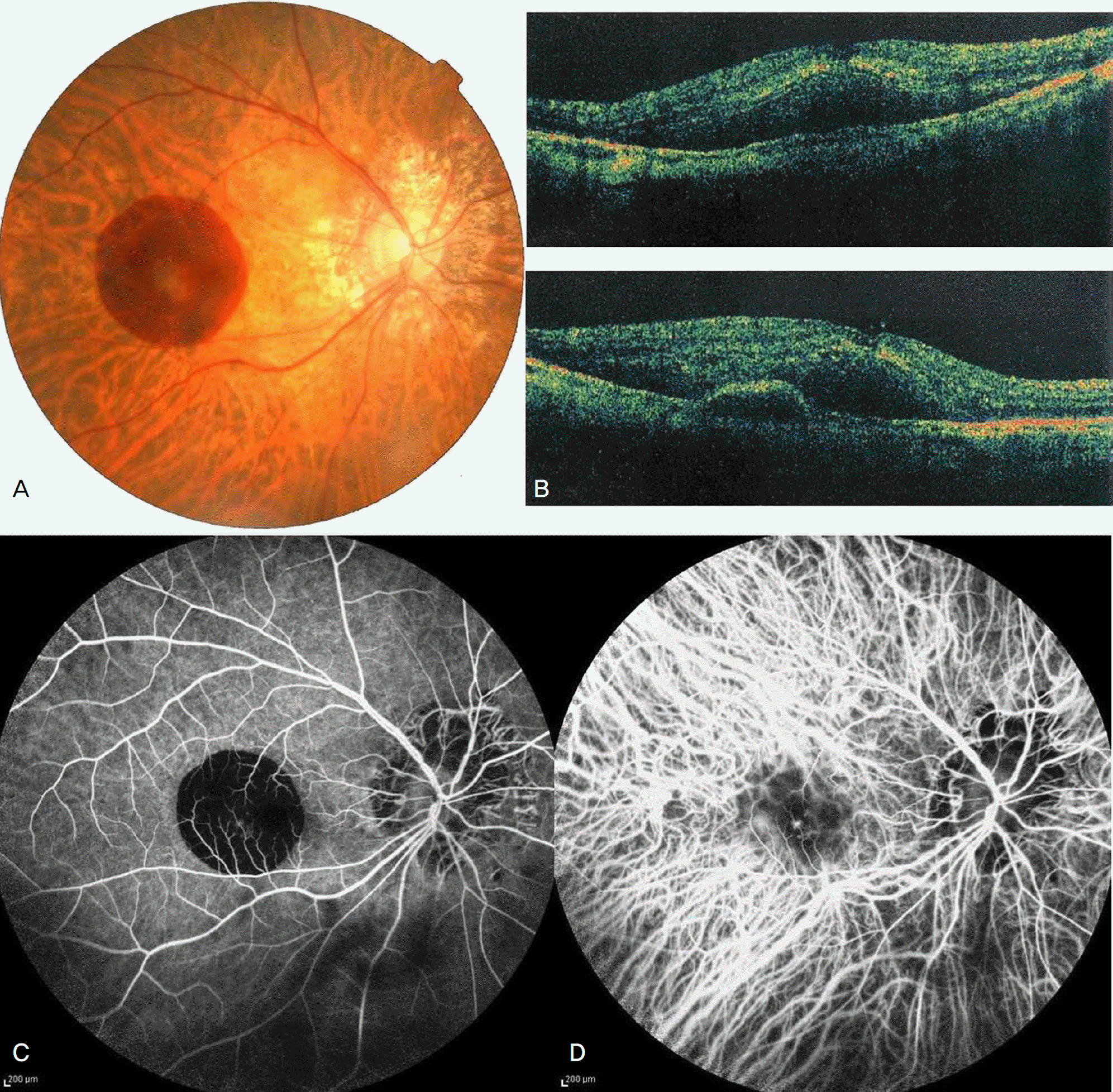
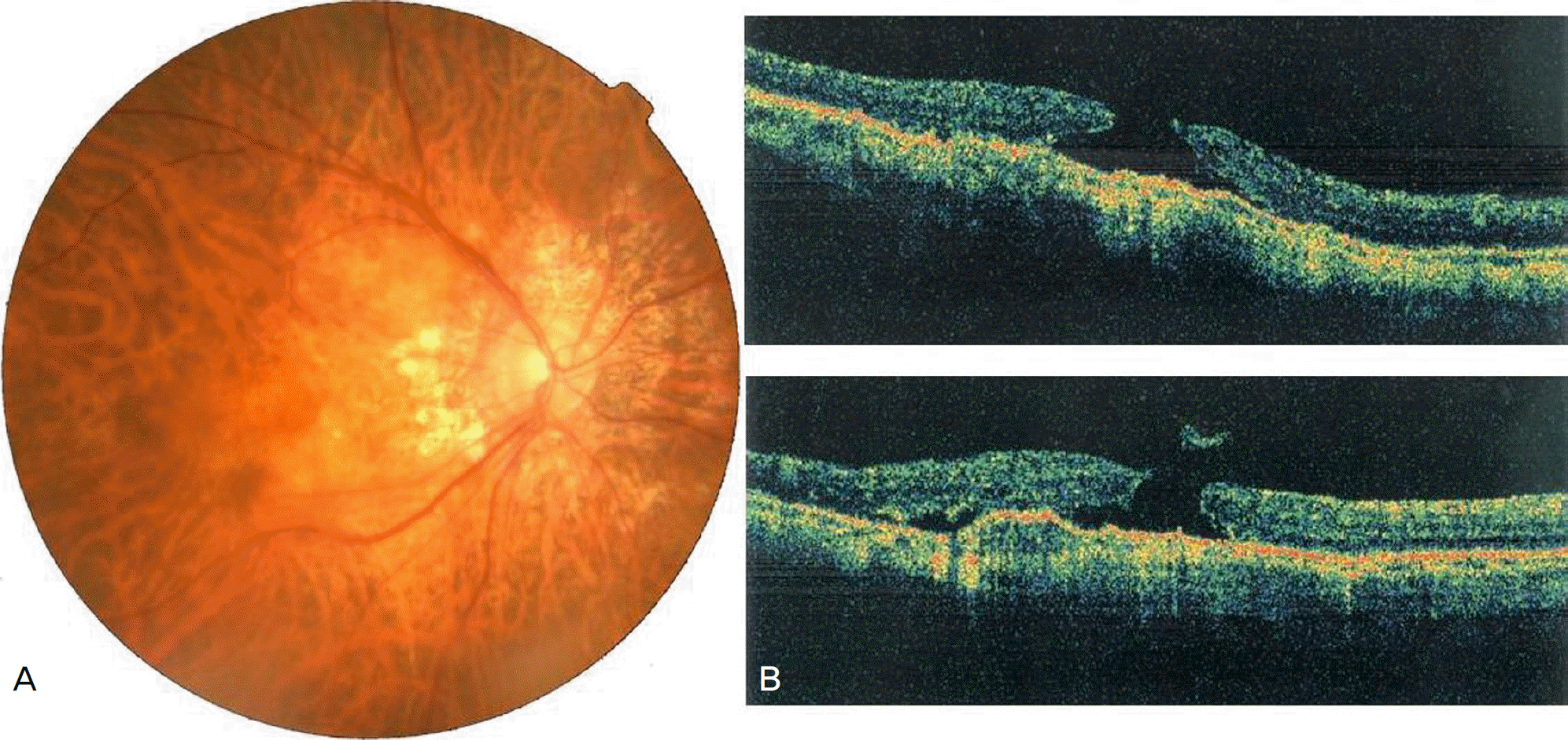
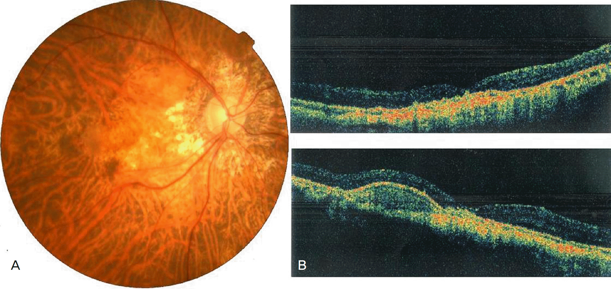
A 73-year-old man was referred for a sudden decline in vision in his right eye. The best corrected visual acuity was 0.15 in the right eye and 0.9 in the left eye. There was a choroidal neovascularization associated with subretinal hemorrhage in the right eye. Optical coherence tomogram (OCT) showed subretinal hemorrhage and pigment retinal epithelium detachment in the right eye. The patient received intravitreal ranibizumab injection. Four weeks after the initial treatment, the best corrected visual acuity of the right eye was improved to 0.3. The patient received a second intravitreal ranibizumab injection. Four weeks after the second injection, the patient presented with further decreased vision in his right eye, with a best corrected visual acuity of 0.02. Although the fundus examination was indistinct, OCT confirmed the presence of a full thickness macular hole. The patient received a third intravitreal ranibizumab injection. Four weeks after receiving the third injection, the patient underwent pars plana vitrectomy with internal limiting membrane peeling and fluid-gas exchange. Four months later, the macula hole was closed completely and visual acuity was 0.1.
Go to : 
References
1. Brown DM, Kaiser PK, Michels M, et al. Ranibizumab versus abdominal for neovascular age-related macular degeneration. N Engl J Med. 2006; 355:1432–44.
2. Rosenfeld PJ, Brown DM, Heier JS, et al. MARINA Study Group. Ranibizumab for neovascular age-related macular degeneration. N Engl J Med. 2006; 355:1419–31.
3. Laud K, Spaide RF, Freund KB, et al. Treatment of choroidal abdominal in pathologic myopia with intravitreal bevacizumab. Retina. 2006; 26:960–3.
4. Fung AE, Rosenfeld PJ, Reichel E. The International Intravitreal Bevacizumab Safety Survey: using the internet to assess drug safety worldwide. Br J Ophthalmol. 2006; 90:1344–9.

5. Shima C, Sakaguchi H, Gomi F, et al. Complications in patients after intravitreal injection of bevacizumab. Acta Ophthalmol. 2008; 86:372–6.

6. Lee GK, Lai TY, Chan WM, Lam DS. Retinal pigment epithelial tear following intravitreal ranibizumab injections for neovascular age-abdominal macular degeneration. Graefes Arch Clin Exp Ophthalmol. 2007; 245:1225–7.
7. Dhalla MS, Blinder KJ, Tewari A, et al. Retinal pigment epithelial tear following intravitreal pegaptanib sodium. Am J Ophthalmol. 2006; 141:752–4.

8. Meyer CH, Mennel S, Schmidt JC, Kroll P. Acute retinal pigment abdominal tear following intravitreal bevacizumab (Avastin) injection for occult choroidal neovascularisation secondary to age related macular degeneration. Br J Ophthalmol. 2006; 90:1207–8.
9. Shah CP, Hsu J, Garg SJ, et al. Retinal pigment epithelial tear after abdominal bevacizumab injection. Am J Ophthalmol. 2006; 142:1070–2.
10. Hoskin A, Bird AC, Sehmi K. Tears of detached retinal pigment epithelium. Br J Ophthalmol. 1981; 65:417–22.

11. Moorfield Macular Study Group. Retinal pigment epithelial abdominals in the elderly: a controlled trial of argon laser photocoagulation. Br J Ophthalmol. 1982; 66:1–16.
12. Gelisken F, Inhoffen W, Partsch M, et al. Retinal pigment epithelial tear after photodynamic therapy for choroidal neovascularization. Am J Ophthalmol. 2001; 131:518–20.

13. Fasolino G, Bamonte G, Patelli F, et al. Retinal pigment epithelial tear after intravitreal injection of triamcinolone acetonide for neovascular pigment epithelial detachment: a mechanical explanation. Acta Ophthalmol Scand. 2007; 85:575–6.

14. Chung EJ, Koh HJ. Retinal detachment with macular hole following combined photodynamic therapy and intravitreal bevacizumab injection. Korean J Ophthalmol. 2007; 21:185–7.

15. Querques G, Souied EH, Soubrane G. Macular hole following abdominal ranibizumab injection for choroidal neovascular membrane caused by age-related macular degeneration. Acta Ophthalmol. 2009; 87:235–7.
16. Bakri SJ, Kitzmann AS. Retinal pigment epithelial tear after intravitreal ranibizumab. Am J Ophthalmol. 2007; 143:505–7.

17. Konstantopoulos A, Williams CP, Newsom RS, Luff AJ. Ocular abdominal associated with intravitreal triamcinolone acetonide. Eye. 2007; 21:317–20.
18. Jo N, Mailhos C, Ju M, et al. Inhibition of platelet-derived growth abdominal B signaling enhances the efficacy of anti-vascular endothelial growth factor therapy in multiple models of ocular neovascularization. Am J Pathol. 2006; 168:2036–53.
19. Husain D, Kim I, Gauthier D, et al. Safety and efficacy of intravitreal injection of ranibizumab in combination with verteporfin PDT on abdominal choroidal neovascularization in the monkey. Arch Ophthalmol. 2005; 123:509–16.
Go to : 
 | Figure 1.(A) Fundus photograph before an intravitreal ranibizumab® injection shows a juxtafoveal grayish subretinal membrane with submacular hemorrhage. (B) Optical coherence tomography reveals a macular edema and a pigment epithelial detachment. There is a mild vitreomacular traction (arrow). (C) Fluorescein angiograph shows a blocked fluorescence due to thick submacular hemorrhage. (D) Indocyanine green angiograph demonstrates a well demarcated area of hyperfluorescence in the juxtafoveal area corresponding to the choroidal neovascularization. |


 PDF
PDF ePub
ePub Citation
 Print
Print


 XML Download