Abstract
Case summary
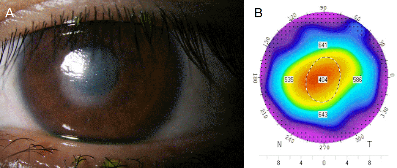
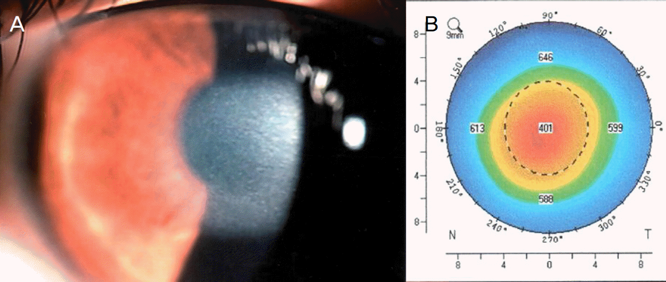
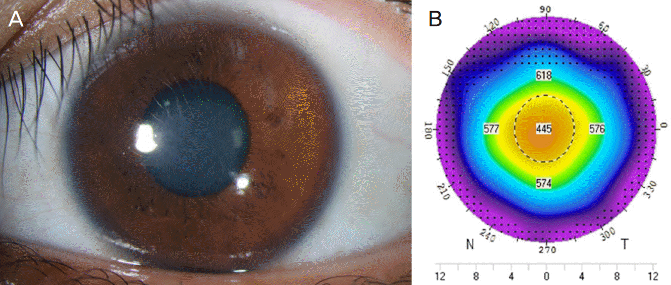
A 21-year-old female visited our clinic complaining of decreased visual acuity in the left eye. The patient had undergone LASEK surgeryten days previously. Before LASEK surgery, the central corneal thickness of the left eye was 540 μ m, and the refractive error was −2.00 Dsph=-0.75 Dcyl ×80A with an estimated ablation depth of 52.2 μ m. At the time of visit (on the tenth day after surgery), the best corrected visual acuity (BCVA) was 0.07, the central corneal thickness was 404 μ m, and the refractive error was +1.00D=+1.25D ×90. Slit-lamp biomicroscopy showed round central corneal haziness, but there were no signs of inflammation. At the third weeks after surgery, the central corneal thickness was 401 μ m and the refractive error was +11.25D=-4.00D ×145. Slit-lamp biomicroscopy showed sustained round central corneal haze. Twenty-two weeks after surgery, the central corneal thickness was 445 μ m and the refractive error was −0.75D=-1.25D ×180. The corneal opacity had disappeared.
Go to : 
References
1. Melki SA, Azar DT. LASIK Complications: etiology, management, and prevention. Surv Ophthalmol. 2001; 46:95–116.
2. Sachdev N, McGhee CN, Craig JP, et al. Epithelial defect, diffuse abdominal keratitis, and epithelial ingrowth following post-LASIK abdominal toxicity. J Cataract Refract Surg. 2002; 28:1463–6.
3. Lim GC, Lin HC, Shen SC, Lin KK. Toxic keratopathy-related corneal dehydration after laser in situ keratomileusis. J Cataract Refract Surg. 2005; 31:1656–8.

4. Mah FS, Romanowski EG, Dhaliwal DK, et al. Role of topical abdominal on the pathogenesis of diffuse lamellar keratitis in abdominal in vivo studies. J Cataract Refract Surg. 2006; 32:264–8.
5. Linebarger EJ, Hardten DR, Lindstrom RL. Diffuse lamellar keratitis: Diagnosis and management. J Cataract Refract Surg. 2000; 26:1072–7.

6. Gil-Cazorla R, Teus MA, de Benito-Llopis L, Fuentes I. Incidence of diffuse lamellar keratitis after laser in situ keratomileusis associated with the IntraLase 15 kHz femtosecond laser and Moria M2 microkeratome. J Cataract Refract Surg. 2008; 34:28–31.

7. Villarrubia A, Palacín E, Gómez del Río M, Martínez P. Description, etiology, and prevention of an outbreak of diffuse lamellar keratitis abdominal LASIK. J Refract Surg. 2007; 23:482–6.
8. Shen YC, Wang CY, Fong SC, et al. Diffuse lamellar keratitis induced by toxic chemicals after laser in situ keratomileusis. J Cataract Refract Surg. 2006; 32:1146–50.

9. Brouzas D, Droutsas D, Charakidas A, et al. Severe toxic effect of Methylene blue 1% on iris epithelium and corneal endothelium. Cornea. 2006; 25:470–1.

10. Fraenkel GE, Cohen PR, Sutton GL, et al. Central focal interface opacity after laser in situ keratomileusis. J Refract Surg. 1998; 14:571–6.

11. Parolini B, Marcon G, Panozzo GA. Central necrotic lamellar abdominal after laser in situ keratomileusis. J Refract Surg. 2001; 17:110–2.
12. Sonmez B, Maloney RK. Central Toxic Keratopathy: Description of a syndrome in laser refractive surgery. Am J Ophthalmol. 2007; 143:420–7.

13. Fantes FE, Hanna KD, Waring Go 3rd, et al. Wound healing after abdominal laser keratomileusis (photorefractive keratectomy) in monkeys. Arch Ophthalmol. 1990; 108:665–75.
14. Smith RJ, Maloney RK. Diffuse lamellar keratitis. A new syndrome in lamellar refractive surgery. Ophthalmology. 1998; 105:1721–6.

15. Moon SW, Kim YH, Lee SC, et al. Bilateral peripheral infiltrative abdominal after LASIK. Korean J Ophthalmol. 2007; 21:172–4.
16. Choi J, Wee WR, Lee JH, Kim MK. High intraocular pressure-induced delayed diffuse lamellar keratitis after laser in situ abdominal(LASIK). J Korean Ophthalmol Soc. 2006; 47:1678–85.
17. Ku M, Shyn KH. 2006 survey for KSCRS members – Current trends in refractive surgery in Korea. J Korean Ophthalmol Soc. 2009; 50:182–8.
Go to : 
 | Figure 1.Anterior segment photograph and corneal thickness map of Pentacam in left eye of a 21 years old female who underwent LASEK 10 days ago. (A) Anterior segment photograph showed central subepithelial circular corneal haziness. (B) The thickness of central cornea was noted 404 μm in Pentacam map. |


 PDF
PDF ePub
ePub Citation
 Print
Print


 XML Download