

 PDF
PDF ePub
ePub Citation
 Print
Print


Abstract
Purpose
The effect of 0.2% cyclosporin A (CsA) as an adjuvant therapy after glaucoma-filtering surgery was the focus of this study.
Methods
A posterior lip sclerotomy was performed in 16 eyes of 8 rabbits, and 0.2% CsA was administered into the right eyes. The left eyes served as controls. The intraocular pressure (IOP) was measured 1, 3, 5, 7, 14, and 28 days after surgery. Hematoxylin-eosin (HE) and anti-bromodeoxyuridine (BrdU) immunocytochemical staining were performed at 1, 2, 4, and 8 weeks.
Results
The IOP at 7 and 14 days after surgery was lower in the 0.2% CsA group and statistically significant (P=0.047, P=0.48; respectively). HE staining did not show any difference between experimental and control eyes, but anti-BrdU staining showed a lower number of positive cells in the experimental eyes at 1 week. The fibroblast proliferation rate was significantly lower 1 week after surgery in the 0.2% CsA group (P=0.003).
Go to : 
References
1. Lu DW, Chang CJ, Chiang CH, et al. Wound modulation after abdominal by different formulations of antimetabolites in rabbits. J Ocul Pharmacol Ther. 2000; 16:529–38.
2. Nuzzi R, Cerruti A, Finazzo C. Cyclosporine C: a study of wound-healing modulation after trabeculectomy in rabbit. Acta Ophthalmol Scand Suppl. 1998; 227:48–9.

3. Turaçli ME, Gündüz K, Aktan G, Sencer H. Topical cyclosporine as a possible new antimetabolite in trabeculectomy. Ophthalmic Surg Lasers. 1996; 27:438–44.
4. Broadway DC, Chang LP. Trabeculectomy, risk factors for failure and the preoperative state of the conjunctiva. J Glaucoma. 2001; 10:237–49.
5. Baek YW, Moon JI, Park CK. Effects of Taxol and Mitomycin C on Morphologic Alteration of Cultured Rabbit Subconjunctival Fibroblasts. J Korea Ophthalmol Soc. 2003; 44:202–7.
6. Shouman AA, Helal A, Marzouk MA, Zaki EM. Methylcellulose, a healing inhibitor factor in an animal model of trabeculectomy. Invest Ophthalmol Vis Sci. 2006; 47:2515–9.
7. Lama PJ, Fechtner RD. Antifibrotics and wound healing in glaucoma surgery. Surv Ophthalmol. 2003; 48:314–46.
8. Park KH, Kim DM. The Effects of Topical Cyclosporin A on Glaucoma Drainage Implant Surgery in Rabbits. J Korean Ophthalmol Soc. 1995; 36:307–15.
9. Garweg JG, Wegmann-Burns M, Goldblum D. Effects of abdominal, mitomycin C, azathioprine and cyclosporin A on human retinal pigmented epithelial, corneal endothelial and conjunctival cell lines. Graefes Arch Clin Exp Ophthalmol. 2006; 244:382–9.
10. Bagci G, Yucel I, Duranoglu Y. The effect of cyclosporin A on abdominal rabbit subconjunctival fibroblast proliferation. Ophthalmologica. 1999; 213:114–9.
11. Cristofanilli M, Pescosolido N, Risuleo G, Scarsella G. A murine cell culture model for post-trabeculectomy anfibrotic treatment: Induction of apoptosis by Cyclosporin. Acta Ophthalmol Scand. 2001; 79:309–12.
11. Akafo SK, Goulstine DB, Rosenthal AR. abdominal post abdominal intraocular pressures. Acta Ophthalmol (Copenh). 1992; 70:312–6.
12. Mills KB. Trabeculectomy: a retrospective long-term follow-up of 444 cases. Br J Ophthalmol. 1981; 65:790–5.
13. Zaidi AA. Trabeculectomy: a review and 4-year follow-up. Br J Ophthalmol. 1980; 64:436–9.
14. Jerndal T, Lundstrom M: 330 trabeculectomies. A long time study (3–5 1/2 years). Acta Ophthalmol (Copenh). 1980; 58:947–56.
15. Ahn JE, Lee YG, Hong YJ. abdominal Follow-up after Trabeculectomy with Mitomycin c. J Korea Ophthalmol Soc. 1998; 39:993–1001.
16. Watson PG, Jakeman C, Ozturk M, et al. The complications of abdominal (a 20-year follow-up). Eye. 1990; 4:425–38.
17. Lattanzio FA Jr, Crouch ER Jr, Mitrev PV, et al. Cyclosporin as an abdominal to glaucoma filtration surgery. J Glaucoma. 2005; 14:441–7.
18. Seetner A, Morin JD. Healing of trabeculectomies in rabbits. Can J Ophthalmol. 1979; 14:121–5.
19. Miller MH, Joseph NH, Ennis KW, et al. An animal model of filtration surgery. Trans Ophthalmol Soc U K. 1985; 104:893–7.
20. Jampel HD, McGuigan LJ, Dunkelberger GR, et al. Cellular abdominal after experimental glaucoma filtering surgery. Arch Ophthalmol. 1988; 106:89–94.
Go to :
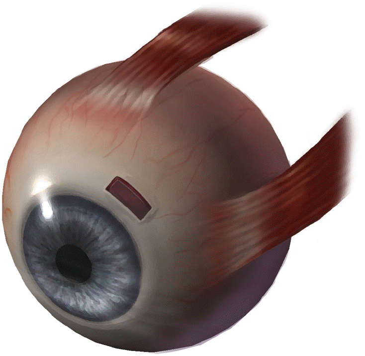 | Figure 1.Glaucoma filtering surgery in a rabbit eye. A fornix-based conjunctival incision was undertaken in the upper-nasal part of the eye (between superior rectus muscle and medial rectus muscle). The anterior chamber was then entered through the filtering site, and a 3×1 mm block of scleral tissue and trabecular meshwork was excised. Peripheral iridectomy was not performed. |
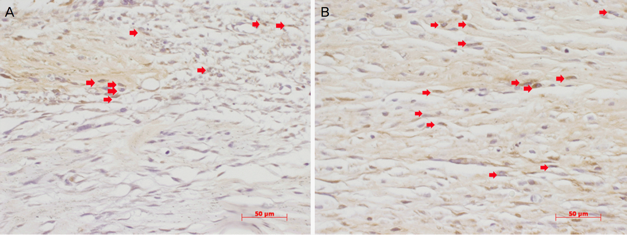 | Figure 2.High magnification photographs of immunocytochemically stained sections (×400). The right photographs are eyes instilled with 0.2% CsA (A). The left photographs are control eyes (B). Many bromodeoxyuridine (BrdU)-positive cells (red arrow) are shown 1 week after surgery (A and B). Fewer BrdU-positive cells existed in experimental eyes (A). The scale bars indicate 50 μm. |
Table 1.
Mean intraocular pressure between two groups (Mean± SD)
Table 2.
Proliferation rates of fibroblasts after BrdU injection in experimental and control eyes (Means± SDs*)
| Days |
Proliferation rate (%) |
P value | |
|---|---|---|---|
| with 0.2% CsA | without 0.2% CsA | ||
| 1 wk | 20.17±1.37 | 30.83±0.22 | .003 |
| 2 wk | 6.92±1.24 | 9.47±1.96 | .138 |
| 1 mo | 2.90±0.60 | 1.96±0.48 | .138 |
| 2 mo | 1.32±0.04 | 1.47±0.32 | 1.000 |
 XML Download