Abstract
Purpose
To evaluate the clinical features of infectious keratitis in the western coastal area of Chungcheongnam-do, Korea.
Methods
We performed bacterial and fungal cultures in patients with findings of infectious keratitis. Any correlations between the culture results and the patients' place of residence, occupation, types of ocular trauma, contact lens wear, previous ocular disease, duration of treatment for complete recovery, time between the onset of symptom and beginning of treatment were evaluated. In addition, we assessed the antibiotic susceptibilities of the cultured organisms.
Results
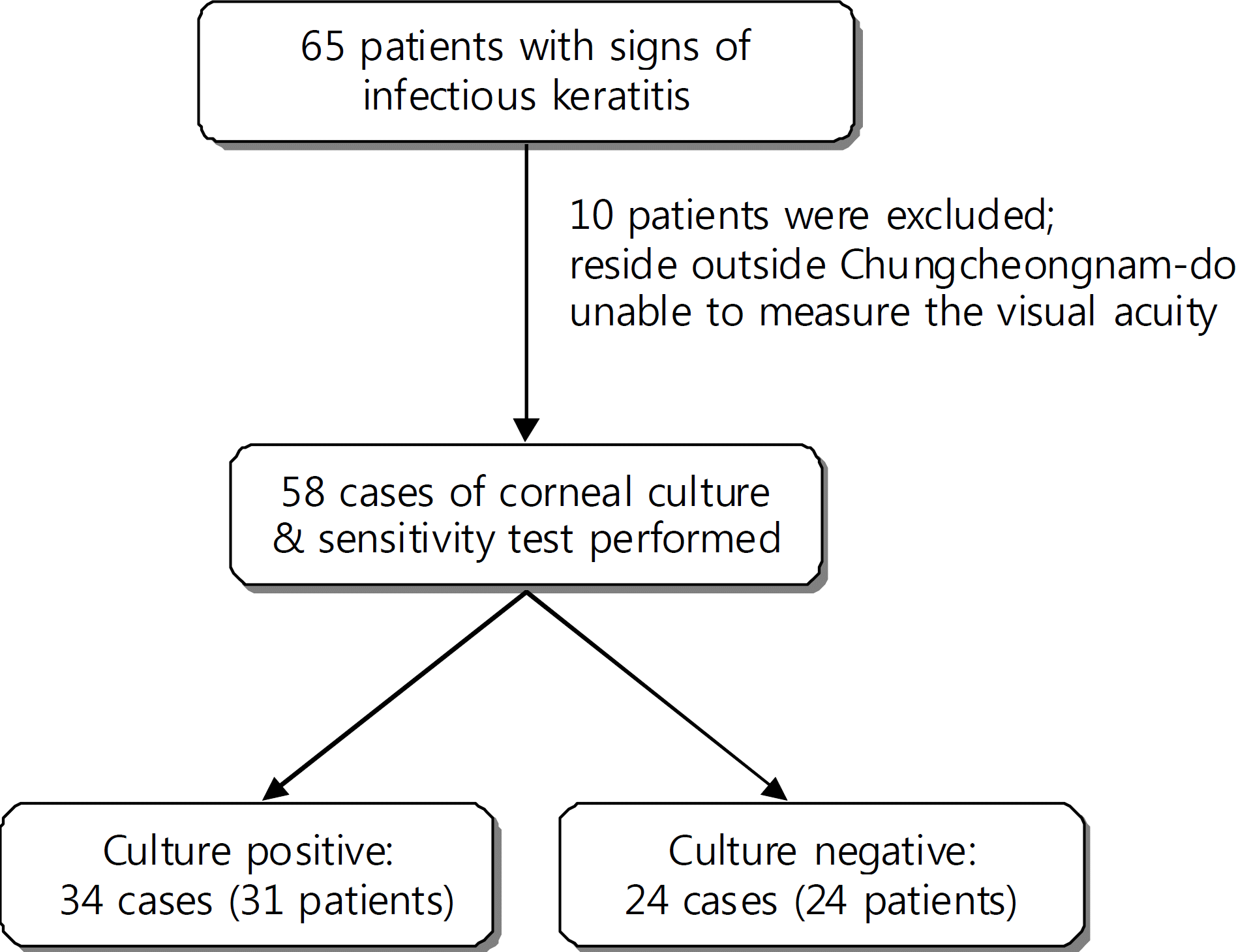
We detected 34 (58.62%) among 58 cultures performed in 55 patients that were positive for organisms; 24 for Gram-positive bacteria, 17 for Gram-negative bacteria, 3 for fungi and 9 for polymicrobial infections. Coagulase-negative staphylococci (CNS) was the most frequent infection. The culture positivity rate was significantly higher (P=0.047) in patients with history of previous ocular disease but no correlations were detected with place of residence, type of ocular trauma or the timing of culture. The average treatment period was 33.95±30.59 days, which extended as the lesion size increased (P=0.003).
Go to : 
References
1. Huang AJ, Wichiensin P, Yang MC. Bacterial keratitis. Krachmer JH, Mannis MJ, Holland EJ, editors. Cornea. 2nd ed.Philadelphia: Elsevier;2005. 1:chap. 81.
2. Asbell P, Stenson S. Ulcerative keratitis. Survey of 30 years' laboratory experience. Arch Ophthalmol. 1982; 100:77–80.
3. Hahn YH, Hahn TW, Choi SH, et al. Epidemiology of infectious abdominal(I): A abdominal study. J Korean Ophthalmol Soc. 1998; 39:1633–51.
4. Hahn YH, Hahn TW, Tchah HW, et al. Epidemiology of infectious keratitis(II): A abdominal study. J Korean Ophthalmol Soc. 2001; 42:247–65.
5. Jones DB. Initial therapy of suspected microbial corneal ulcers. II. Specific antibiotic therapy based on corneal smears. Surv Ophthalmol. 1979; 24:105–16.
6. Green M, Apel A, Stapleton F. Risk factors and causative organisms in microbial keratitis. Cornea. 2008; 27:22–7.

7. Wright TM, Afshari NA. Microbial keratitis following corneal transplantation. Am J Ophthalmol. 2006; 142:1061–2.

8. Ly CN, Pham JN, Badenoch PR, et al. Bacteria commonly isolated from keratitis specimens retain antibiotic susceptibility to abdominal and gentamicin plus cephalothin. Clin Experiment Ophthalmol. 2006; 34:44–50.
9. Benson WH, Lanier JD. Current diagnosis and treatment of corneal ulcers. Curr Opin Ophthalmol. 1998; 9:45–9.

10. Alfonso E, Crider J. Ophthalmic infections and their anti-infective challenges. Surv Ophthalmol. 2005; 50:S1–6.

11. Smitha S, Lalitha P, Prajna VN, Srinivasan M. Susceptibility trends of pseudomonas species from corneal ulcers. Indian J Med Microbiol. 2005; 23:168–71.

Go to : 
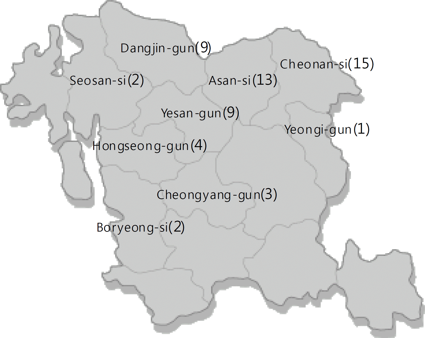
 | Figure 2.Distribution of patients with infectious keratitis in Chungcheongnam-do. The number of patients are marked in parenthesis. |
Table 1.
Causative organisms of microbial keratitis
| Organisms |
Culture positive keratitis (n=34) |
|||
|---|---|---|---|---|
| No. of isolates | Prevalence (%)* | |||
| Bacteria | 41 | 120.59 | ||
| Gram(+) cocci | 18 | 52.94 | ||
| Coagulase negative staphylococcus (CNS) | 11 | 32.35 | ||
| Staphylococcus aureus | 1 | 2.94 | ||
| Streptococcus pneumoniae | 3 | 8.82 | ||
| Viridans group streptococcus | 3 | 8.82 | ||
| Gram(+) rod | 6 | 17.65 | ||
| Coryneform gram-positive rods | 5 | 14.71 | ||
| Bacillus spp. | 1 | 2.94 | ||
| Gram(−) cocci | 0 | 0 | ||
| Gram(−) rod | 17 | 50.00 | ||
| Pseudomonas aeruginosa | 5 | 14.71 | ||
| Pseudomonas pseudoalcaligenes | 1 | 2.94 | ||
| Acinetobacter spp. | 1 | 2.94 | ||
| Serratia spp. | 3 | 8.82 | ||
| Enterobacter aerogenes | 1 | 2.94 | ||
| Morganella morganii | 1 | 2.94 | ||
| Stenotrophomonas maltophilia | 1 | 2.94 | ||
| Achromobacter xylosoxidans | 1 | 2.94 | ||
| Burkholderia cepacia | 1 | 2.94 | ||
| Citrobacter freundii | 1 | 2.94 | ||
| Ochrobactum anthropi | 1 | 2.94 | ||
| Fungus | 3 | 8.82 | ||
| Filamentary | 1 | 2.94 | ||
| Trichothecium spp. | 1 | 2.94 | ||
| Yeast | 2 | 5.88 | ||
| Candida | 2 | 5.88 | ||
| Total | 44 | 129.41 | ||
Table 2.
Comparisons between culture positive and negative groups
Table 3.
Percentage of antibiotics resistance in Gram positive and negative bacteria


 PDF
PDF ePub
ePub Citation
 Print
Print


 XML Download