Abstract
Purpose
To estimate the utility of the enhanced ectasia display mode of Pentacam in discriminating keratoconus and keratoconus suspect from normal cornea.
Methods
Corneal topography was measured using the Pentacam in keratoconus, keratoconus suspect and a normal control group. A best-fit sphere (BFS) and an enhanced best-fit sphere (EBFS) were used as reference surfaces for corneal elevation measurements, and measured values from both the anterior and posterior surfaces were compared among the three groups. Receiver operating characteristic (ROC) curves were used to identify the optimal posterior corneal elevation cutoff points for maximal sensitivity and specificity in discriminating keratoconus and keratoconus suspect from the normal control group.
Results
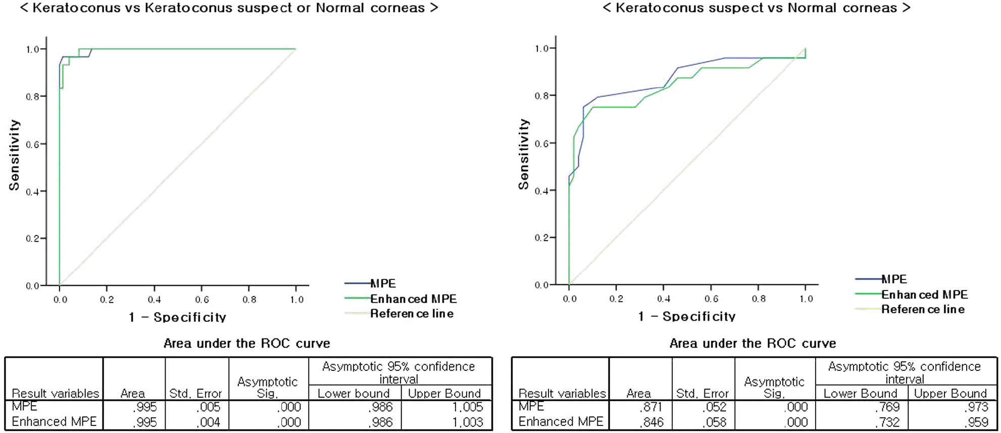
Mean anterior and posterior corneal elevation were statistically higher in keratoconus than in keratoconus suspect and normal corneas. The optimal cutoff point of posterior elevation was 23 μ m for the keratoconus group, and this value was associated with a sensitivity and a specificity of 96.7% and 98.6%, respectively for keratoconus. The optimal cutoff point of enhanced posterior elevation was 43 μ m for the keratoconus group, and this value was associated with a sensitivity and a specificity of 96.7% and 95.5%, respectively.
Go to : 
References
1. Krachmer JH, Feder RS, Belin MW. Keratoconus and related noninflammatory corneal thinning disorders. Surv Ophthalmol. 1984; 28:293–322.

3. Lee LR, Hirst LW, Readshaw G. Clinical detection of unilateral keratoconus. Aust N Z J Ophthalmol. 1995; 23:129–33.

4. Rabinowitz YS, Nesburn AB, McDonnell PJ. Videokeratography of the fellow eye in unilateral keratoconus. Ophthalmology. 1993; 100:181–6.

5. Rabinowitz YS. Videokeratographic indices to aid in screening for keratoconus. J Refract Surg. 1995; 11:371–9.

6. Meada N, Klyce SD, Smolek MK, Thompson HW. Automated abdominal screening with corneal topography analysis. Invest Ophthalmol Vis Sci. 1994; 35:2749–57.
7. Mamalis N, Montgomry S, Anderson C, Miller C. Radial keratotomy in a patient with keratoconus. Refract Corneal Surg. 1991; 7:374–6.

8. Ellis W. Radial keratotomy in a patient with keratoconus. J Cataract Refract Surg. 1992; 18:406–9.

9. Seiler T, Quurke AW. Iatrogenic keratectasia after LASIK in a case of forme fruste keratoconus. J Cataract Refract Surg. 1998; 24:1007–9.

10. Binder PS, Lindstrom RL, Stulting RD, et al. Keratoconus and corneal ectasia after LASIK. J Cataract Refract Surg. 2005; 31:2035–8.

11. Rao SN, Raviv T, Majmudar PA, Epstein RJ. Role of Orbscan II in screening keratoconus suspects before refractive corneal surgery. Ophthalmology. 2002; 109:1642–6.

12. Choi HJ, Kim MK, Lee JL. Diagnostic criteria for keratoconus using Orbscan II slit scanning topography/pachymetry system. J Korean Ophthalmol Soc. 2004; 45:928–35.
13. Lee SU, Lee CH, Lee JE, Lee JS. Corneal Topographic Study Using Orbscan II between Keratoconus and Keratoconus Suspect. J Korean Ophthalmol Soc. 2007; 48:1599–606.

14. de Sanctis U, Loiacono C, Richiardi L, et al. Sensitivity and specificity of posterior corneal elevation measured by Pentacam in discriminating keratoconus/subclinical keratoconus. Ophthalmology. 2008; 115:1534–9.

15. Miháltz K, Kovács I, Takács A, Nagy ZZ. Evaluation of Keratometric, Pachymetric, and Elevation Parameters of Keratoconic Corneas With Pentacam. Cornea. 2009; 28:976–80.

16. Belin MW, Khachikian SS, Ambrósio R Jr, Salomão M. Keratoconus/ecatasia detection with the oculus pentacam: Belin/Ambrósio abdominal ectasia display. Highlights of Ophthalmology. 2007; 35:5–12.
17. Zadnik K, Barr JT, Edrington TB, et al. Baseline findings in the Collaborative Longitudinal Evaluation of Keratoconus (CLEK) study. Invest Ophthalmol Vis Sci. 1998; 39:2537–46.
Go to : 
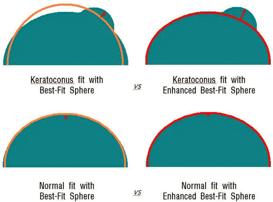
 | Figure 1.Schematic comparing how removal of the exclusion zone will affect the Best-fit sphere. The elevation difference between using a standard best-fit sphere and the enhanced best-fit sphere will be significant for a conical cornea, while the difference is minimal in a normal cornea. |
 | Figure 2.Receiver operator characteristic (ROC) curves for keratoconus versus keratoconus suspect or normal corneas (Left). ROC curves for keratoconus suspect versus normal corneas (Right). MPE=maximal posterior elevation on the thinnest point. |
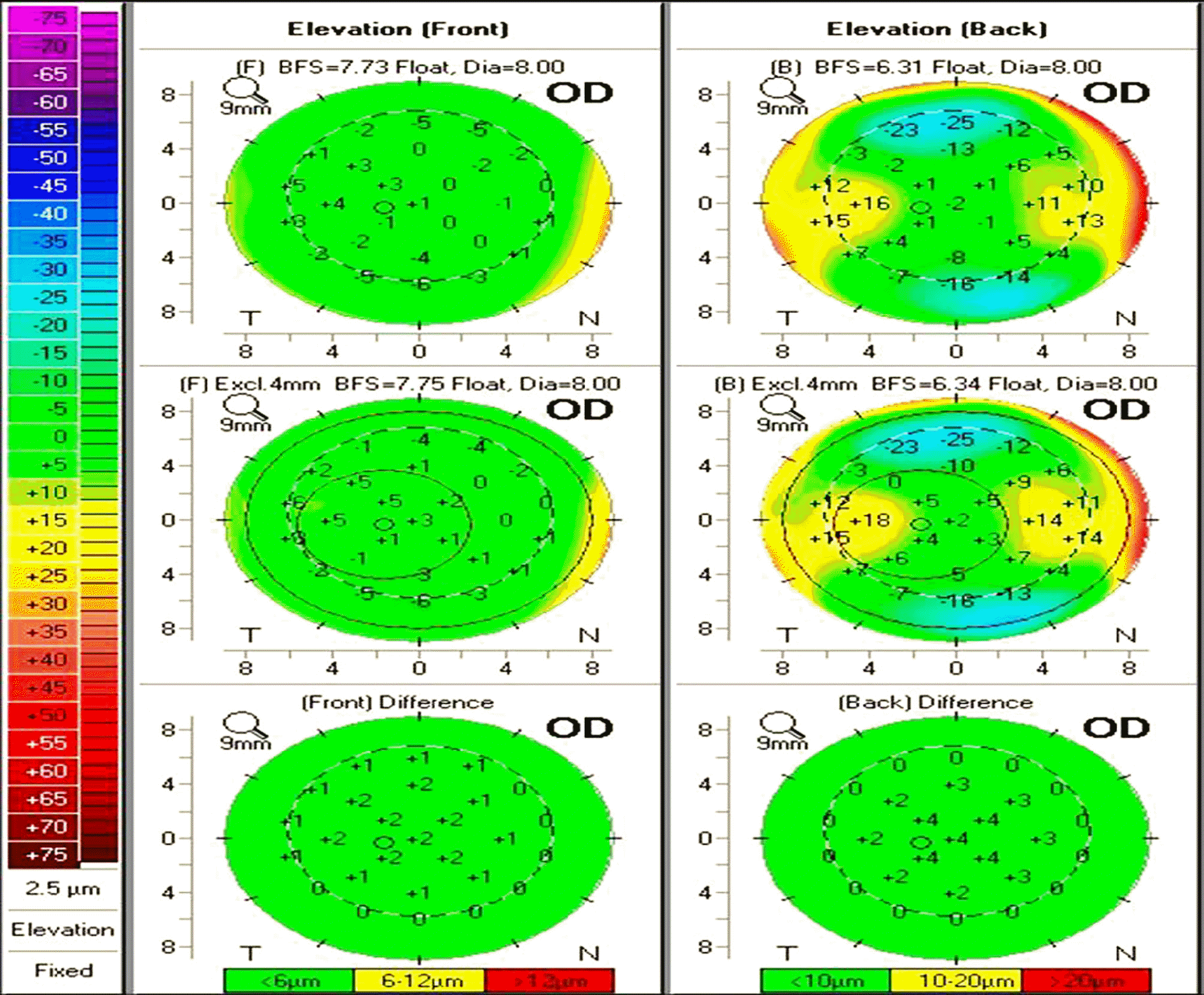
 | Figure 3.Enhanced ectasia display with elevation data presented in normal group. There is little change in relative elevation, or the appearance of the elevation map when comparing the best-fit sphere to the enhanced best-fit sphere. Corneal surface dose not show much change from the baseline to the exclusion elevation map (the map is all green). |
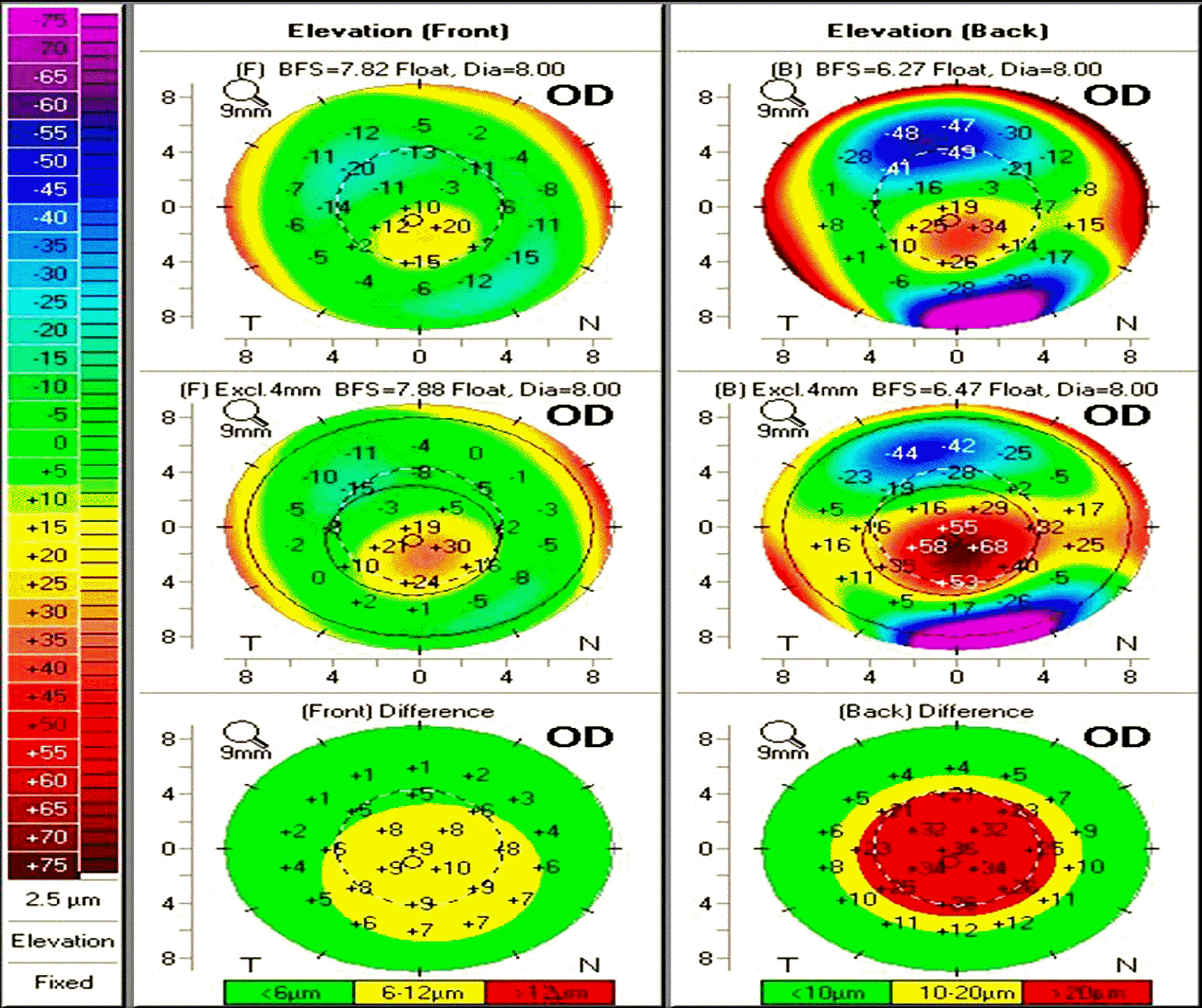
 | Figure 4.Enhanced ectasia display with elevation data presented in keratoconus group. There is significant change in relative elevation, and the appearance of the elevation map when comparing the best-fit sphere to the enhanced best-fit sphere. Corneal posterior surface show much change from the baseline to the exclusion elevation map (central area of red). |
Table 1.
Demographic data of subjects
Table 2.
Comparison of the mean value of the anterior cornea indices
| Type | Normal | Keratoconus suspect | Keratoconus |
|---|---|---|---|
| BFS* radius (mm) | 8.02±0.28 | 7.76±0.23‡ | 7.43±0.43‡§ |
| MAE† (μm) | 2.48±1.52 | 6.33±3.94 | 29.90±16.80‡§ |
| Enhanced BFS radius (mm) | 8.06±0.28 | 7.79±0.24‡ | 7.49±0.39‡§ |
| Enhanced MAE (μm) | 5.78±2.74 | 10.38±5.06 | 46.40±32.07‡§ |
| BFS change (mm) | 0.03±0.02 | 0.03±0.03 | 0.06±0.15 |
| Elevation change (μm) | 3.30±1.78 | 4.04±3.83 | 16.50±17.22‡§ |
Table 3.
Comparison of the mean value of the posterior cornea indices
| Type | Normal | Keratoconus suspect | Keratoconus |
|---|---|---|---|
| BFS* radius (mm) | 6.52±0.24 | 6.31±0.24‡ | 6.05±0.47‡§ |
| MPE† (μm) | 3.86±3.08 | 12.13±8.26 | 69.00±33.56‡§ |
| Enhanced BFS radius (mm) | 6.60±0.26 | 6.41±0.24‡ | 6.33±0.35‡ |
| Enhanced MPE (μm) | 14.16±6.68 | 28.29±13.09 | 125.13±67.95‡§ |
| BFS change (mm) | 0.07±0.10 | 0.10±0.05 | 0.28±0.20‡§ |
| Elevation change (μm) | 10.30±4.63 | 16.17±6.81 | 56.13±37.19‡§ |
Table 4.
Specificity, sensitivity identified by cutoff points of posterior corneal elevation selected a priori
| Cutoff point (μm) |
Standard BFS* |
Cutoff point (μm) |
Enhanced BFS |
||
|---|---|---|---|---|---|
| Specificity (%) | Specificity (%) | Sensitivity (%) | Specificity (%) | ||
| KC† vs KS‡ or NC§ | |||||
| 13.0 | 100 | 85.1 | 34.5 | 100 | 90.5 |
| 15.0 | 100 | 86.5 | 36.5 | 100 | 91.9 |
| 17.5 | 96.7 | 89.2 | 37.5 | 96.7 | 91.9 |
| 19.5 | 96.7 | 91.9 | 39.5 | 96.7 | 93.2 |
| 21.0 | 96.7 | 95.9 | 41.5 | 96.7 | 94.6 |
| 23.0 | 96.7 | 98.6 | 43.0 | 96.7 | 95.9 |
| 28.0 | 93.3 | 100 | 44.5 | 93.3 | 95.9 |
| KS vs NC | |||||
| 2.5 | 95.8 | 34.0 | 8.5 | 95.8 | 18.0 |
| 3.5 | 91.7 | 54.0 | 12.5 | 91.7 | 44.0 |
| 4.5 | 83.3 | 60.0 | 16.5 | 79.2 | 68.0 |
| 5.5 | 83.3 | 62.0 | 20.5 | 75.0 | 80.0 |
| 6.5 | 79.2 | 88.0 | 23.5 | 75.0 | 90.0 |
| 7.5 | 75.0 | 94.0 | 25.5 | 62.5 | 98.0 |
| 9.0 | 62.5 | 94.0 | 31.0 | 41.7 | 100 |
| KC vs NC | |||||
| 8.5 | 100 | 94.0 | 22.0 | 100 | 86.0 |
| 11.0 | 100 | 96.0 | 23.5 | 100 | 90.0 |
| 14.0 | 100 | 100 | 24.5 | 100 | 96.0 |
| 20.0 | 96.7 | 100 | 27.5 | 100 | 98.0 |
| 28.0 | 93.3 | 100 | 33.5 | 100 | 100 |
| 33.0 | 90.0 | 100 | 40.5 | 96.7 | 100 |
| 35.0 | 83.3 | 100 | 47.0 | 93.3 | 100 |


 PDF
PDF ePub
ePub Citation
 Print
Print


 XML Download