

 PDF
PDF ePub
ePub Citation
 Print
Print


Abstract
Purpose
To evaluate two surgical methods-amniotic membrane transplantation (AMT) and split-conjunctival grafts (SCG)-for double-head pterygium, with regard to the postoperative outcome and recurrence rate.
Methods
In a total of 16 eyes (14 patients), 7 eyes (6 patients) receiving amniotic membrane transplantation and 9 eyes (8 patients) receiving split-conjunctival grafts were compared to evaluate recurrence and complications.
Results
Within the amniotic membrane transplantation group, two eyes (29%) had corneal recurrence, and 3 eyes (43%) had conjunctival recurrence. The mean follow-up period was 21.9±3.5 months, and all recurrences were on the nasal side. The average period preceding the corneal recurrences was 7.2±1.8 months. Within the split-conjunctival grafts group, the mean fol-low-up was 13.6±2.1 months, and neither the corneal nor conjunctival recurrences were observed. In addition, the eyes of this group were more aesthetically stable, with only one eye exhibiting pseudo-pterygium at the donor site.
Conclusions
In cases of double-head pterygium without contraindication of conjunctival autograft, the split-conjunctival grafts produced fewer recurrences and showed enhanced cosmetic results, as compared to the amniotic membrane transplantation, indicating that the split-conjunctival grafts is the superior choice over amniotic membrane transplantation.
Go to : 
References
1. Kenyon KR, Wagoner MD, Hettinger ME. Conjunctival autograft transplantation for advanced and recurrent pterygium. Ophthalmology. 1985; 92:1461–70.

2. Prabhasawat P, Barton K, Burkett G, Tseng SC. Comparison of abdominal autografts, amniotic membrane grafts, and primary closure for pterygium excision. Ophthalmology. 1997; 104:974–85.
3. Sanchez-Thorin JC, Rocha G, Yelin JB. Meta-analysis on the abdominal rates after bare sclera resection with and without mitomycin C use and conjunctival autograft placement in surgery for primary pterygium. Br J Ophthalmol. 1998; 82:661–5.
4. Ma DH, See LC, Liau SB, Tsai RJ. Aminotic membrane graft for abdominal pterygium: comparision with conjunctival autograft and topical mitomycin C treatment. Br J Ophthalmol. 2000; 84:973–8.
5. Hirst LW. Prospective study of primary pterygium surgery using pterygium extended removal followed by extended conjunctival transplantation. Ophthalmology. 2008; 115:1663–72.
6. Solomon A, Pires RT, Tseng SC, et al. Amniotic membrane abdominal after extensive removal of primary and recurrent pterygia. Ophthalmology. 2001; 108:449–60.
7. Avisar R, Snir M, Weinberger D. Outcome of double-head pterygium surgery. Cornea. 2003; 22:501–3.
8. Maheshwari S. Split-conjunctival grafts for double-head pterygium. Indian J Ophthalmol. 2005; 53:53–5.
9. Wu WK, Wong VW, Chi SC, Lam DS. Surgical management of dou-ble-head pterygium by using a novel technique: conjunctival rotational autograft combined with conjunctival autograft. Cornea. 2007; 26:1056–9.
10. Rubinfeld RS, Pfister RR, Stein RM, et al. Serious complications of topical mitomycin C after pterygium surgery. Ophthalmology. 1992; 99:1647–54.
11. Safianik B, Ben-Zion I, Garzozi HJ. Serious corneoscleral complications after pterygium excision with mitomycin C. Br J Ophthalmol. 2002; 86:357–8.
12. Ti SE, Tan DT. Tectonic corneal lamellar grafting for severe scleral melting after pterygium surgery. Ophthalmology. 2003; 110:1126–36.
13. Wan Norliza WM, Raihan IS, Azwa JA, Ibrahim M. Scleral melting 16 years after pterygium excision with topical Mitomycin C adjuvant therapy. Cont Lens Anterior Eye. 2006; 29:165–7.
14. Jang JH, Choi TH. The effect of amniotic membrane transplantation for pterygium excision. J Korean Ophthalmol Soc. 2005; 46:597–604.
15. Katircioglu YA, Altiparmak UE, Duman S. Comparison of three methods for the treatment of pterygium: amniotic membrane graft, conjunctival autograft and conjunctival autograft plus mitomycin C. Orbit. 2007; 26:5–13.
16. Kim YI, Paek KU, Park HS. Ocular surface reconstruction with abdominal membrane transplantation in pterygium. J Korean Ophthalmol Soc. 1999; 40:1178–83.
17. Kim MJ, Tchah HW. Treatment of pterygium with amniotic abdominal transplantation. J Korean Ophthalmol Soc. 1998; 39:59–64.
18. Tananuvat N, Martin T. The results of amniotic membrane transplantation for primary pterygium compared with conjunctival autograft. Cornea. 2004; 23:458–63.
19. Hirst LW. The treatment of pterygium. Surv Ophthalmol. 2003; 48:145–80.
20. John T. Pterygium excision and conjunctival mini-autograft: Preliminary report. Eye. 2001; 15:292–6.
21. Dupps WJ Jr, Jeng BH, Meisler DM. Narrow-strip conjunctival abdominal for treatment of pterygium. Ophthalmology. 2007; 114:227–31.
22. Coroneo MT, Di Girolam N, Wakefield D. The pathogenesis of pterygia. Curr Opin Ophthalmol. 1999; 10:282–8.
23. Yoon KC, Heo H, Jeong IY, Park YG. The use of fibrin glue for abdominal autotransplantation in pterygium. J Korean Ophthalmol Soc. 2006; 47:198–204.
24. Koranyi G, Seregard S, Kopp ED. Cut and paste: a no suture, small abdominal approach to pterygium surgery. Br J Ophthalmol. 2004; 88:911–4.
26. Kim HH, Mun HJ, Park YJ, et al. Conjunctivolimbal autograft using a fibrin adhesive in pterygium surgery. Korean J Ophthalmol. 2008; 22:147–54.
27. Dolezalova V. Is the occurrence of a temporal pterygium really so rare? Ophthalmologica. 1977; 174:88–91.
Go to :
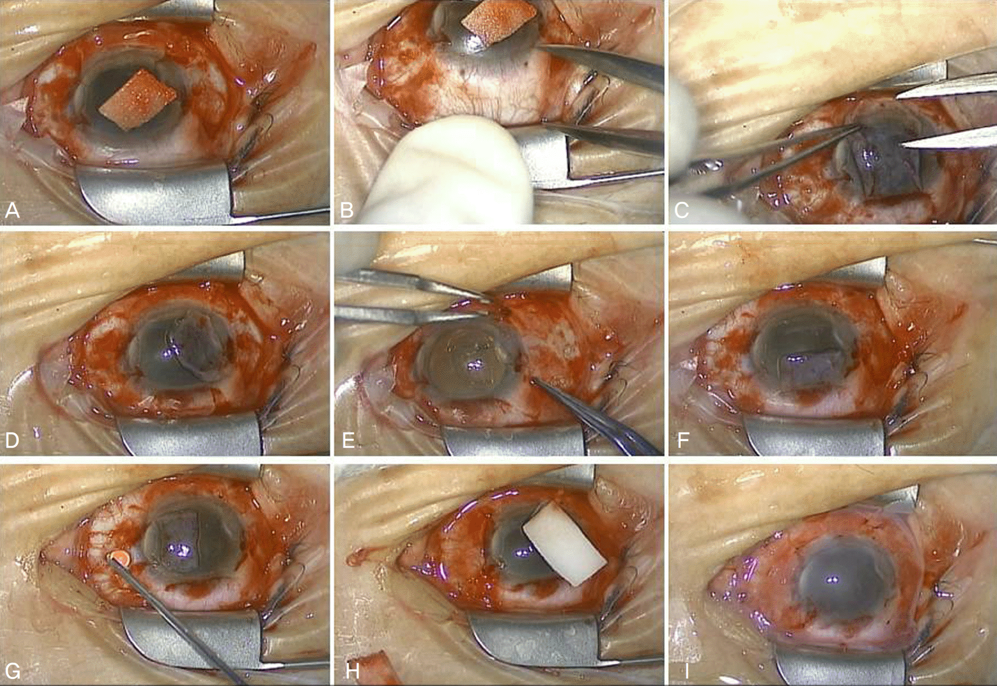 | Figure 1.Surgical procedure of split-conjunctival grafts for double-head pterygium. (A) The temporal and nasal side pterygium is along with adjacent fibrovascular tissue. (B, C) Conjunctival graft (6 mm) is dissected from the fornix leaving the underlying Tenon's capsule intact, flipped over the cornea. (D, E) From the donor conjunctiva, the distal 3 mm is excised and attached to the temporal bare sclera area using tissue adhesives. (F, G, H) The remaining graft is excised all the way down to the limbus, and attached to the nasal bare sclera area using tissue adhesives. (I) To protect and stabilize the conjunctiva autograft, temporary amniotic membrane transplantation is applied onto the whole eye with 10–0 nylon suture left in place for 5∼7 days. |
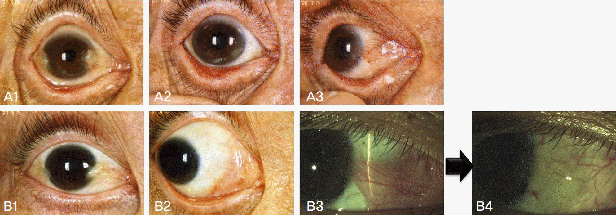 | Figure 2.Pre and postoperative photographs for amniotic membrane transplantation group. (A, B1) Preoperative photograghs of double-head pterygium. (A, B2) The transplanted amniotic membrane seems to blend in with stable ocular surface at 3 months postoperatively. (A3) Conjunctival recurrence at 5 months postoperatively. (B3, B4) Shows corneal recurrence at 9 months postoperatively, and the following stabilization after limbal-conjunctival autograft. |
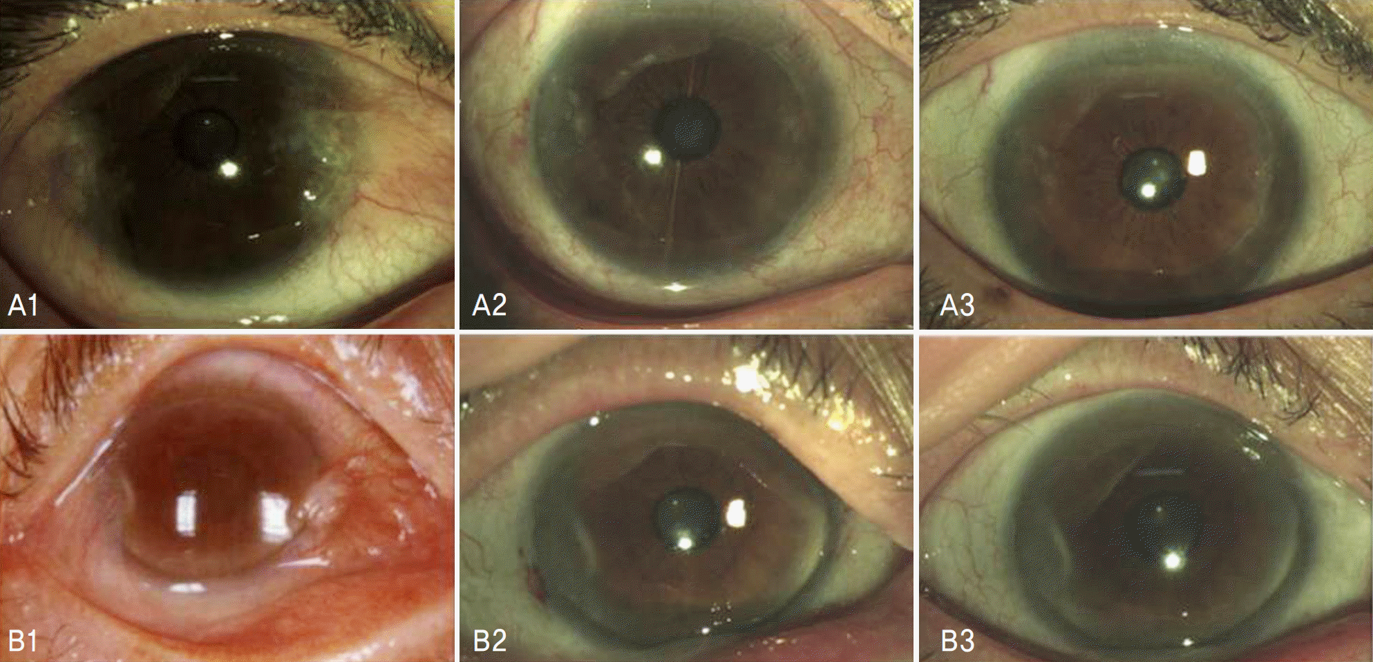 | Figure 3.Pre and postoperative photogragh for split-conjunctival grafts group. (A, B1) Preoperative photograghs of double-head pterygium. (A, B2) Well-positioned graft and quiet conjunctival surface are observed in subjects at 1 month postoperatively. (A, B3) Normal appearance is seen and recurrence of pterygium is not recognized in subjects at 12 months postoperatively. |
Table 1.
Demographic data of patients in the two study groups
| AMT* | Split-conjunctival graft | |
|---|---|---|
| Number of eyes (patients) | 7 (6) | 9 (8) |
| Primary / Recurrent (eyes) | 5 / 2 | 7 / 2 |
| Age (years) (range) | 60.1±7.9 (48∼72) | 59.0±9.9 (44∼78) |
| Gender (Male:Female) | 4: 3 | 3: 6 |
| Follow-up (months) (range) | 21.9±3.5 (18∼27) | 13.6±2.1 (12∼18) |
Table 2.
Summary of surgical results within amniotic membrane transplantation group
| Case | Age/Sex | Primary/ | Follow-up | VA* change | Corneal astigmatism change |
|---|---|---|---|---|---|
| Recurrent | (months) | (Diopter) | |||
| 1 | 48/M | Primary | 24 | 0.7 → 1.0 | 0.5 → 0.25 |
| 2 | 66/M | Primary | 18 | 0.15 → 0.5 | 4 → 0.75 |
| 3 | 72/M | Recurrent | 27 | 0.06 → 0.2 | 13.25 → 4.25 |
| 4 | 61/F | Primary | 19 | 0.4 → 0.4 | Error → 3 |
| 5 | 61/F | Recurrent | 18 | 0.06 → 0.4 | Error → 6.25 |
| 6 | 60/F | Primary | 24 | 0.7 → 0.8 | 0.75 → 1.0 |
| 7 | 53/M | Primary | 23 | 0.9 → 0.9 | 6.25 → 0.5 |
Table 3.
Summary of surgical results within split-conjunctival grafts group
Table 4.
Recurrence and complications of the two study groups
| AMT* | Split-conjunctival graft | |
|---|---|---|
| G2 (conjunctival recurrence) | 3/7 (43%) | 0/9 (0%) |
| G3 (corneal recurrence) | 2/7 (29%) | 0/9 (0%) |
| Location of recurrence | Nasal: 5/5 (100%) | · |
| Mean recurrent period (months) (range) | 7.2±1.8 (5∼9) | · |
| Complication | ||
| Vascularization at donor site† | 0 | 1 |
| Subgraft hemorrhage | 2 | 2 |
Table 5.
Previous reports on surgical treatment for double-head pterygium
| Author(s) | Pterygium | Number | Surgery technique | Mean follow-up | Recurrence |
|---|---|---|---|---|---|
| type | of eyes | (months) | rate‡ (%) | ||
| Solomon et al6 | Primary | 11 | Extensive pterygium excision with AMT† | 12.8±4.3 | 1/11 (9%) |
| Avisar et al7 | Primary | 10 | Bare sclera technique with | 36.3±3.8 | 0/10 (0%) |
| Recurrent | 3 | 0.02% MMC* (5 minutes) | 28.4±2.7 | 1/3 (33%) | |
| Maheshwari et al8 | Primary | 7 | Split-conjunctival graft | 17.7±6.0 | 0/7 (0%) |
| Wu et al9 | Primary | 20 | Conjunctival rotational autograft combined | 22.6 | 7/20 (35%) |
| with conjunctival autograft |
 XML Download