

 PDF
PDF ePub
ePub Citation
 Print
Print


Abstract
Purpose
Recently, biodegradable collagen matrix has been used as a possible substitute for antimetabolite in trabeculectomy in order to control the responsiveness of the wound healing process. This paper reports a case of encapsulation of the biodegradable collagen matrix after trabeculectomy.
Case summary
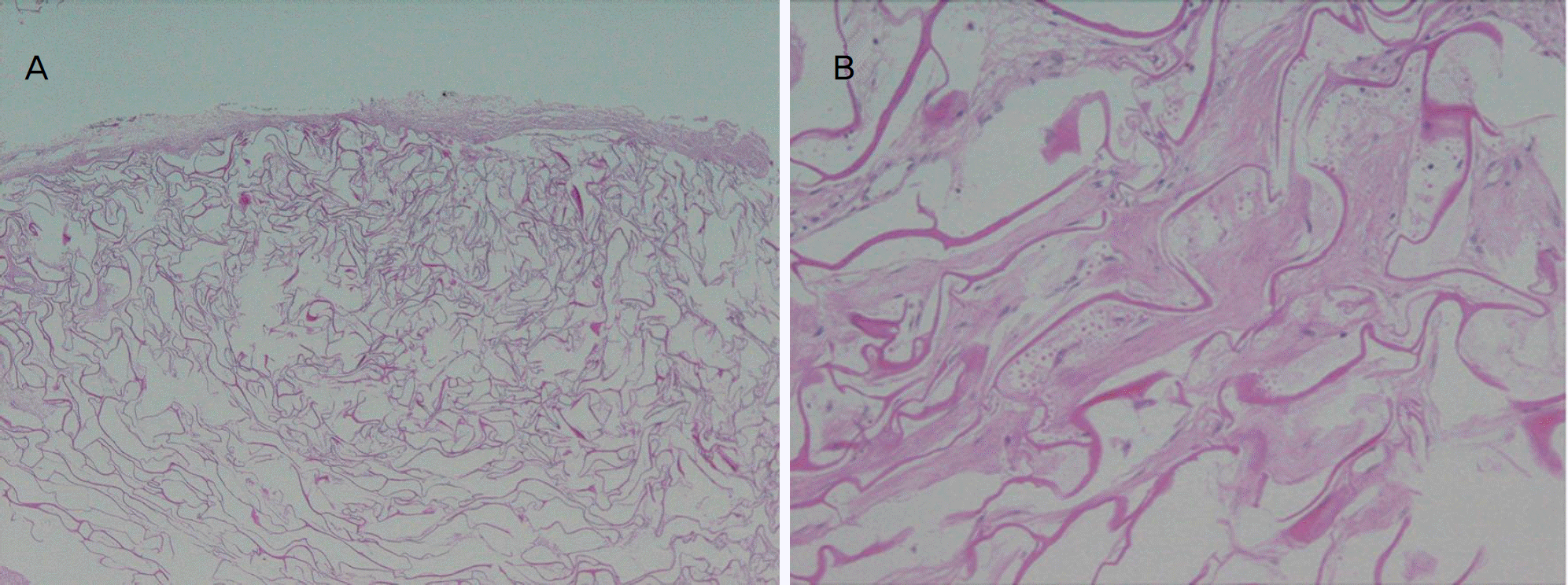
We conducted a fornix-based trabeculectomy on a 58-year-old man with medically uncontrollable steroid-in-duced glaucoma. We implanted biodegradable collagen matrix onto the sclera flap beneath the conjunctiva. Immediately after the surgery, we observed a localized bleb with high elevation. In the three months of follow-up, the bleb became encapsulated, and an increase in intraocular pressure was noted. During the wound revision, encapsulated material surrounded by thick fibrous membrane was found and removed from the subconjunctival space, followed by biopsy. Biopsy results demonstrated that amorphous collagenous material was surrounded by spindle and inflammatory cells.
References
1. Teng CC, Chi HH, Katzin HM. Histology and mechanism of abdominal operations. Am J Ophthalmol. 1959; 47:16–34.
2. Addicks EM, Quigley HA, Green WR, Robin AL. Histologic characteristics of filtering blebs in glaucomatous eyes. Arch Ophthalmol. 1983; 101:795–8.

3. Azuara-blanco A, Katz LJ. Dysfunctional filtering blebs. Surv Ophthalmol. 1998; 43:93–126.
4. Kitazawa Y, Kawase K, Matsuhita H, Minobe M. Trabeculectomy with Mitomycin. Arch Ophthalmol. 1991; 109:1693–8.
5. Skuta GL, Beeson CC, Higginbotham EJ, et al. Intraoperative abdominal versus postoperative 5-fluorouracil in high risk glaucoma filtering surgery. Ophthalmology. 1992; 99:438–44.
6. Cairns JE. Trabeculectomy-preliminary report of a new method. Am J Ophthalmol. 1968; 66:673–9.
7. DeBry PW, Perkins TW, Heatley G, et al. Incidence of late-onset bleb-related complications following trabeculectomy with mitomycin. Arch Ophthalmol. 2002; 120:297–300.
8. Hsu WC, Ritch R, Krupin T, Chen HS. Tissue bioenginnering for surgical bleb defects: an animal study. Graefes Arch Clin Exp Ophthalmol. 2008; 246:709–17.
9. Panek WC, Holland GN, Lee DA, Christensen RE. Glaucoma in patients with uveitis. Br J Ophthalmol. 1990; 74:223–7.
10. Shigeeda T, Tomidokoro A, Chen YN, et al. abdominal follow-up of initial trabeculectomy with mitomycin C for primary openangle glaucoma in Japanese patients. J Glaucoma. 2006; 15:195–9.
11. Yaldo MK, Stamper RL. Long term effects of mitomycin on abdominal bleb, Lack of fibrovascular response following severe inflammation. Arch Ophthalmol. 1993; 111:824–6.
12. Young MJ, Borras T, Walter M, Ritch R. Potential applications of tissue engineering to glaucoma. Arch Ophthalmol. 2005; 123:1725–31.
13. Chen HS, Ritch R, Krupin T, Hsu WC. Control of filtering bleb structure through tissue bioengineering: An animal model. Invest Ophthalmol Vis Sci. 2006; 47:5310–4.
14. Aptel F, Dumas S, Denis P. Ultrasound biomicroscopy and abdominal coherence tomography imaging of filtering blebs after deep sclerectomy with new collagen implant. Eur J Ophthalmol. 2009; 19:223–30.
15. Castner DG, Ratner BD. Biomedical surface science: Foundations to frontiers. Surface Science. 2002; 500:28–60.
Figure 1.
Dome-shaped, tense, opalescent, thick-walled encapsulated bleb is noted in the superior conjunctival area at postoperative 3 months. IOP ranged from 30 to 48 mm Hg during the 3-month follow-up period.
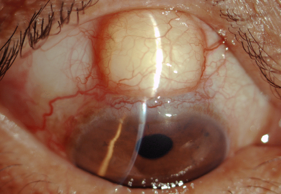
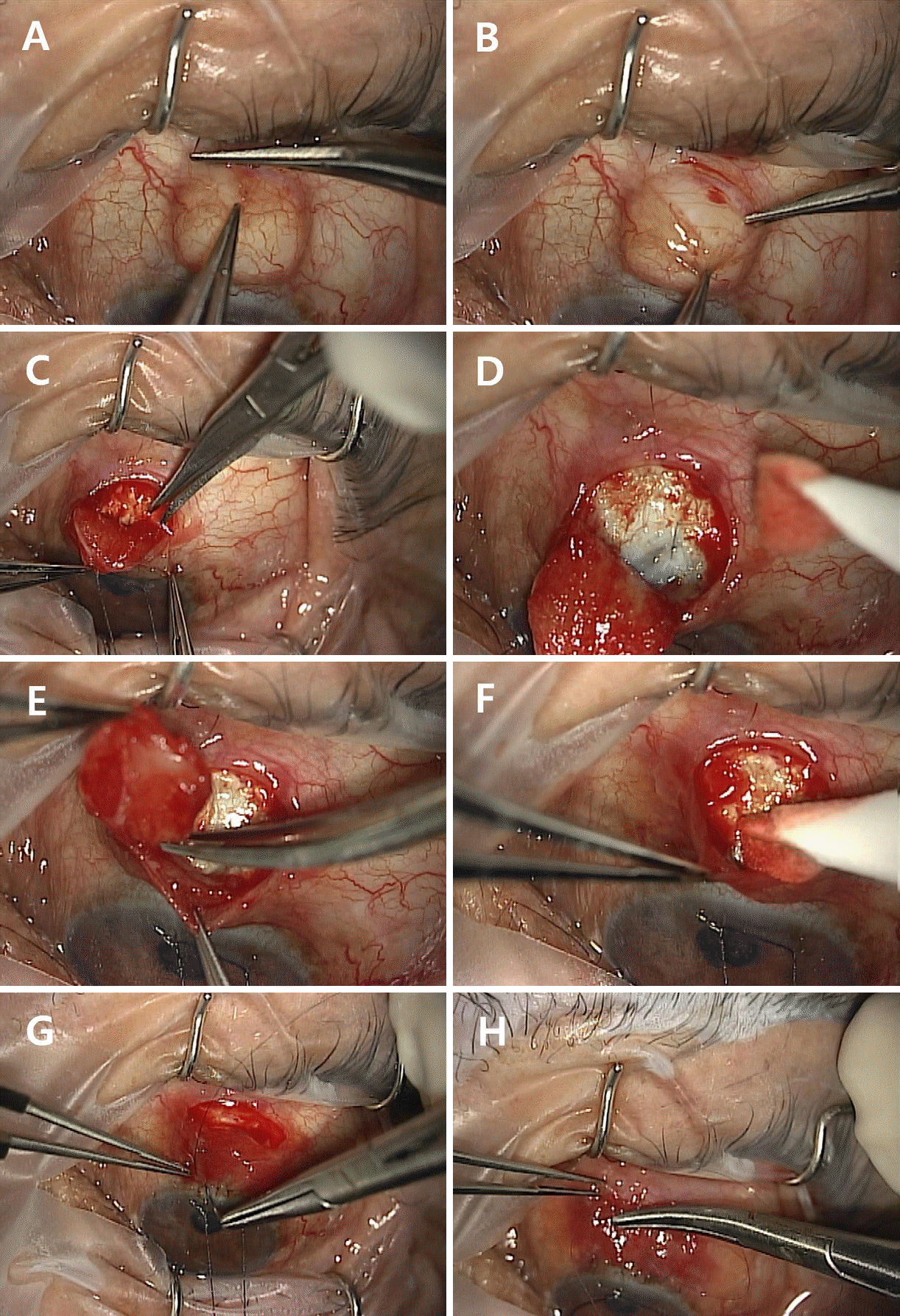
Figure 2.
Bleb revision procedure. (A) Conjunctiva is incised at the forniceal area. (B) Encapsulated tense opaque biomaterial is noted and dissected from the overlying conjunctiva. (C, D) Encapsulated material is dissected from the previous scleral flap. (E, F) Encapsulated material is removed from the subconjunctival space. (G, H) The conjunctiva is sutured.
 XML Download