

 PDF
PDF ePub
ePub Citation
 Print
Print


Abstract
Purpose
To investigate the correlation between central corneal thickness (CCT) and peripapillary retinal nerve fiber layer (RNFL) thickness in the eyes of patients with normal tension glaucoma (NTG) at the initial examination and to examine the difference in the degrees of damage of RNFL thickness depending on the CCT.
Methods
Fifty-one eyes of 36 patients with NTG were included in the study, and 51 eyes of 30 people without the disease were used as a control group. CCT and peripapillary RNFL thickness were measured in each eye by ultrasonic pachymetry and optical coherence tomography(OCT), respectively. Patients from the normal NTG group who underwent OCT more than three times inthree years were identically assigned to two groups based on CCT: thin (< 553.6 μ m) and thick (≥ 553.6 μ m). Thus, a comparison of the changes in the thickness of retinal nerve fiber layer was performed between the two groups.
References
1. Quigley HA, Miller NR, George T. Clinical evaluation of nerve fiber layer atrophy as an indicator of glaucomatous optic nerve damage. Arch Ophthalmol. 1980; 98:1564–71.

2. Levene RZ. Low tension glaucoma: a critical review and new material. Surv Ophthalmol. 1980; 24:621–64.
3. Kim C, Kim TK. Comparison of risk factors for bilateral and unilateral eye involvement in normal-tension glaucoma. Invest Ophthalmol Vis Sci. 2009; 50:1215–50.
4. Hollows FC, Graham PA. Intraocular pressure, glaucoma and glaucoma suspects in a defined population. Br J Ophthalmol. 1966; 50:570–86.
5. Kahn HA, Leibowbitz HM, Ganley JP, et al. The Framigham eye study. 1. Outline and major prevalence findings. Am J Epidemiol. 1977; 106:17–32.
6. Medeiros FA, Sample PA, Zangwill LM, et al. Corneal thickness as a risk factor for visual field loss in patients with preperimetric glaucomatous optic neuropathy. Am J Ophthalmol. 2003; 136:805–13.
7. Herndon LW, Weizer JS, Stinnett SS. Central corneal thickness as a risk factor for advanced glaucoma damage. Arch Ophthalmol. 2004; 122:17–21.
8. Brandt JD, Beiser JA, Kass MA, Gordon MO. Central corneal thickness in the ocular hypertension treatment study (OHTS). Ophthalmology. 2001; 108:1779–88.
9. Gordon MO, Beiser JA, Brandt JD, et al. The ocular hypertension treatment study: baseline factors that predict the onset of primary open-angle glaucoma. Arch Ophthalmol. 2002; 108:1779–88.
10. Kass MA, Hart WM Jr, Gorgon M, Miller JP. Risk factors favoring the development of glaucomatous visual field loss in ocular hypertension. Surv Ophthalmol. 1980; 25:155–62.
11. Burganski-Eliash Z, Wollstein G, Chu T, et al. Optical coherence tomography machine learning classifiers for glaucoma detection: a preliminary study. Invest Ophthalmol Vis Sci. 2005; 46:4147–52.
12. Lalezary M, Medeiros FA, Weinreb RN, et al. Baseline optical coherence tomography predicts the development of glaucomatous change in glaucoma suspects. Am J Ophthalmol. 2006; 142:576–82.
13. Medeiros FA, Zangwill LM, Bowd C, et al. Evaluation of retinal nerve fiber layer, optic nerve head, and macular thickness aberrations for glaucoma detection using optical coherence aberrations. Am J Ophthalmol. 2005; 139:44–55.
14. Lee ES, Kim CY, Ha SJ, et al. Central corneal thickness of Korean patients with glaucoma. Ophthalmology. 2007; 114:927–30.
15. Lee EJ, Kim TW, Park KH, et al. Ability of Stratus OCT to detect progressive retinal nerve fiber layer atrophy in glaucoma. Invest Ophthalmol Vis Sci. 2009; 50:622–8.
16. Emera BY, Tingey DP, Probst LE, Motolko MA. Central corneal thickness in low-tension glaucoma. Can J Ophthalmol. 1999; 34(3):19–24.
17. Ehlers N, Bramsen T, Sperling S. Applanation tonometer and central corneal thickness. Acta Ophthalmol. 1975; 53:34–43.
18. Doughty MJ, Zaman ML. Human corneal thickness and its aberrations on intraocular pressure measures: a review and meta-analysis approach. Surv Ophthalmol. 2000; 44:367–408.
19. Copt RP, Thomas R, Mermoud A. Corneal thickness in ocular hypertension, primary open-angle glaucoma, and normal tension glaucoma. Arch Ophthalmol. 1999; 117:14–6.
20. Kanamori A, Nakamura M, Escano MF, et al. Evaluation of the glaucomatous damage on retinal nerve fiber layer thickness aberrations by optical coherence tomography. Am J Ophthalmol. 2003; 135:513–20.
21. Sommer A, Katz J, Quigley HA, et al. Clinically detectable nerve fiber atrophy precedes the onset of glaucomatous field loss. Arch Ophthalmol. 1991; 109:77–83.
22. Jonas JB, Holbach L. Central corneal thickness and thickness of the lamina cribrosa in human eyes. Invest Ophthalmol Vis Sci. 2005; 46:1275–9.
23. Mumcuoglu T, Twonsend KA, Wollstein G, et al. Assessing the relationship between central corneal thickness and retinal nerve fiber layer thickness in healthy subjects. Am J Ophthalmol. 2008; 146:561–6.
Figure 1.
Correlation between central corneal thickness and average peripapillary retinal nerve fiber layer (RNFL) thicknessin normal group (Pearson correlation coefficient=-0.26, p=0.07).
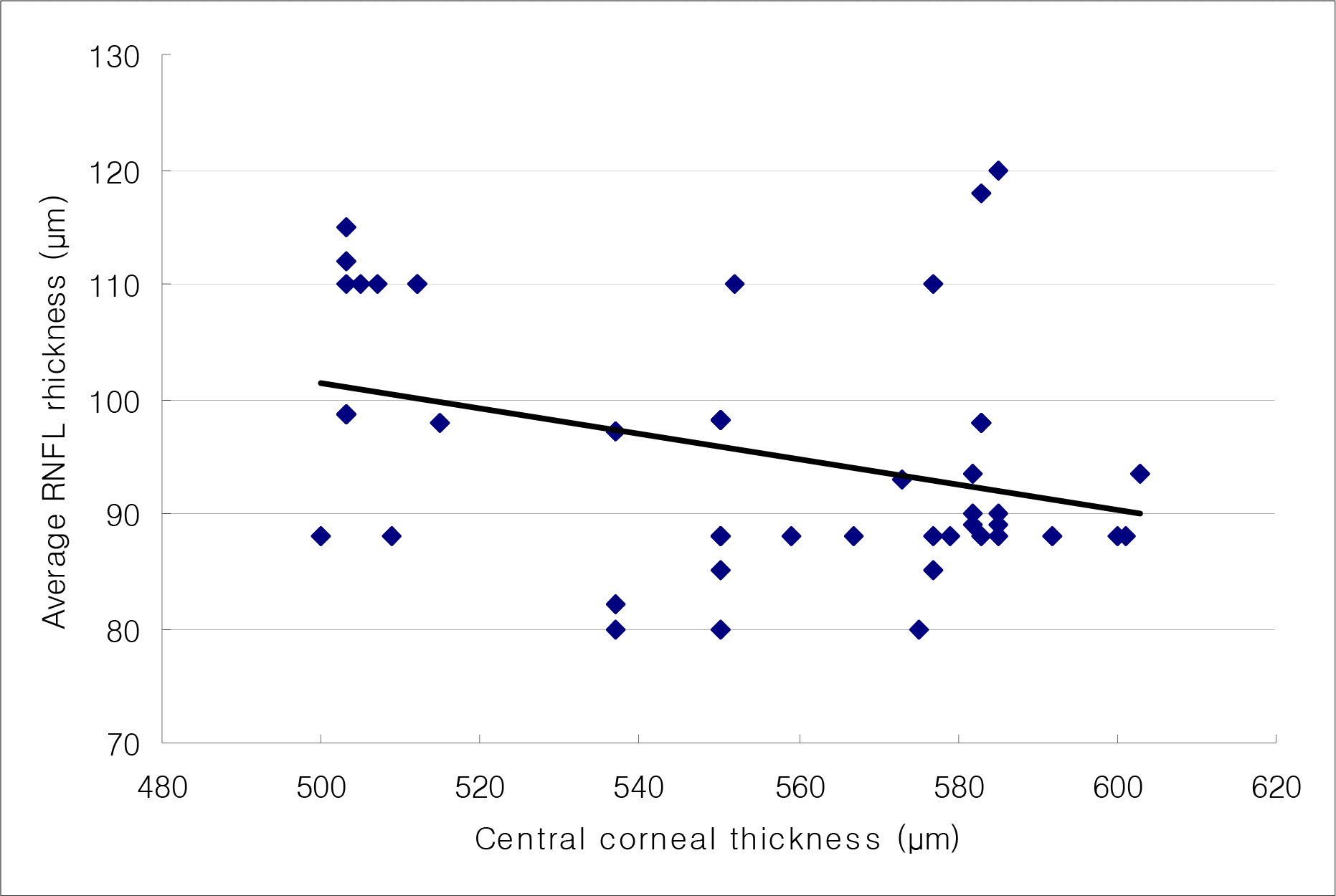
Figure 2.
Correlation between central corneal thickness and average peripapillary retinal nerve fiber layer (RNFL) thickness in normal tension glaucoma group (Pearson correlation coefficient=0.68, p<0.01).
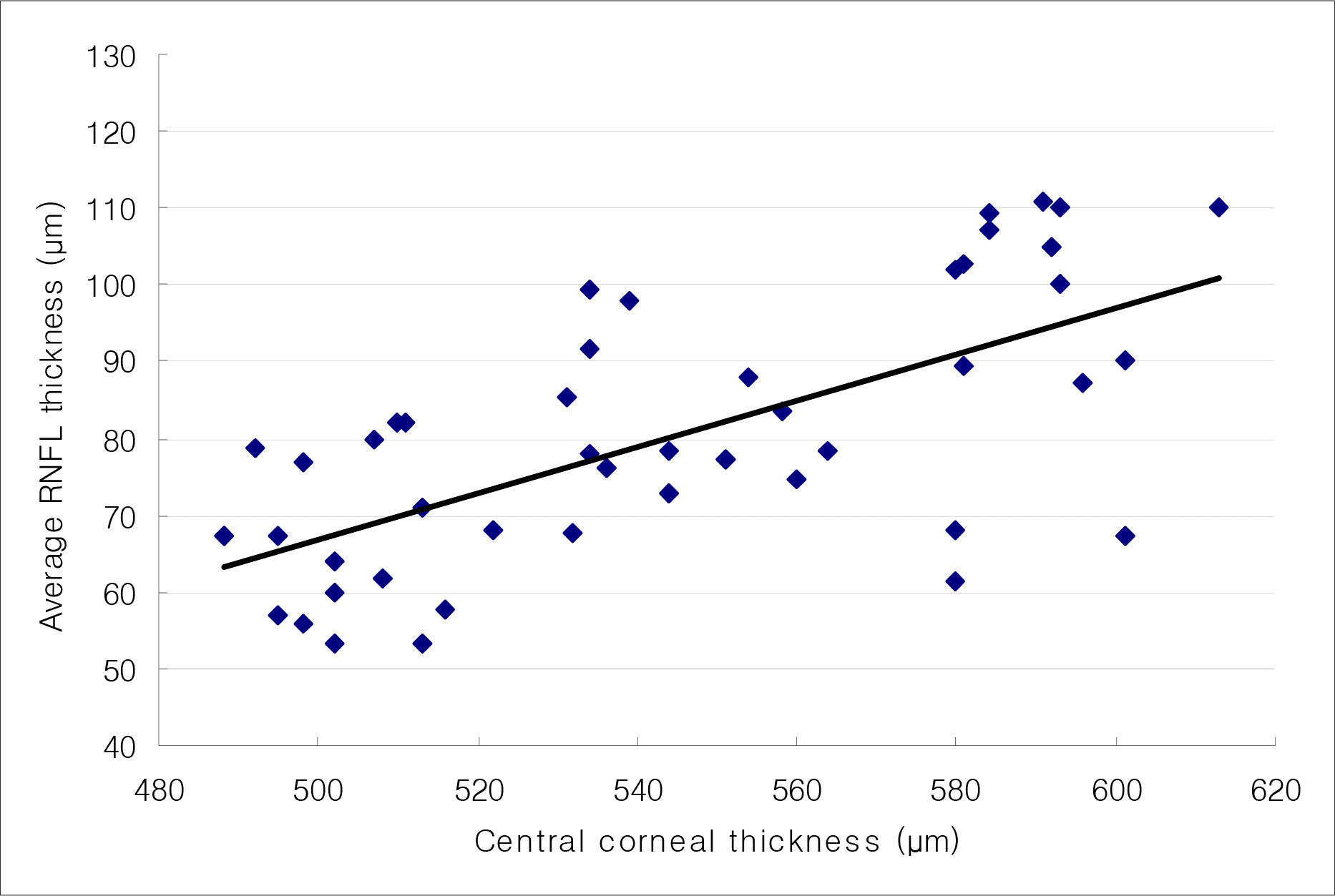
Table 1.
Comparison of clinical characteristic sand central corneal thickness between normal group and normal tension glaucoma group
| | Normal (n=51 eyes) | Normal tension glaucoma (n=51 eyes) | p-value* |
|---|---|---|---|
| Age (years) (Mean± SD) | 52.80±8.70 | 57.13±9.57 | 0.069 |
| Spherical Equivalent (diopter) (Mean± SD) | −0.41±2.24 | −0.35±1.71 | 0.882 |
| Intraocular Pressure (mmHg) (Mean± SD) | 16.53±2.53 | 13.88±1.53 | <0.05 |
| Central Corneal Thickness (μm) (Mean± SD) | 552.86±32.71 | 543.19±37.24 | 90.167 |
Table 2.
Comparison of peripapillary retinal nerve fiber layer between normal group and normal tension glaucoma group at initial examination
| Peripapillary RNFL thickness | Normal (n=51 eyes) | Normal tension glaucoma (n=51 eyes) | p-value* |
|---|---|---|---|
| Superior (μm) | 115.43±18.32 | 94.14±22.89 | <0.001 |
| Inferior (μm) | 119.88±20.15 | 95.04±27.06 | <0.001 |
| Nasal (μm) | 72.30±6.75 | 65.67±12.98 | <0.001 |
|
Temporal (μm) |
74.84±13.06 |
59.90±11.92 |
<0.001 |
| Average (μm) | 95.50±10.38 | 79.95±16.49 | <0.001 |
Table 3.
Correlation between central corneal thickness and peripapillary retinal nerve fiber layer thickness in normal group and normal tension glaucoma group
| |
Normal (n=51 eyes) |
Normal tension glaucoma (n=51 eyes) |
||
|---|---|---|---|---|
| | R* | p-value† | R* | p-value† |
| Superior (μm) | −0.23 | 0.11 | 0.48 | <0.01 |
| Inferior (μm) | 0.00 | 0.98 | 0.63 | <0.01 |
| Nasal (μm) | 0.05 | 0.73 | 0.44 | <0.01 |
|
Temporal (μm) |
0.13 |
0.36 |
0.28 |
0.04 |
| Average (μm) | −0.26 | 0.07 | 0.68 | <0.01 |
Table 4.
Patients characteristics and corrected intraocular pressure grouped by central corneal thickness
| | Group 1 (CCT<553.6 μm)(n=19 eyes) | Group 2 (CCT≥553.6 μm)(n=17 eyes) | p-value* |
|---|---|---|---|
| Age (years) (Mean± SD) | 58.89±10.46 | 60.47±7.45 | 0.610 |
| CCT (μm) (Mean± SD) | 513.74±14.48 | 579.59±21.17 | >0.01 |
| Initial IOP (mmHg) (Mean± SD) | 17.05±0.85 | 17.47±0.72 | 0.122 |
| Corrected IOP, Initial (mmHg) (Mean± SD) | 18.08±0.85 | 15.76±1.17 | >0.01 |
| Average IOP (mm Hg) (Mean± SD) | 13.58±1.07 | 14.82±1.07 | >0.01 |
| Corrected IOP, Average (mmHg) (Mean± SD) | 14.97±0.92 | 12.64±1.25 | >0.01 |
| Follow up (months) (Mean± SD) | 39.74±2.05 | 39.76±2.17 | 0.969 |
Table 5.
Comparison of the number of OCT and an interval of OCT between thinner central corneal thickness group (Group 1) and thicker central corneal thickness group (Group 2)
| | Group 1 (n=19) | Group 2 (n=17) | p-value* |
|---|---|---|---|
| No. of OCT (Mean ± SD) | 3.53 ± 0.51 | 3.47 ± 0.51 | 0.224 |
| Interval of OCT (months) (Mean ± SD) | 10.68± 2.24 | 11.06 ± 1.30 | 0.549 |
| Interval of OCT (from initial to last, months) (Mean ± SD) | 38.89 ± 1.37 | 38.18 ± 1.88 | 0.195 |
Table 6.
Comparison of peripapillary retinal nerve fiber layer thickness between thinner central corneal thickness group (Group 1) and thicker central corneal thickness group (Group 2)
| Peripapillary RNFL thickness |
Group 1 (n=19) |
Group 2 (n=17) |
p-value* |
||||||
|---|---|---|---|---|---|---|---|---|---|
| Initial exam | Last exam. | Change | Initial exam | Last exam. | Change | Initial exam | Last exam. | Change | |
| Superior (μm) | 98.11±15.83 | 87.37 ±18.37 | −10.74±14.13 | 102.47±27.61 | 93.59 ±23.20 | −9.00±9.08 | 0.572 | 0.376 | 0.668 |
| Inferior (μm) | 90.95±23.93 | 80.47 ±21.06 | −10.47±13.50 | 112.88±9.78 | 104.77±8.39 | −8.12±6.23 | 0.01 | 0.001 | 0.515 |
| Nasal (μm) | 63.26±16.79 | 58.32 ±14.26 | −4.95±6.99 | 69.35±14.48 | 65.29±12.86 | −4.06±3.36 | 0.255 | 0.134 | 0.626 |
| Temporal (μm) | 61.84±10.75 | 57.79±9.00 | −3.95±4.13 | 70.18±16.72 | 64.18±12.86 | −6.00±4.30 | 0.081 | 0.155 | 0.157 |
| Average (μm) | 78.52±11.20 | 72.07 ±11.71 | −6.39±7.28 | 88.77±12.74 | 82.30 ±11.62 | −5.47±4.73 | 0.015 | 0.007 | 0.661 |
Table 7.
Multivariate hazard ratios and 95% confidence intervals for the progression of retinal nerve fiber layer damage
| Baseline factors | Hazard ratio | 95% Confidence interval | p-value* |
|---|---|---|---|
| Thin central corneal thickness | 5.36 | 0.63∼45.47 | 0.124 |
| Sex (Male) | 0.84 | 0.21∼3.39 | 0.80 |
| Age (per 1 year older) | 0.93 | 0.87∼1.00 | 0.06 |
| Controlled intraocular pressure (per 1mmHg higher) | 0.84 | 0.35∼2.04 | 0.71 |
 XML Download