

 PDF
PDF ePub
ePub Citation
 Print
Print


Abstract
Purpose
To report a patient with Coats' disease who presented with a premacular membrane that was peeled off after laser photocoagulation.
Case summary
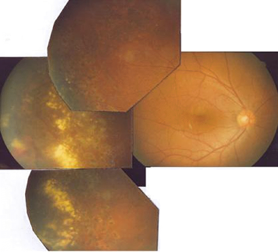
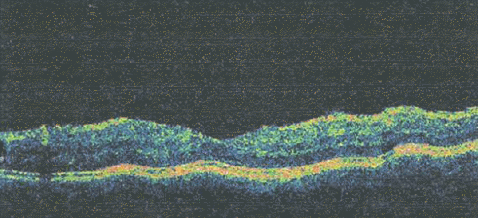
A 17-year-old male presented with decreased visual acuity of the right eye, and showed serous elevation, subretinal hemorrhage, telangiectasis and thick premacular membrane upon fundus examination. Upon diagnosis with Coats' disease, the telangiectatic area was treated with argon laser photocoagulation. Two weeks later, the premacular membrane was peeled off spontaneously and the decrease of macular thickness was verified by optical coherence tomography (OCT). The patient's visual acuity was improved to 1.0.
References
1. Coats G. Forms of retinal disease with massive exudation. Roy London Ophthalmol Hospital Report. 1908; 17:440–525.
2. Deutsch TA, Rabb MF, Jampol LM. Spontaneous regression of retinal lesion in Coats' disease. Can J Ophthalmol. 1982; 17:169–72.
3. Tarkkanen A, Laatikainen L. Coats' disease: clinical, angiographic, histopathological findings and clinical management. Br J Ophthalmol. 1983; 67:766–76.

4. Woods AC, Duke JR. Coats's disease. Review of the literature abdominal criteria, clinical findings, and plasma lipid studies. Br J Ophthalmol. 1963; 47:385–412.
5. Morales AG. Coats' disease: natural history and results of treatment. Am J Ophthalmol. 1965; 60:855–65.
6. Egerer I, Tasman W, Tomer T. Coats' disease. Arch ophthalmol. 1974; 92:109–12.
7. Ridley ME, Shieles JA, Brown GC, Tasman W. Coats' disease, evaluation of management. Ophthalmology. 1982; 89:1381–7.
8. Sumers KD, Jampol LM, Goldberg MF, Huamonte FU. Spontaneous separation of epiretinal membranes. Arch Ophthalmol. 1980; 98:318–20.
9. Wolfensberger TJ, Holz FG, Gregor ZJ. Juvenile Coats' disease associated with epiretinal membrane formation. Retina. 1995; 15:261–3.
10. Lefaut BA, Priem H, Laey JJ. Premacular fibrosis in juvenile Coats' disease with spontaneous peeling after photocoagulation of the congenital vascular anomalies. Bull Soc Belge Ophthalmol. 1996; 261:79–84.
11. Sugimoto M, Sasoh M, Ito Y. A case of Coats' disease with a peeling of premacular fibrosis after photocoagulation. Acta Ophthalmol Scand. 2002; 80:96–7.
Figure 1.
Fundus photograph of the patient's right eye at the initial examination. Premacular membrane is seen.
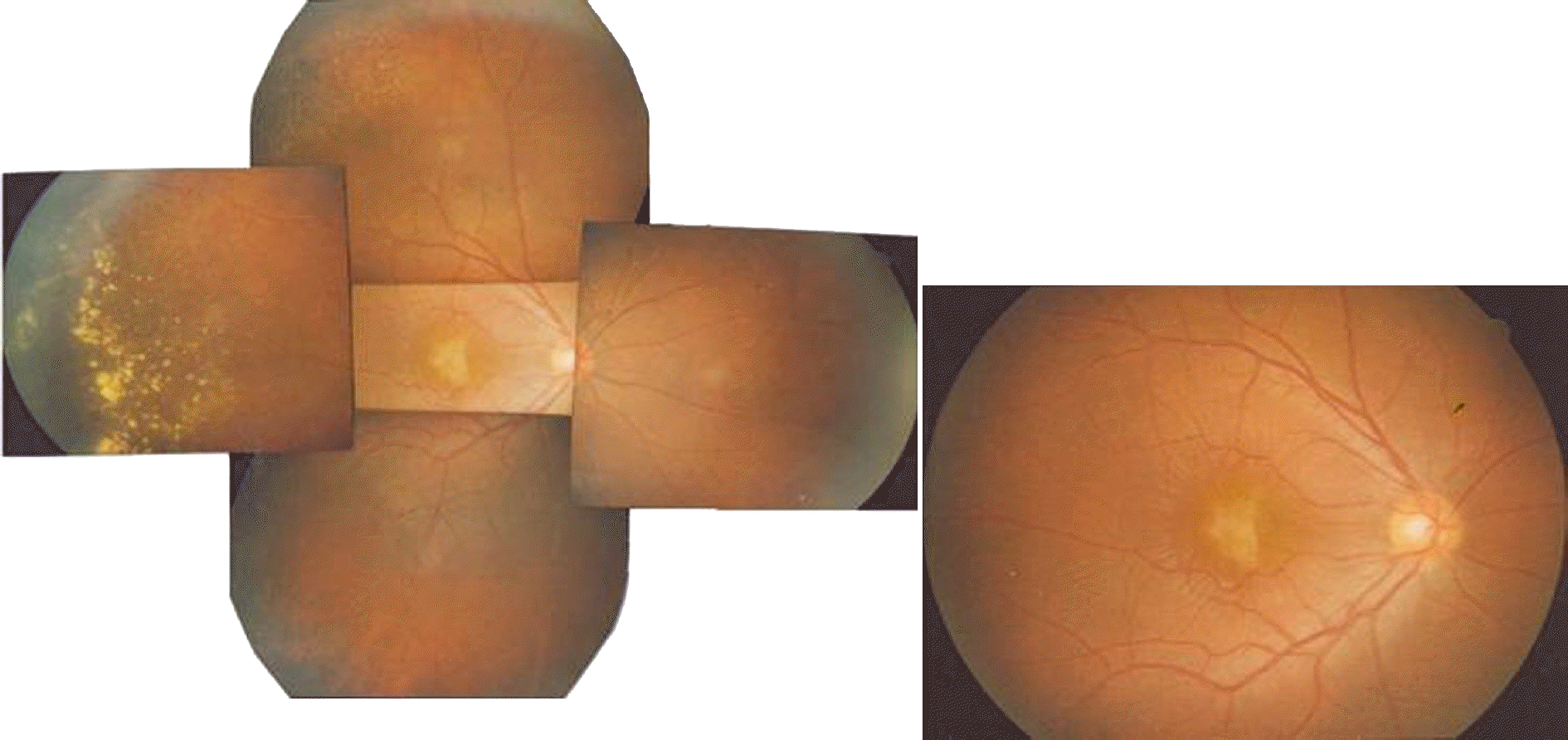
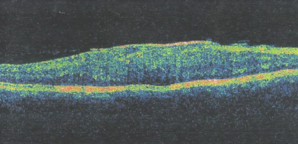
Figure 2.
The OCT finding of the patient's right eye at the initial examination. The macular thickness of the patient was 528 μm with premacular membrane.
 XML Download