

 PDF
PDF ePub
ePub Citation
 Print
Print


Abstract
Case summary
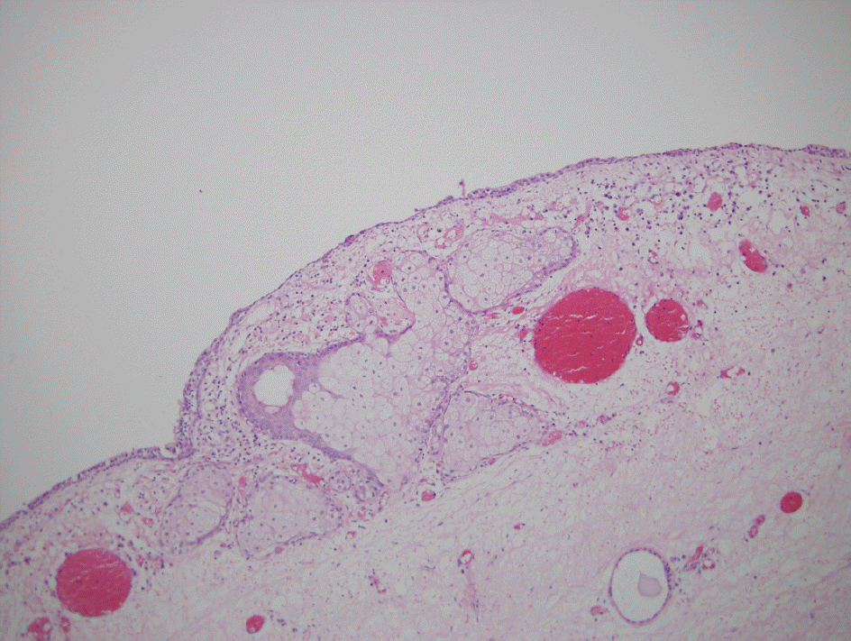
A 41-year-old man complained of swelling of the left lower eyelid and left periocular pain for a week. Examination revealed 3 mm of proptosis with superotemporal displacement of the left eye. Orbital CT revealed a 32×27×33-mm well-defined giant cyst with a fat-fluid level in the superonasal aspect of the left orbit. Orbital MRI showed bone remodeling around the cyst, consistent with a dermoid cyst. The cyst was approached via lateral orbitotomy and transcaruncular incision but was ruptured just prior to the end of the dissection and was totally excised using a cryoprobe to freeze the ruptured site. Upon histopathological examination, the cyst was misdiagnosed as a conjunctival cyst because there was no dermal appendage but was rediagnosed as a conjunctival dermoid cyst after the tissue sample was examined more thoroughly. After surgery, the patient presented with diplopia due to esodeviation and was prescribed prismatic lenses.
Go to : 
References
1. Shields JA, Augsburger JJ, Donoso LA. Orbital dermoid cyst of conjunctival origin. Am J Ophthalmol. 1986; 101:726–9.

2. Shields JA, Kaden IH, Eagle RC Jr, Shields CL. Orbital dermoid cysts: clinicopathologic correlations, classification, and management. The 1997 Josephine E. Schueler Lecture. Ophthal Plast Reconstr Surg. 1997; 13:265–76.
3. Jakobiec FA, Bonanno PA, Sigelman J. Conjunctival adnexal cysts and dermoids. Arch Ophthalmol. 1978; 96:1404–9.
4. Shields JA, Shields CL. Orbital cysts of childhood-classification, clinical features, and management. Surv Ophthalmol. 2004; 49:281–99.
5. Martinez LM, Cohen KL. Conjunctival dermoid cyst seen on abdominal as a chronically red eye. Arch Ophthalmol. 1998; 116:1109–11.
6. Dutton JJ, Fowler AM, Proia AD. Dermoid cyst of conjunctival origin. Ophthal Plast Reconstr Surg. 2006; 22:137–9.
7. Goldstein MH, Soparkar CN, Kersten RC, et al. Conjunctival cyst of the orbit. Ophthalmology. 1998; 105:2056–60.
8. Colombo F, Holbach LM, Naumann GO. Conjunctival cyst and conjunctival dermoid of the orbit. Orbit. 2000; 19:13–9.
9. Boynton JR, Searl SS, Ferry AP, et al. Primary nonkeratinized abdominal (‘conjunctival') orbital cysts. Arch Ophthalmol. 1992; 110:1238–42.
10. Gloor P, Horio B, Klassen M, Eagle RC Jr. Conjunctival cyst. Arch Ophthalmol. 1996; 114:1020–1.
11. Soll SM, Lisman RD, Harrison W, Weiner M. Conjunctival abdominal cyst. Ophthal Plast Reconstr Surg. 1994; 10:216–9.
Go to :
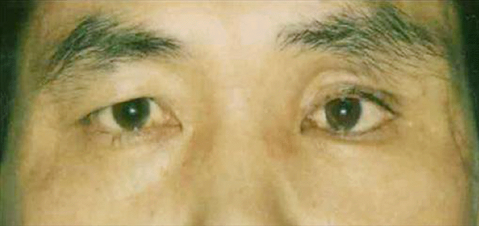
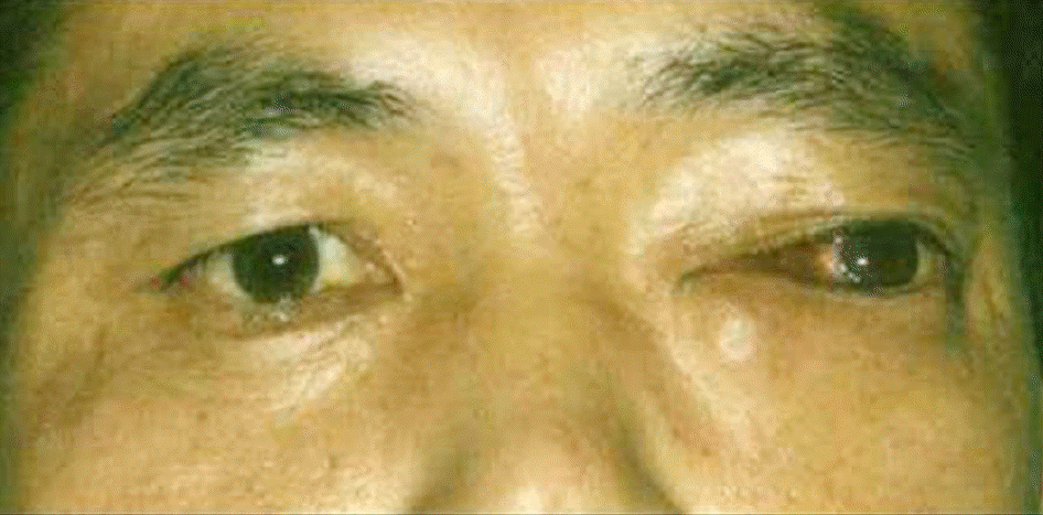 | Figure 1.External photograph of a 41-year-old man with progressive proptosis and lateral displacement of the left eye. |
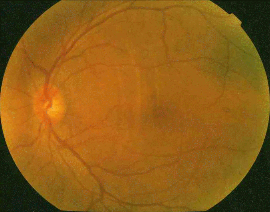 | Figure 2.Fundus photograph of the left eye showing choroidal folds at the posterior pole due to globe compression. |
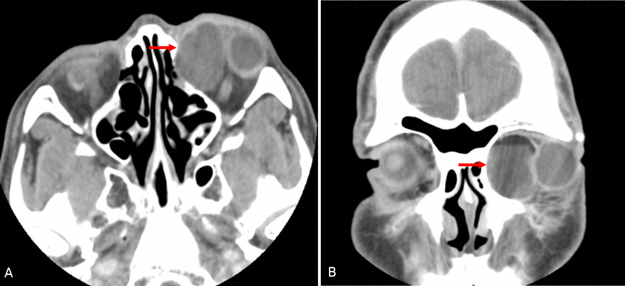 | Figure 3.Preoperative axial (A) and coronal views (B) of orbital CT scan showing a well-defined unilocular giant cyst (red arrow), measuring 32×27×33 mm, occupying the superonasal area of the left orbit. |
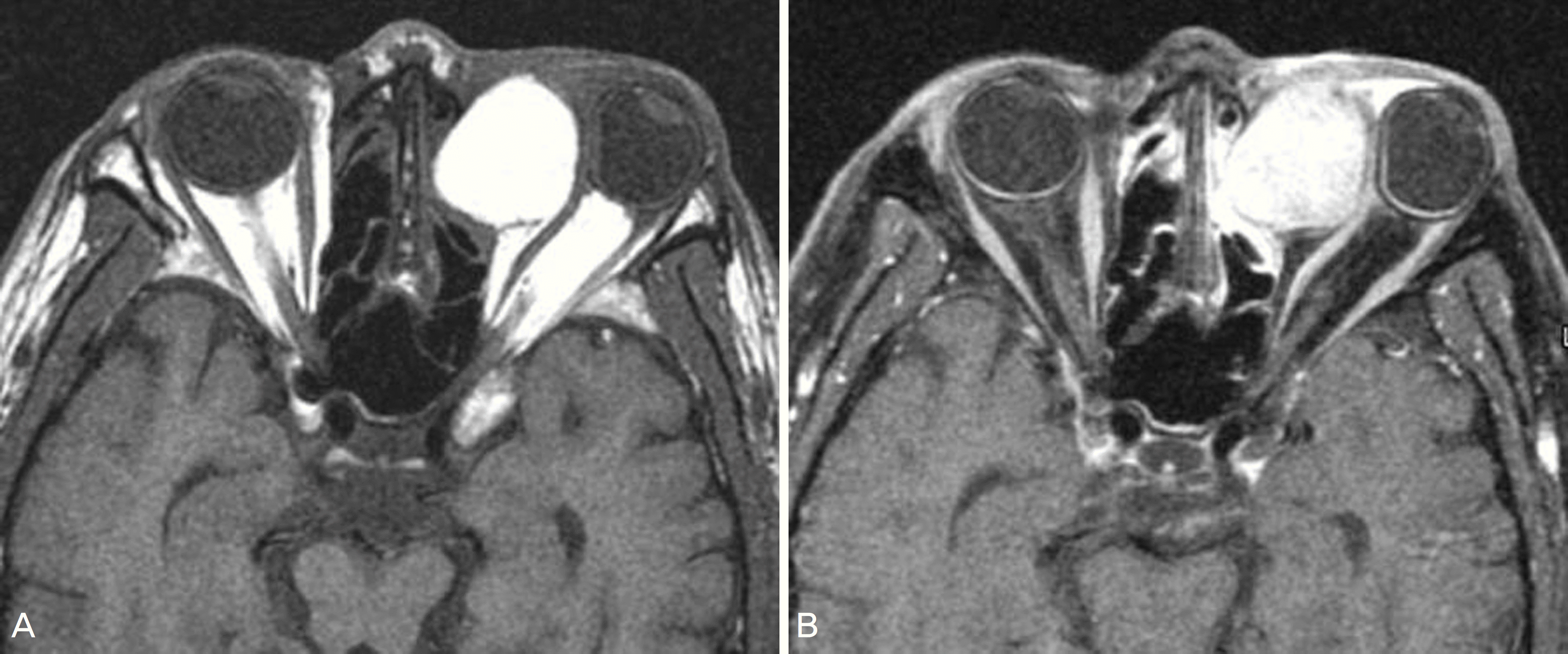 | Figure 4.Preoperative axial view of orbital MRI reveals a well-defined cyst of the nasal orbit showing high signal intensity on T1-(left) and relatively homogenous enhanced on post-contrast fat suppressed T1-weighted image (right). Globe compression and bone remodeling are seen. |
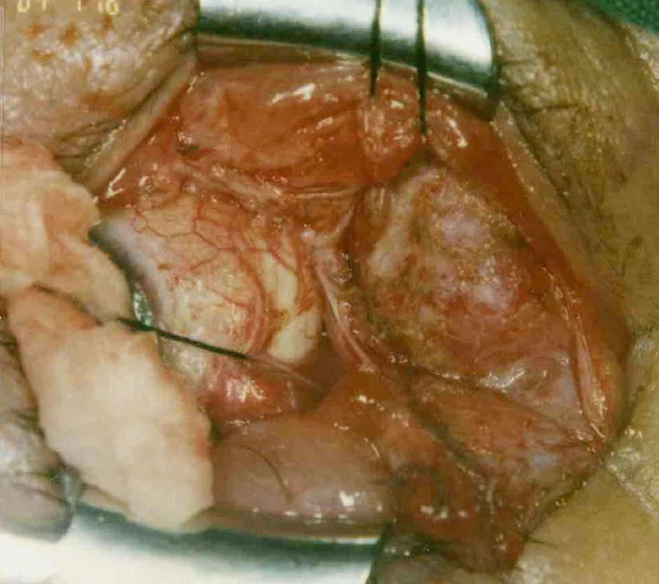 | Figure 5.Surgical photograph demonstrating the conjunctival dermoid cyst in the left orbit. |
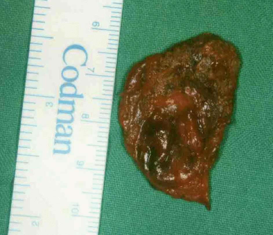 | Figure 6.Gross finding of the completely excised conjunctival dermoid cyst which was accidentally perfo-rated and aspirated during operation. |
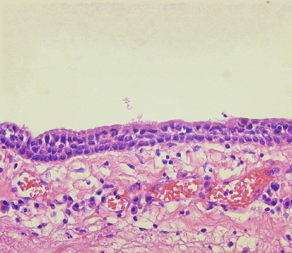 | Figure 7.Microscopic examination of cyst wall shows a triple layer of nonkeratinizing stratified epithelium with underlying loose connective tissue. Definite goblet cells and dermal appendages are not seen (hematoxylin and eosin stain, ×400). |
 XML Download