

 PDF
PDF ePub
ePub Citation
 Print
Print


Abstract
Purpose
To investigate changes in corneal astigmatism and refractive power in intermittent exotropia after lateral rectus recession with or without medial rectus resection.
Methods
We compared visual acuity, spherical equivalent, refractive power, astigmatism from cycloplegic refraction, and Orbscan corneal topography in two groups consisting of 40 eyes from 20 patients who underwent bilateral lateral rectus recession (Group 1) and 33 eyes from 33 patients who underwent monocular medial rectus resection with lateral rectus recession (Group 2) immediately preoperatively and at 1 and 4 weeks postoperatively.
Results
In Group 1, the refractive power changed +0.12 D on average in the horizontal median and −0.08 D on average in the vertical median at 1 week postoperatively. The refractive power changed +0.07 D on average in the horizontal median and −0.04 D on average in the vertical median at 4 weeks postoperatively. No significant change in the corneal astigmatic axis was detected. In Group 2, the refractive power changed +0.4 D on average in the horizontal median and −0.19D on average in the vertical median, and the corneal astigmatic axis significantly shifted by +0.51 D in the ‘with-the-rule astigmatism’ direction at 1 week postoperatively (p=0.02). However, the refractive power changed +0.2 D on average in the horizontal median and −0.09 D on average in the vertical median, and the corneal astigmatic axis changed +0.2 D at 4 weeks postoperatively, although these values were not statistically significant.
References
1. Lieberman DM, Grierson JW. The lids influence on corneal shape. Cornea. 2000; 19:336–42.

2. Read SA, Collins MJ, Carney LG. The influence of eyelid abdominal on normal corneal shape. Invest Ophthalmol Vis Sci. 2007; 48:112–9.
3. Preslan MW, Cioffi G, Min YI. Refractive error changes abdominal strabismus surgery. J Pediatr Ophthalmol Strabismus. 1992; 29:300–4.
4. Dottan SA, Hoffman P, Oliver MD. Astigmatism after strabismus surgery. Ophthalmic Surg. 1988; 19:128–9.
5. Rajavi Z, Mohammad Rabei H, Ramezani A, et al. Refractive effect of the horizontal rectus muscle recession. Int Ophthalmol. 2008; 28:83–8.
6. Baldwin WR, Mills D. A longitudinal study of corneal abdominal and total astigmatism. Am J Optom Physiol Opt. 1981; 58:206–11.
7. Dobson V, Fulton AB, Sebris SL. Cycloplegic refractions of infants and young children: the axis of astigmatism. Invest Ophthalmol Vis Sci. 1984; 25:83–7.
8. Nardi M, Rizzo S, Pellegrini G, Lepri A. Effect of strabismus abdominal on corneal topography. J Pediatr Ophthalmol Strabismus. 1997; 34:244–6.
9. Thompson WE, Reinecke RD. The changes in refractive status following routine strabismus surgery. J Pediatr Ophthalmol Strabismus. 1980; 17:372–4.
10. Kitthaweesin K, Singhakul S. Effect of horizontal strabismus abdominal on the astigmatism. J Med Assoc Thai. 2007; 90:744–7.
11. Bagheri A, Farahi A, Guyton DL. Astigmatism induced by abdominal recession of both horizontal rectus muscles. J AAPOS. 2003; 7:42–6.
12. Yoo JM, Ryoo MH. The changes of astigmatism following abdominal strabismus surgery. J Korean Ophthalmol Soc. 1990; 31:337–85.
13. Lee YC, Yoon JW, Lee WY. Refractive changes following abdominal surgery. J Korean Ophthalmol Soc. 1994; 35:704–18.
14. Lee JH, Choi DG. The changes of refractive error and corneal abdominal after horizontal strabismus surgery. J Korean Ophthalmol Soc. 1995; 36:1221–7.
15. Denis D, Bardot J, Volot F, et al. Effects of strabismus surgery on refraction in children. Ophthalmologica. 1995; 209:136–40.
16. Kwitko S, Feldon S, McDonnell PJ. Corneal topographic changes following strabismus surgery in Grave's disease. Cornea. 1992; 11:36–40.
17. Snir M, Nissenkorn I, Buckman G, et al. Postoperative refractive changes in children with congenital esotropia: a preliminary study. Ophthalmic Surg. 1989; 20:57–62.
18. Kwito S, Sawusch MR, McDonnell PJ, et al. Effect of extraocular muscle surgery on corneal topography. Arch Ophthalmol. 1991; 109:873–8.
19. Asejczyk-Widlicka M, Pierscionek BK. The elasticity and rigidity of the outer coats of the eye. Br J Ophthalmol. 2008; 92:1415–8.
20. Asejczyk-Widlicka M, Sródka DW, Kasprzak H, et al. Modelling the elastic properties of the anterior eye and their contribution to maintenance of image quality: the role of the limbus. Eye. 2007; 21:1087–94.
21. Hainsworth DP, Bierly JR, Schmeisser ET, et al. Corneal abdominal changes after extraocular muscle surgery. J AAPOS. 1999; 3:80–6.
22. Abrahamsson M, Fabian G, Sjöstrand J. Changes in astigmatism between the ages of 1 and 4 years: a longitudinal study. Br J Ophthalmol. 1988; 72:145–9.
Figure 1.
Changes in mean corneal astigmatism using Orbscan at preoperatively and at 1 and 4 weeks postoperatively. (A) patients who underwent bilateral lateral rectus recession. (B) patients who underwent unilateral medial rectus resection with lateral rectus recession. * P<0.05, paired T test.
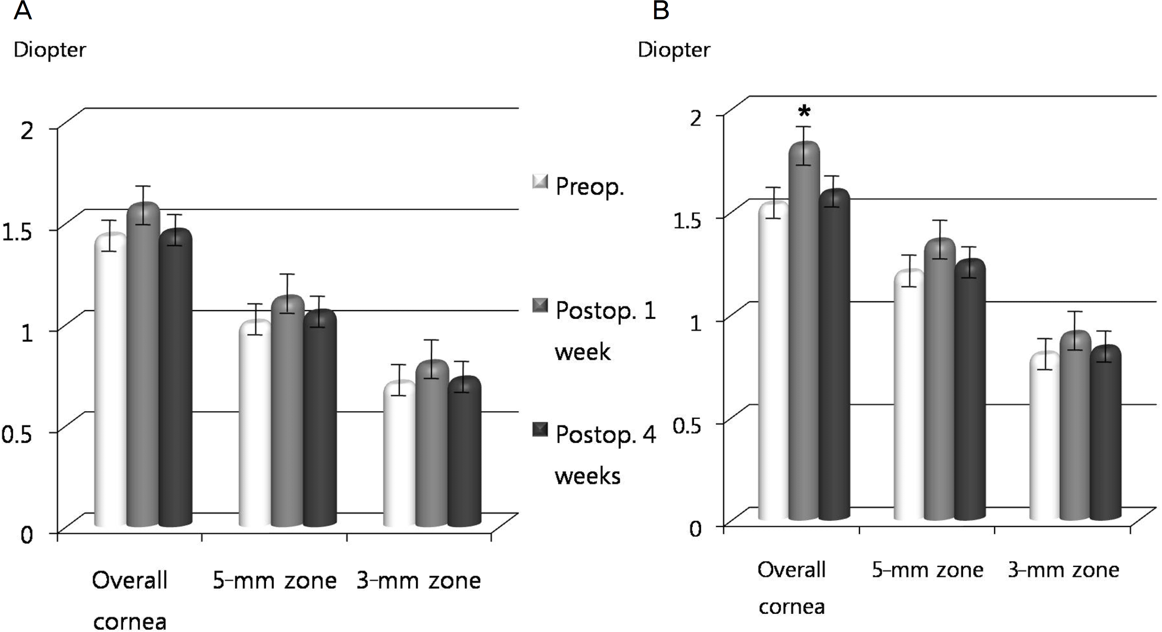
Figure 2.
Changes in mean horizontal and vertical refractive power at preoperatively and at 1 and 4 weeks postoperatively. (A) Patients who underwent bilateral lateral rectus recession. (B) patients who underwent unilateral medial rectus resection and lateral rectus recession. *P<0.05, paired T test. Preop.=preoperative; Postop.= postoperative.
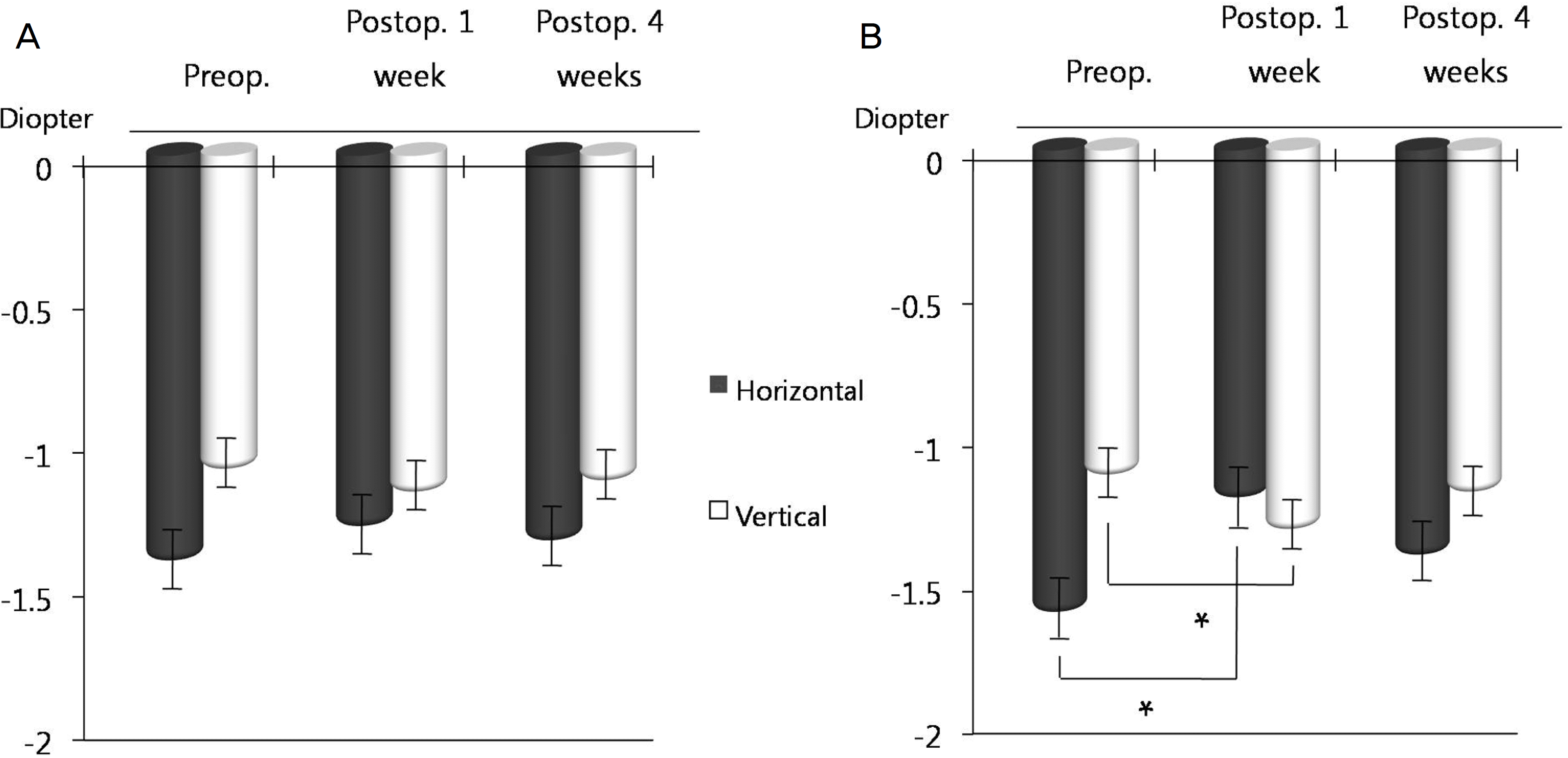
Figure 3.
The graphs show a negative relationship in refractive power and corneal astigmatism change between preoperatively and 1 week postoperatively (A, C) and between 1 week and 4 weeks postoperatively (B, D) by Spearman correlation. Pre.=preoperative; Post.=postoperative.
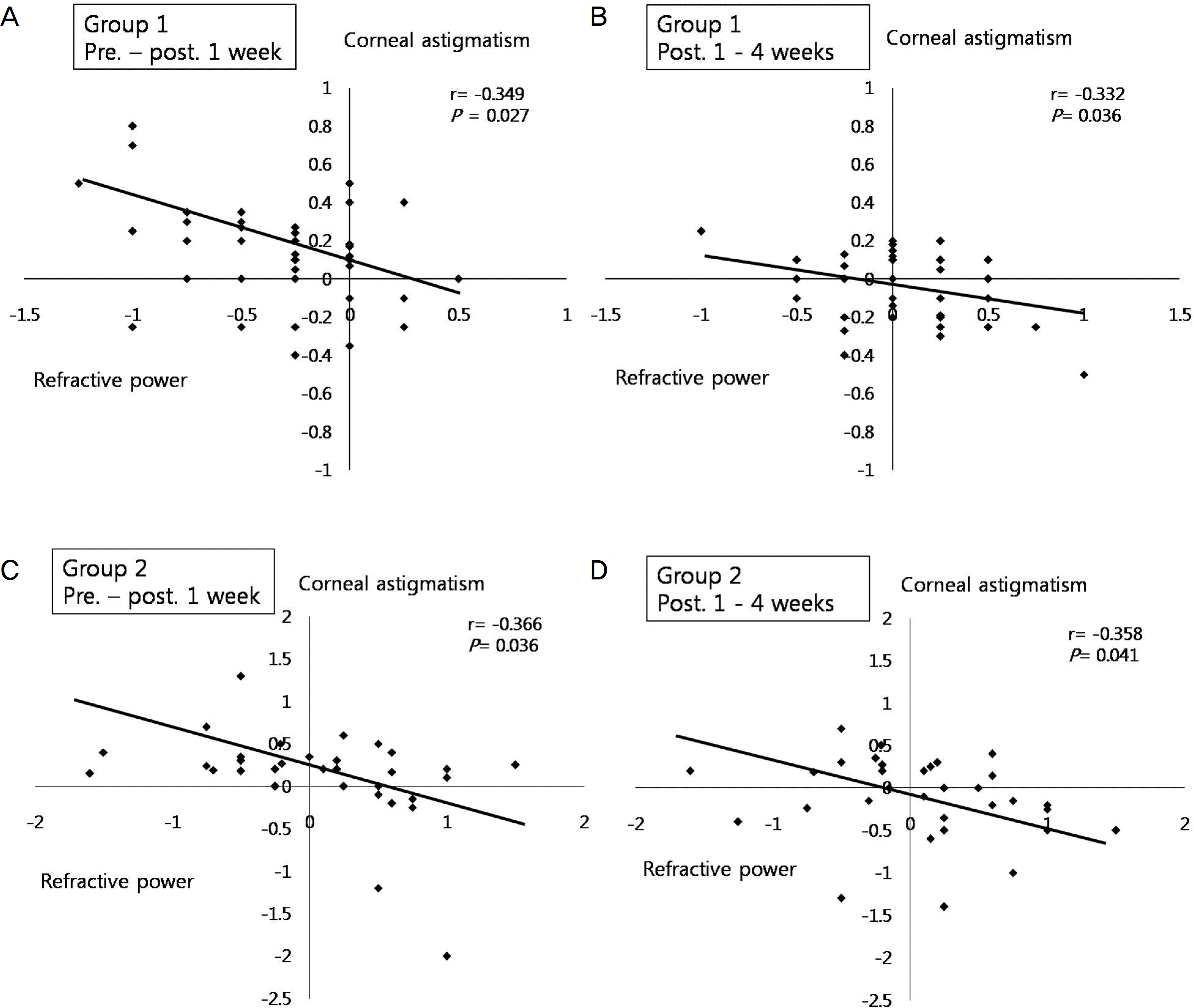
Table 1.
Surgical guideline used in our study
| Method | 25PD‡ | 30PD | 35PD | 40PD | 45PD | 50PD | |
|---|---|---|---|---|---|---|---|
| BLR Rc* | LR Rc (mm) | 6 | 7 | 7.5 | 7 | 8 | 9 |
| Unilateral R&R† | MR Rs§ (mm) | 3 | 3 | 3.5 | 4 | 4.5 | 5 |
| LR Rc (mm) | 5 | 6 | 6.5 | 7 | 7.5 | 8 |
Table 2.
Comparison of group 1 and group 2
| Group 1* | Group 2† | P-value‡ | |
|---|---|---|---|
| Age (years, mean± SD§) | 7.8 ± 2.4 (6∼13) | 8.7 ± 3.4 (5∼12) | 0.65 |
| Sex (men/women) | 22/18 | 17/16 | |
| Laterality (OD/OS) | 20/20 | 19/14 | |
| LogMAR visual acuity | |||
| BCVA∏ | 0.05±0.97 (0.2∼0.0) | 0.06±0.51 (0.2∼0.0) | 0.74 |
| UCVA∏ | 0.12±0.64 (0.3∼0.0) | 0.11±0.49 (0.3∼0.0) | 0.81 |
| Deviation angle (prism diopter, mean± | SD) | ||
| Near | 33.7 ± 8.3 (25∼50) | 36.1 ± 5.9 (25∼50) | 0.61 |
| Far | 31.3± 7.9 (20∼45) | 29.5 ± 8.6 (18∼50) | 0.69 |
| Spherical equivalent (mean± SD) | –1.12 ± 2.19 | –1.29 ± 2.20 | 0.43 |
| (−4.25∼+2.0) | (−6.38∼+3.75) | ||
| Spherical | –0.78 ± 2.20 | –0.75 ± 1.94 | 0.55 |
| (−5.25∼+3.5) | (−3.25∼+3.5) | ||
| Cylinder | –0.66 ± 0.48 | –0.88 ± 1.48 | 0.21 |
| (−1.75∼+0.75) | (−3.5∼+1.0) |
 XML Download