

 PDF
PDF ePub
ePub Citation
 Print
Print


Abstract
Purpose
To evaluate the incidence and clinical features of age-related macular degeneration (AMD) in Korea
Methods
Web-based (www.armd-nova.or.kr) registration was conducted for AMD patients aged 50 or more who were newly diagnosed by retinal specialists in Korea from August 20, 2005 to August 20, 2006. Patient data including ophthalmologic examination, fundus photography, fluorescein angiogram and/or indocyanin green angiogram (ICG), past medical history, behavioral habit, combined systemic diseases were uploaded.
Results
Among finally enrolled 1,141 newly diagnosed AMD patients, 690 patients (60.5%) were male and 451 patients (39.5%) were female. The average age of AMD patients was 69.7±8.0. Early AMD was observed in 190 patients and 951 patients had late AMD. Classic choroidal neovascular membrane (CNVM) was observed in 18.6% of exudative AMD patients and 63.4 % had occult CNVM. Subfoveal CNVM was observed in 80.4% of the patients with CNVM. Among the 580 exudative AMD eyes that performed indocyanin green angiography (ICG), 184 eyes (31.7%) had polypoidal choroidal vasculopathy (PCV) and 36 eyes (6.2%) showed retinal angiomatous proliferation (RAP). Age, male gender, smoking, diabetes and hypertension significantly increased the risk of the AMD among Koreans.
Conclusions
Because of the low rate of participation by retinal specialists, definite incidence of AMD was not obtainable. However, the estimated 1-year AMD incidence in the Pusan area of Korea is at least 0.4%. In contrast to Western people, 31.7% of exudative AMD cases were revealed to be PCV and 6.2% were revealed to be RAP. This discrepancy between ethnic groups should be considered in the diagnosis and treatment modality selection of Korean AMD patients.
Go to : 
References
1. Friedman DS, O'Colmain BJ, Munoz B, et al. Prevalence of agerelated macular degeneration in the United States. Arch Ophthalmol. 2004; 122:564–72.

2. Roh MI, Kim JH, Byeon SH, et al. Estimated prevalence and risk factor for age-related maculopathy. Yonsei Med J. 2008; 49:931–41.
3. Song SJ, Youm DJ, Chang Y, Yu HG. Age-Related Macular Degeneration in a screened Korean population: prevalence, risk factors and subtypes. Ophthalmic Epidemiol. 2009; 16:304–10.
4. Yannuzzi LA, Sorenson J, Spaide RF, Lipson B. Idiopathic abdominal choroidal vasculopathy (IPCV). Retina. 1990; 10:1–8.
5. Kwok AK, Lai TY, Chan CW, et al. Polypoidal choroidal abdominal in Chinese patients. Br J Ophthalmol. 2002; 86:892–7.
6. Sho K, Takahashi K, Yamada H, et al. Polypoidal choroidal abdominal: incidence, demographic features, and clinical characteristics. Arch Ophthalmol. 2003; 121:1392–6.
7. Wen F, Chen C, Wu D, Li H. Polypoidal choroidal vasculopathy in elderly Chinese patients. Graefes Arch Clin Exp Ophthalmol. 2004; 242:625–9.
8. Maruko I, Iida T, Saito M, et al. Clinical characteristics of abdominal age-related macular degeneration in Japanese patients. Am J Ophthalmol. 2007; 144:15–22.
9. Yannuzzi LA, Freund KB, Takahashi BS. Review of retinal abdominalmatous proliferation or type 3 neovascularization. Retina. 2008; 28:375–84.
10. Kawasaki R, Wang JJ, Ji GJ, et al. Prevalence and risk factors for age-related macular degeneration in an adult Japanese population: the Funagata study. Ophthalmology. 2008; 115:1376–81.
11. Li Y, Xu L, Jonas JB, et al. Prevalence of age-related maculopathy in the adult population in China: the Beijing eye study. Am J Ophthalmol. 2006; 142:788–93.
12. Krishnaiah S, Das T, Nirmalan PK, et al. Risk factors for age-abdominal macular degeneration: findings from the Andhra Pradesh eye disease study in South India. Invest Ophthalmol Vis Sci. 2005; 46:4442–9.
13. Gupta SK, Murthy GV, Morrison N, et al. Prevalence of early and late age-related macular degeneration in a rural population in abdominal India: the INDEYE feasibility study. Invest Ophthalmol Vis Sci. 2007; 48:1007–11.
14. Miyazaki M, Kiyohara Y, Yoshida A, et al. The 5-year incidence and risk factors for age-related maculopathy in a general Japanese population: the Hisayama study. Invest Ophthalmol Vis Sci. 2005; 46:1907–10.
15. Klein R, Klein BE, Linton KL. Prevalence of age-related maculopathy. The Beaver Dam Eye Study. Ophthalmology. 1992; 99:933–43.
16. Vingerling JR, Dielemans I, Hofman A, et al. The prevalence of age-related maculopathy in the Rotterdam Study. Ophthalmology. 1995; 102:205–10.
17. Smith W, Mitchell P, Leeder SR. Smoking and age-related maculopathy. The Blue Mountains Eye Study. Arch Ophthalmol. 1996; 114:1518–23.
18. Klein R, Knudtson MD, Cruickshanks KJ, Klein BE. Further abdominals on the association between smoking and the long-term incidence and progression of age-related macular degeneration: the Beaver Dam Eye Study. Arch Ophthalmol. 2008; 126:115–21.
Go to :
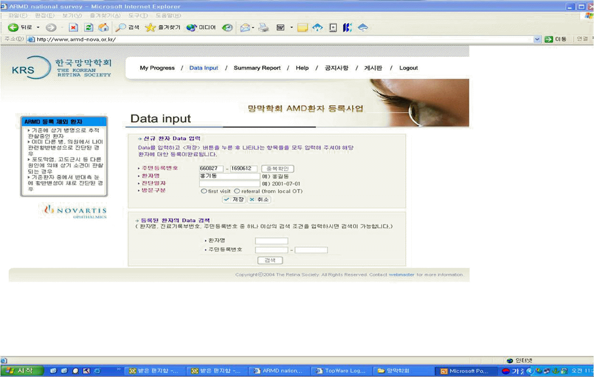 | Figure 1.Registry homepage (www.armd-nova.or.kr) of the Korean Retina Society for uploading the data of patients with age-related macular degeneration. |
Table 1.
Demographics and clinical features of age-related macular degeneration (AMD) in Korea
Table 2.
Age distribution and subtypes of age-related macular degeneration (AMD) in Korea
| Age | Early AMD | Exudative AMD | Geographic atrophy | Total |
|---|---|---|---|---|
| 50∼59 | 22 | 106 | 1 | 129 (11.3) |
| 60∼69 | 85 | 336 | 13 | 434 (38.0) |
| 70∼79 | 66 | 361 | 20 | 447 (39.2) |
| 80∼ | 17 | 111 | 3 | 131 (11.5) |
| total | 190 | 914 | 37 | 1141 |
Table 3.
Phenotypic characteristics of the age-related macular degeneration (AMD) in Korea
Table 4.
Location of choroidal neovascularization in korean age-related macular degeneration (AMD) patients
| location | Predominantly classic CNV* | Minimally classic CNV | Occult CNV | Total (%) |
|---|---|---|---|---|
| Subfovea | 140 (15.9) | 129 (14.7) | 593 (67.5) | 862 (80.4) |
| Juxtafovea | 36 (35.3) | 17 (16.7) | 45 (44.1) | 98 (9.1) |
| Extrafovea | 28 (23.9) | 26 (22.2) | 58 (49.6) | 112 (10.5) |
| Total (%) | 204 (18.6) | 172 (15.7) | 696 (63.4) | 1072 (100.0) |
Table 5.
Subgroup analysis of choroidal neovascularization in age-related macular degeneration (AMD) patients; Analysis of 580 eyes of exudative AMD patients who underwent indocyanin green angiogram
| Lesions | Number (%) |
|---|---|
| Choroidal neovascular membrane | 360 (62.1) |
| Polypoidal choroidal vasculopathy | 184 (31.7) |
| Retinal angiomatous proliferation | 36 (6.2) |
| Total | 580 (100.0) |
Table 6.
Laterality of the korean patients with age-related macular degeneration (AMD)
| Phenotype | Early AMD |
Late AMD |
||
|---|---|---|---|---|
| Exudative AMD | Geographic atrophy | |||
| Laterality | unilateral | 73 (38.4) | 728 (79.6) | 26 (70.3) |
| bilateral | 117 (61.6) | 186 (20.4) | 11 (29.7) | |
| Total | 190 | 914 | 37 | |
Table 7.
Comparison of characteristics between normal control and early and late age-related macular degeneration (AMD) patients
| Risk factors | Control (n=3,135) | Early AMD (n=190) | Late AMD (n=951) | P-value |
|---|---|---|---|---|
| Age (years) | 65.04±4.31 | 68.77±7.63 | 69.88±8.10 | <0.001 |
| Gender (M/F) | 1698/1437 (54.2%) | 85/105 (44.7%) | 605/346 (63.6%) | <0.001 |
| Smoking* | 1039/2096 (33.14%) | 58/132 (30.5%) | 422/529 (44.3%) | 0.022 |
| Drinking (yes/no) | 1248/1887 (39.8%) | 62/128 (32.6%) | 384/567 (40.4%) | 0.388 |
| Diabetes (yes/no) | 132/3003 (4.2%) | 44/146 (22.2%) | 141/810 (14.8%) | <0.001 |
| Hypertension (yes/no) | 396/2739 (12.6%) | 69/121 (36.3%) | 297/654 (31.2%) | <0.001 |
| BMI (kg/m2) | 24.27±2.89 | 23.04±2.93 | 23.41±2.80 | <0.001 |
| Cerebral or Cardiovascular disease (yes/no) | 256/2879 (8.2%) | 21/169 (11.1%) | 91/860 (9.6%) | 0.086 |
Table 8.
Associations with early and late age-related macular degeneration (AMD)
| AMD vs. Control OR (95% CI) | P-value | Early AMD vs. Control OR (95% CI) | P-value | Late AMD vs. Control OR (95% CI) | P-value | |
|---|---|---|---|---|---|---|
| Age (yrs) | ||||||
| 50∼60 | 1.0 (referent) | 1.0 (referent) | 1.0 (referent) | |||
| 61∼70 | 7.84 (6.17–9.98) | 0.000 | 11.20 (6.94–18.09) | 0.000 | 7.65 (5.88–9.95) | 0.000 |
| >71 | 44.04(33.53–57.84) | 0.000 | 35.94 (21.22–60.85) | 0.000 | 46.90 (35.07–62.72) | 0.000 |
| Male gender | 1.55 (1.22–1.97) | 0.000 | 0.74 (0.46–1.20) | 0.23 | 1.85 (1.43–2.40) | 0.000 |
| Smoking* | 2.66 (2.09–3.38) | 0.000 | 3.76 (2.32–6.08) | 0.000 | 2.68 (2.07–3.46) | 0.000 |
| Alchol (2–3 times/week) | 0.99 (0.81–1.20) | 0.882 | 1.14 (0.74–1.75) | 0.55 | 0.98 (0.79–1.21) | 0.85 |
| Diabetes (yes/no) | 3.22 (2.39–4.34) | 0.000 | 5.71 (3.55–9.16) | 0.000 | 2.93 (2.13–4.04) | 0.000 |
| Hypertension (yes/no) | 2.36 (1.91–2.92) | 0.000 | 3.14 (2.13–4.61) | 0.000 | 2.27 (1.82–2.85) | 0.000 |
| CVA History† (yes/no) | 0.89 (0.66–1.21) | 0.46 | 0.99 (0.54–1.80) | 0.97 | 0.89 (0.64–1.24) | 0.49 |
| BMI (kg/m2) | ||||||
| <18.5 | 1.0 (referent) | 1.0 (referent) | 1.0 (referent) | |||
| 18.5∼22.9 | 1.20 (0.80–1.80) | 0.39 | 0.58 (0.29–1.16) | 0.13 | 1.32 (0.84–2.06) | 0.23 |
| 23∼25 | 1.35 (0.89–2.05) | 0.16 | 0.50 (0.24–1.03) | 0.06 | 1.56 (0.99–2.47) | 0.058 |
| >25 | 9.74 (6.10–15.55) | 0.000 | 2.89 (1.30–6.42) | 0.009 | 11.56 (6.93–19.30) | 0.000 |
 XML Download