Abstract
Purpose
To examine the effects, complications, and safeties of sclera allograft and amniotic membrane transplantation with fibrin glue as surgical treatment methods for scleromalacia.
Methods
The study included 14 eyes of 14 scleromalacia patients who needed surgical treatment. Among them, seven eyes of seven patients whose scleral defect was small (<6 mm) were operated on using only fibrin glue and no suturing, while seven eyes of seven patients whose defect was large (>6 mm) were operated on using fibrin glue and minimum suturing. Amniotic membrane transplantation was performed at the site of the conjunctival defect.
Go to : 
References
1. Hong SB, Oh SJ, Oh JH. The effects and complications of Mitomycin-C for prevention of recurrence after pterygium operation. J Korean Ophthalmol Soc. 1998; 39:2013–8.
2. Moriarty AP, Crawford GJ, Mcallister IL, Constable IJ. Severe corneosclera infection: a complication of beta irradiation sclera necrosis following pterygium excision. Arch Ophthalmol. 1993; 111:947–51.
3. Kwak JY, Chang HG. Autogenous temporalis fascia grafting and conjunctival flap transposition in scleromalacia after pterygium excision. J Korean Ophthalmol Soc. 2004; 45:180–6.
4. Kim JH, Lee HB, Yoon DK. Scleral grafts for the cases of scleral perforation, scleral ectasia and scleral necrosis. J Korean Ophthalmol Soc. 1978; 19:55–64.
5. Kim JH. Scleral grafting on necrotic scleritis following pterygium excision. J Korean Ophthalmol Soc. 1982; 23:29–39.
6. Rhee HS, Kim MS, Kim JH. Scleral graft on necrotic scleritis abdominal pterygium excision. J Korean Ophthalmol Soc. 1987; 28:565–9.
7. Lin CP, Tsai MC, Wu YH, Shih MH. Repair of a giant scleral abdominal with preserved sclera and tissue adhesive. Ophthalmic Surg Lasers. 1996; 27:995–9.
8. Kim SY, Chung WS, Hahn DK. Surgical management of scleral necrosis. J Korean Ophthalmol Soc. 1995; 36:7–12.
9. Oh JH, Kim JC. Repair of scleromalacia using preserved sclera graft with amniotic membrane transplantation. Cornea. 2003; 22:288–93.
10. Na YS, Joo MJ, Kim JH. Results of sclera allografting on sclera abdominal following pterygium excision. J Korean Ophthalmol Soc. 2005; 46:402–9.
11. Ozcan AA, Bilgic E, Yagmur M, Ersöz TR. Surgical management of scleral defects. Cornea. 2005; 24:308–11.

12. Sangwan VS, Jain V, Gupta P. Structural and functional outcome of scleral patch graft. Eye. 2007; 21:930–5.

13. Sohn YH, Yu SL, Uhm KB. Surgical treatment of sclera abdominal as a complication of pterygium excision. J Korean Ophthalmol Soc. 1995; 36:1323–30.
14. Lee CO, Jong SH, Lee JJ. Autologous simple conjunctival graft and conjunctiva/tenon graft on focal scleromalacia. J Korean Ophthalmol Soc. 1997; 38:1737–41.
15. Breslin CW, Katz JI, Kaufmann HE. Surgical management of abdominal scleritis: sclera reinforcement with autogenous periosteum. Arch Ophthalmol. 1977; 95:2038–40.
16. Mauriello JA Jr, Fiore PM, Pokorny KS, Cinotti DJ. Use of split-thickness dermal graft in the surgical treatment of corneal and scleral defects. Am J Ophthalmol. 1998; 105:244–7.

17. Cohen RA, McDonald MB. Fixation of conjunctival autografts with an organic tissue adhesive. Arch Ophthalmol. 1993; 111:1167–8.

18. Koranyi G, Seregard S, Kopp ED. Cut and paste: a no suture, small incision approach to pterygium surgery. Br J Ophthalmol. 2004; 88:911–4.

19. Uy HS, Reyes JM, Flores JD, Lim-Bon-Siong R. Comparison of fibrin glue and sutures for attaching conjunctival autografts after pterygium excision. Ophthalmology. 2005; 112:667–71.

20. Yoon KC, Heo H, Jeong IY, Park YG. The use of fibrin glue for conjunctival autotransplantation in pterygium. J Korean Ophthalmol Soc. 2006; 47:198–204.
22. Kaufman HE, Insler MS, Ibrahim-Elzembely HA, Kaufman SC. Human fibrin tissue adhesive for sutureless lamellar keratoplasty and sclera patch adhesion: a pilot study. Ophthalmology. 2003; 110:2168–72.
23. Stenkula S, Gislason I, Törnquist R. Primary sclera atrophy and retinal detachment. Acta Ophthalmol (Copenh). 1981; 59:350–8.
24. Mizano K, Hayasaka S. Penetrating keratoplasty with use of abdominal and sclera strip in acute corneal perforations. Ophthalmic Surg. 1982; 13:475–7.
Go to : 
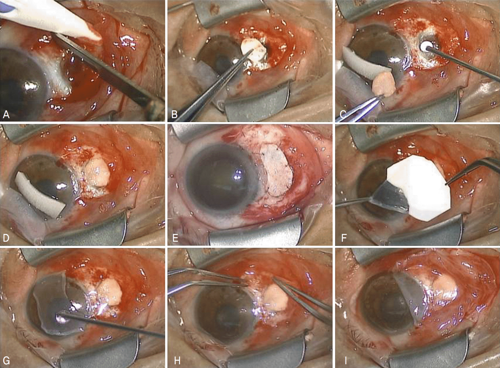
 | Figure 1.(A) The scleromalacia lesion is cleaned. (B) The preserved sclera is cut according to the size and shape of the lesion. (C, D) Fibrin glue is applied to the lesion and then the sclera graft is placed and adhered to the lesion. (E) Large sclera defect (>6 mm) is operated using fibrin glue and minimum suturing (F) The amniotic membrane is peeled off from the carrier paper from the storage medium. (G) Thrombin solution is administered on the exposed sclera, and fibrinogen solution is applied on stromal side of amniotic membrane. (H) The amniotic graft is immediately transferred onto the exposed sclera. (I) The amniotic patch is then secured to the conjunctiva by continuous sutures with 10–0 nylon. |
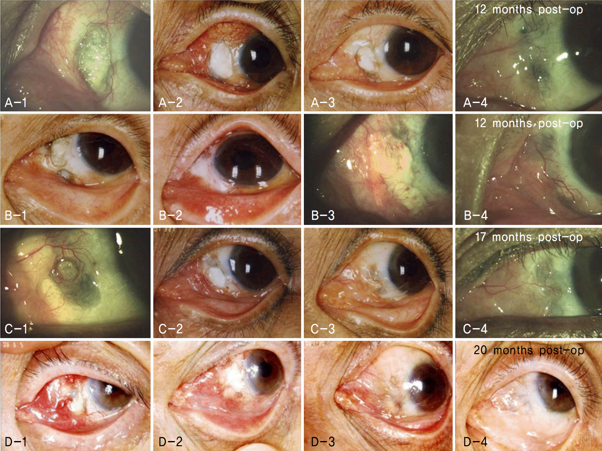
 | Figure 2.(A, B, C, D-1) Preoperative photograph showing sclera thinning with exposed uveal tissue at the site of previous pterygium excision. (A, B, C, D-2) 1 month postoperative photograph. In all cases conjunctival re-epithelization is shown but completely vascularized over the exposed scleral graft is not yet shown. (A, B, C, D-3) 6 months postoperative photograph. Sclera graft is completely covered by fibrovascular patch and in 2 cases partial reabsorption of scleral graft are shown. (A, B, C, D-4) Last follow-up photograph showing stable ocular surface but in all cases partial reabsorption of sclera graft are shown. |
Table 1.
Clinical outcome and complication of preserved sclera graft and amniotic membrane transplantation with fibrin glue for repair of scleromalacia
Table 2.
Previous reports of preserved sclera graft with conjunctival flap or amniotic membrane transplantation for repair of scleromalacia
| Author(s) | Number of eyes | Previous history | Surgery technique | Mean follow-up (months) | Results | Complication (eyes) |
|---|---|---|---|---|---|---|
| Kim et al8 | 16 | Pterygium surgery with | Lamellar sclera grafting with | 12 | Stable ocular | Corneal erosion (6) |
| MMC* (9) | conjunctival flap (13) | surface | Conjunctival cyst | |||
| With Strontium (5) | Simple conjunctival flap (3) | (15, 93.8%) | formation (2) | |||
| Thermal burn (1) | Sclera graft melting (1) | |||||
| Cryotherapy (1) | Partial absorption of | |||||
| conjunctival flap (1) | ||||||
| Oh and Kim9 | 8 | Pterygium surgery with | Preserved sclera allograft | 24 | Stable ocular | None |
| MMC (6) | with AMT§ | surface | ||||
| Cataract op. (2) | ||||||
| Na et al10 | 5 P | Pterygium excision with | Preserved sclera allograft | 19 | Stable ocular | None |
| MMC or irradiation | with conjunctival | surface | ||||
| advancement (4) or with | ||||||
| AMT (1) | ||||||
| Ozcan et al11 | 8 | Intraocular surgery (6) | Corneoscleral graft (3) | 13.1±3.9 | Stable ocular | Recurrence (1) |
| Trauma (1) | Scleral graft (4) | surface | ||||
| Both (1) | Fascia lata (1) | |||||
| Sangwan et al12 | 13 | Pterygium surgery (6) | Scleral patch graft with | 24.3 | Stable ocular | Endophthalmitis (1) |
| Injury (3) | conjunctival flap (10) or | surface | Graft dehiscence (1) | |||
| None (3) | with AMT (3) | (10, 76.9%) | Necrotizing scleritis (1) | |||
| Cataract surgery and | ||||||
| TSCP† (1) | ||||||
| OSSN‡ excision (1) | ||||||
| This study | 14 P | Pterygium surgery (10) | Preserved scleral allograft | 14.6±4.4 | Stable ocular | None |
| With MMC (4) | with AMT with fibrin glue | surface |


 PDF
PDF ePub
ePub Citation
 Print
Print


 XML Download