

 PDF
PDF ePub
ePub Citation
 Print
Print


Abstract
Purpose
To evaluate the long-term results of orbital fat decompression with or without lateral wall decompression in Grave's ophthalmopathy.
Methods
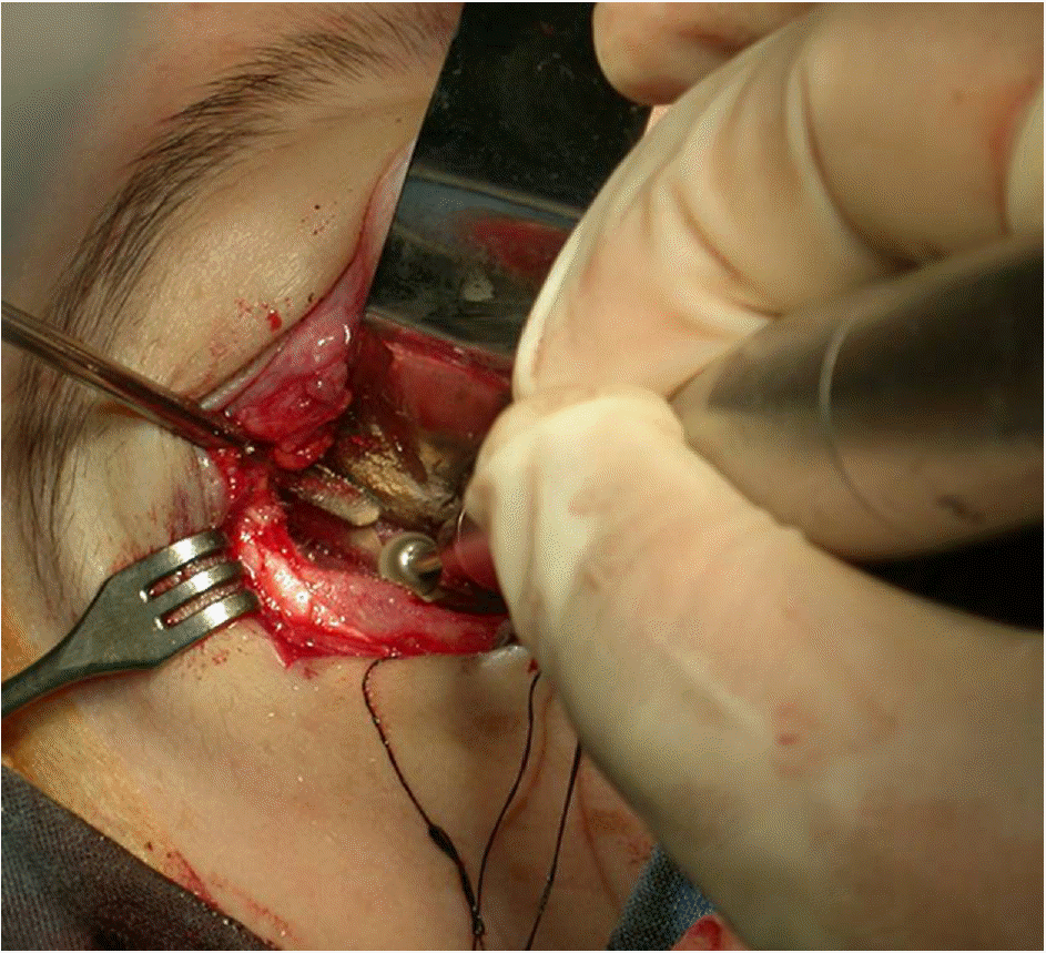
The authors studied retrospectively the 24 thyroid-related orbitopathy patients (32 eyes) who had undergone fat removal decompression or combined orbital decompression (lateral wall decompression with fat removal) based on average exophthalmometric values for Koreans. The patients were followed for over six months postoperatively, from December 2000 to December 2008.
Results
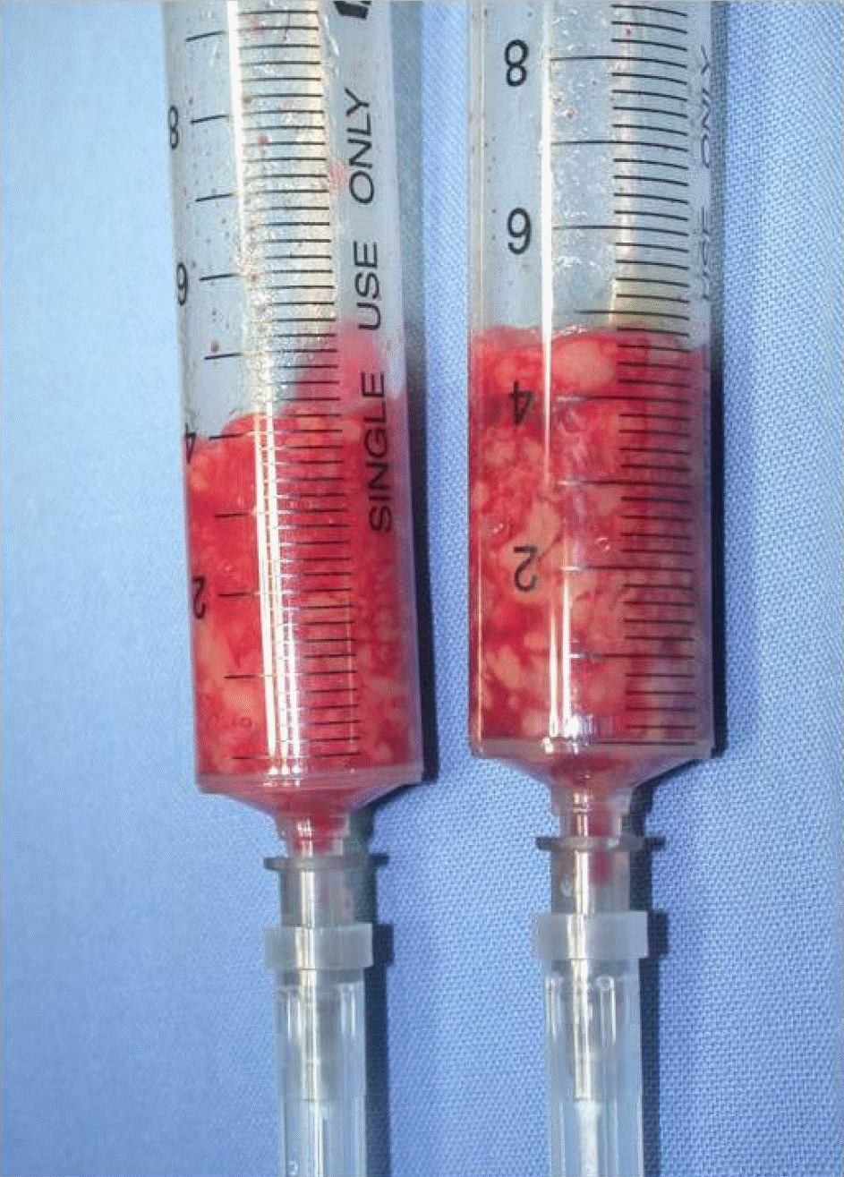
The average amount of exophthalmos reduction was 4.13±2.60 mm in the fat removal decompression group and 5.79±2.02 mm in the combined orbital decompression group, respectively (p<0.001). The final postoperative BCVA (best corrected visual acuity) in patients with compressive optic neuropathy improved in all cases. Intraocular pressure decreased significantly in all groups after operation (p<0.001). Except for one patient who had newly onset diplopia, which was transient, no patient's diplopia worsened.
Go to : 
References
1. Lyons CJ, Rootman J. Orbital decompression for disfiguring exophthalmos in thyroid orbitopathy. Ophthalmology. 1994; 101:223–30.

2. Garrity JA, Fatourechi V, Bergstralh EJ, et al. Results of abdominal orbital decompression in 428 patients with severe Graves' ophthalmopathy. Am J Ophthalmol. 1993; 116:533–47.
3. Leone CR, Piest KL, Newman RJ. Medial and lateral wall deabdominal for thyroid ophthalmopathy. Am J Ophthalmol. 1989; 108:160–6.
4. Trokel S, Kazim M, Moore S. Orbital fat removal. Decompression for Graves orbitopathy. Ophthalmology. 1993; 100:674–82.
5. Kook KH, Kim YK, Lee SY. Exophthalmometric values of Korean using Hertel and Naugle exophthalmometers. J Korean Ophthalmol Soc. 2003; 44:10–5.
6. Walsh TE, Ogura JH. Transantral orbital decompression for abdominal exophthalmos. Laryngoscope. 1957; 67:544–68.
7. Ogura JH, Lucente FE. Surgical results of orbital decompression for malignant exophthalmos. Laryngoscope. 1974; 84:637–44.
8. Ogura JH, Thawley SE. Orbital decompression of exophthalmos. Otolaryngol Clin North Am. 1980; 13:29–38.
9. Shorr N, Neuhaus RW, Baylis HI. Ocular motility problems after orbital decompression for dysthyroid ophthalmopathy. Ophthalmology. 1982; 89:323–8.
10. De Santo LW. The total rehabilitation of Graves' ophthalmopathy. Laryngoscope. 1980; 90:1652–78.
11. Liao SL, Chang TC, Lin LL. Transcaruncular orbital deabdominal-an alternate procedure for Graves' ophthalmopathy with compressive optic neuropathy. Am J Ophthalmol. 2006; 141:810–9.
12. Goldberg RA, Rootman J, Stuart B. Orbital surgery: a conceptual approach. Philadelphia, Pennsylvania: Lippincott-Raven;1995. p. 362–3.
13. Goldberg RA, Shorr N, Cohen MS. The medial orbital strut in the prevention of post-decompression dystopia in dysthyroid ophthalmopathy. Ophthal Plast Reconstr Surg. 1992; 8:32–4.
14. Goldberg RA, Perry JD, Hortaleza V, Tong JT. Strabismus after balanced medial plus lateral wall versus lateral wall only orbital decompression for dysthyroid orbitopathy. Ophthal Plast Reconstr Surg. 2000; 16:271–7.
15. Liao SL, Shih MJ, Chang TC, Lin LL. Transforniceal lateral deep bone decompression—a modified technique to prevent abdominal diplopia in patients with disfiguring exophthalmos due to dysthyroid orbitopathy. J Formos Med Assoc. 2006; 105:611–6.
16. Ben-Simon GJ, Wang L, McCann JD, Goldberg RA. Primary gaze diplopia in patients with thyroid-related orbitopathy abdominal deep lateral orbital decompression with intraconal fat debulking: a retrospective analysis of treatment outcome. Thyroid. 2004; 14:379–83.
17. Ben-Simon GJ, Syed HM, Lee S, et al. Strabismus after deep lateral wall orbital decompression in thyroid-related orbitopathy abdominals using automated Hess screen. Ophthalmology. 2006; 113:1050–5.
18. Stabile JR, Trokel SM. Increase in orbital volume obtained by decompression in dried skulls. Am J Ophthalmol. 1983; 95:327–31.
19. Adenis JP, Robert PY, Lasudry JG, Dalloul Z. Treatment of proptosis with fat removal orbital decompression in Graves' ophthalmopathy. Eur J Ophthalmol. 1998; 8:246–52.
20. Baek SH, Lee TS. Prediction of late enophthalmos by volumetric analysis of blowout fractures using orbital CT. J Korean Ophthalmol Soc. 1999; 40:3239–45.
Go to :
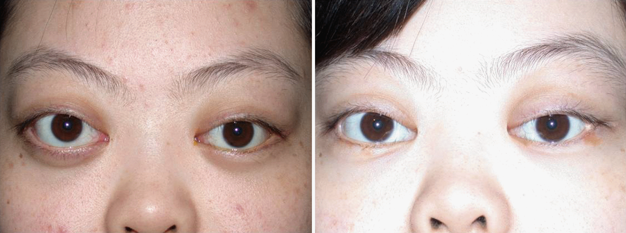 | Figure 3.(Left) Exophthalmos of the right eye is noted preoperatively. (Right) Exophthalmos is significantly reduced by lateral wall and fat decompression of the right eye. |
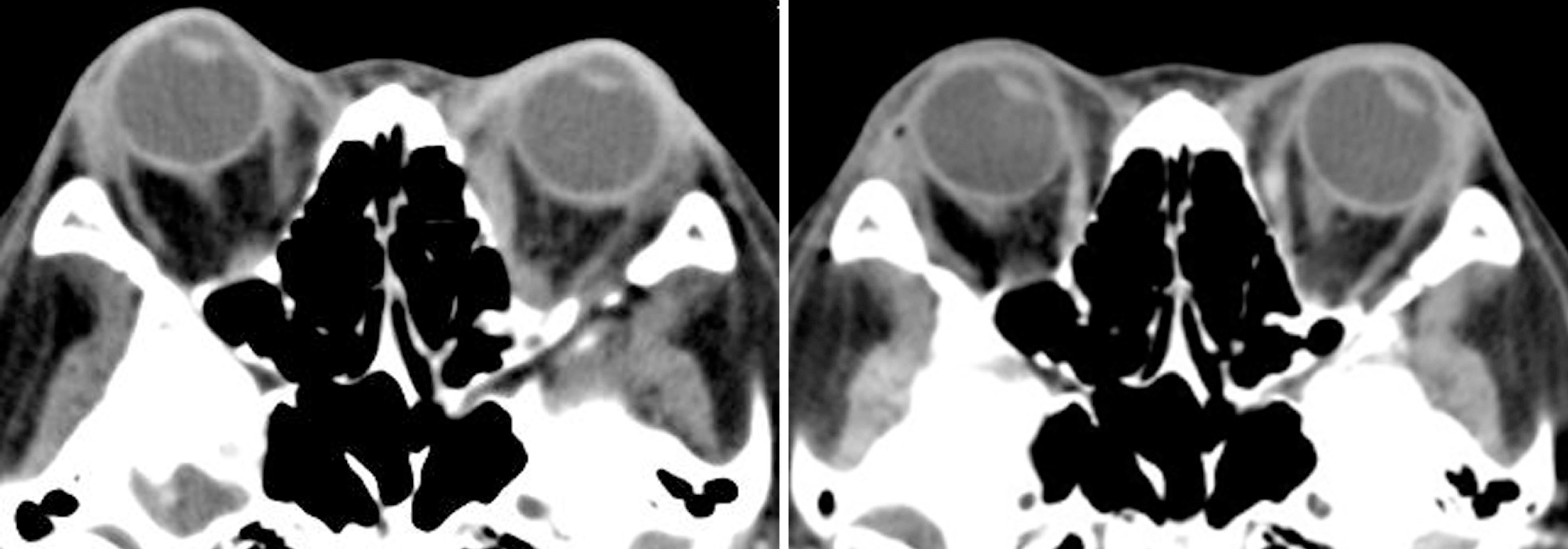 | Figure 4.Pre-(left) and post-operative (right) orbit CT scans show a reduction of exophthalmos of the right orbit. |
Table 1.
Classification of patients by amount of preop Hertel value of exophthalmos
| Number (Orbit) | Gender (M:F) | Mean age (yr*) | Duration (mon†) | F/U‡ (mon†) | |
|---|---|---|---|---|---|
| Group1 | 14 (20) | 5:9 | 37.75±11.39 | 12.85±9.25 | 25.25±13.75 |
| Group2 | 10 (12) | 3:7 | 38.25±9.95 | 12.67±9.19 | 22.33±10.99 |
Table 2.
Results of surgical decompression, visual acuity, and intraocular pressure
| Group 1 | Group 2 | |
|---|---|---|
| Preoperative proptosis (mm) | 21.93±3.23 | 23.08±2.58 |
| Postoperative proptosis (mm) | 17.80±2.57 | 17.29±1.86 |
| p-value‡ | <0.001 | <0.001 |
| Fat removal amount (ml) | 4.85±2.12 | 4.12±2.06 |
| Decreased proptosis (mm) | 4.13±2.60 | 5.79±2.02 |
| Preoperative BCVA* (log MAR) | 0.29±0.31 | 0.31±0.44 |
| Postoperative BCVA* (log MAR) | 0.25±0.25 | 0.29±0.43 |
| p-value‡ | 0.154 | 0.882 |
| Preoperative IOP† (mmHg) | 18.30±4.24 | 16.25±1.91 |
| Postoperative IOP† (mmHg) | 16.25±3.09 | 13.75±1.22 |
| p-value‡ | <0.001 | <0.001 |
Table 3.
Visual field changes in each group
| Improved | No change | Aggravated | |
|---|---|---|---|
| Group1 | 7 (35.0%) | 12 (63.2%) | 1* |
| Group2 | 4 (33.3%) | 8 (66.7%) | 0 |
Table 4.
Diplopia changes in each group
| Preoperative diplopia | New-onset diplopia | Improved | No change | Aggravated | |
|---|---|---|---|---|---|
| Group 1 | 4 (20.0%) | 1* | 3 | 16 | 0 |
| Group 2 | 4 (33.3%) | 0 | 3 | 9 | 0 |
 XML Download