Abstract
Purpose
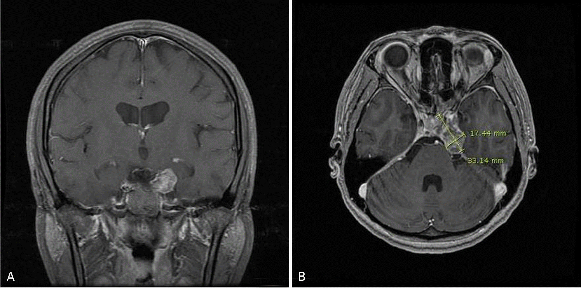
To report a patient with an eyeball movement disorder due to a chondrosarcoma in the left cavernous sinus.
Case summary
A 33-year-old woman visited our clinic complaining of diplopia she hand for one month. She had been undergone two operation for the removal of chondrosarcoma in the left cavernous sinus, and she had left esotropia of 25 prism diopters in the primary position and limited abduction of approximately −1.5 in the left eye. Therefore she was treated with radiotherapy for six weeks. Three weeks after radiotherapy she had esotropia of 2 prism diopters in the primary position and limited abduction of approximately −1 in the left eye. Six weeks later, only limited abduction of approximately −0.5 in the left eye was present.
References
1. Richards BW, Jones FR, Younge BR. Causes and prognosis in 4,278 cases of paralysis of the oculomotor, trochlear, and abdominal cranial nerves. Am J Ophthalmol. 1992; 113:489–96.
2. Patel SV, Mutyala S, Leske DA, et al. Incidence, associations and evaluation of six nerve palsy using a population-based method. Ophthalmology. 2004; 111:369–75.
3. Han ER, Lim KH. Clinical features of the sixth cranial nerve palsy. J Korean Ophthalmol Soc. 2008; 49:1323–9.

4. Peters GB, Bakri SJ, Krohel GB. Cause and prognosis nontraumatic sixth nerve palsies in young adults. Ophthalmology. 2002; 109:1925–8.
5. Falconer MA, Bailey IC, Duchen LW. Surgical treatment of abdominal and chondroma of the skull base. J Neurosurg. 1968; 29:261–75.
6. Bae JH, Kim HC, Cho YU, Son KR. Mesenchymal abdominal of the orbit; A case report and review of the literature. J Korean Ophthalmol Soc. 1991; 32:599–603.
7. Suh JG, Lee DG, Kim SM. A case of extraskeletal mesenchymal chondrosarcoma of the orbit. J Korean Ophthalmol Soc. 1981; 22:681–4.
8. Sekhar LN, Pranatartiharan R, Chanda A, Wright DC. Chordomas and chondrosarcoma of the skull base: results and complications of surgical management. Neurosurg Focus. 2001; 10:E2.
9. Volpe NJ, Liebsch NJ, Munzenrider JE, Lessell S. Neuro-oph-thalmologic findings in chordoma and chondrosarcoma of the skull base. Am J Ophthalmol. 1993; 115:97–104.

10. Hassounah M, Al-Mefty O, Akhtar M, et al. Primary cranial and intracranial chondrosarcoma. A survey. Acta Neurochir (Wien). 1985; 78:123–32.
11. Hug EB, Loredo LN, Slater JD, et al. Proton radiation therapy for chordomas and chondrosarcomas of the skull base. J Neurosurg. 1999; 91:432–9.

12. Moster ML, Savino PJ, Sergott RC, et al. Isolated sixth-nerve abdominal in younger adults. Arch Ophthalmol. 1984; 102:1328–30.
13. Tiffin PA, MacEwen CJ, Craig EA, Clayton G. Acquired palsy of the oculomotor, trochlear and abducens nerves. Eye. 1996; 10:377–84.

14. Veth R, Schreuder B, van Beem H, et al. Cryosurgery in aggressive, benign, and low-grade malignant bone tumours. Lancet Oncol. 2005; 6:25–34.

15. Brown RV, Sage MR, Brophy BP. CT and MRI findings in abdominals with chordomas of the petrous apex. Am J Neuroradiol. 1990; 11:121–4.
Figure 1.
At the initial visit, the patient had approximately 25 prism diopters of left esodeviation (middle). Full adduction was present (left), but there was limitation of abduction of approximately −1.5 in the left eye (right).

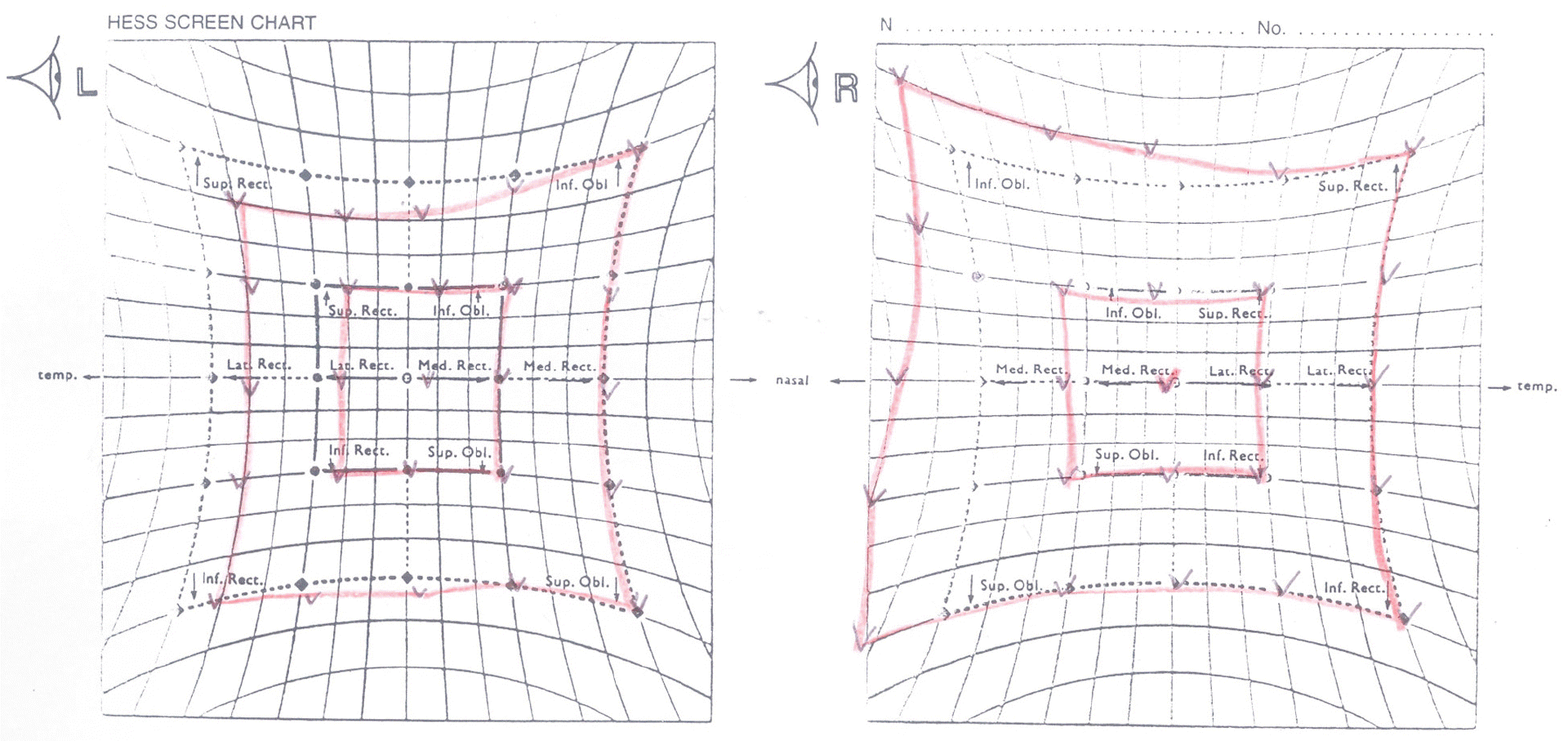
Figure 2.
Hess screen test. It showed a hypofunction of lateral rectus muscle of left eye and a hyperfunction of medial rectus muscle of right eye.


 PDF
PDF ePub
ePub Citation
 Print
Print


 XML Download