Abstract
Purpose
To report a case of choroidal metastasis of breast cancer that was treated with modified photodynamic therapy.
Case summary
A 45-year-old woman visited our clinic with blurred vision of the right eye, which began 1 month before. The patient previously suffered from a low back pain for 1 year. The best corrected visual acuity was 20/20 in both eyes. Fundus examination revealed an elevated yellowish mass-like lesion at the superonasal area in the right eye. Ultrasonography of the right eye showed a highly echogenic choroidal mass with moderate to high internal reflectivity. Fluorescein angiography showed hypofluorescence during the prearterial and arteriovenous phase, and well circumscribed hyperfluorescence during the venous phase. Radiologic examination was performed upon suspicion of metastasis. The examination revealed breast cancer with lung, spine and ovary metastasis. Subsequently, biopsy of the breast mass revealed an invasive ductal carcinoma. Based on these results, the patient was diagnosed with choroidal metastasis from breast carcinoma. The patient received systemic chemotherapy, and modified photodynamic therapy (PDT) was performed on the metastatic choroidal mass. Six days after modified PDT, the mass size was unchanged, and serous retinal detachment developed at the macula and inferior retina. However, 22days after treatment, the mass size markedly decreased and the serous retinal detachment was improved.
References
1. Bloch RS, Gartner S. The incidence of ocular metastatic abdominal. Arch Ophthalmol. 1971; 85:673–75.
2. Ferry AP, Font RL. Carcinoma metastatic to the eye and orbit. I.A clinocopathologic study of 227 cases. Arch Ophthalmol. 1974; 92:276–86.
3. Mejia-Novelo A, Alvarado-Miranda A, Morales-Vázquez F, et al. Ocular metastases from breast carcinoma. Med Oncol. 2004; 21:217–21.
4. Shields CL, Shields JA, Gross NE, et al. Survey of 520 eyes with uveal metastases. Ophthalmology. 1997; 104:1265–76.

5. Freedman MI, Folk JC. Metastatic tumors to the eye and orbit: patient survival and clinical characteristics. Arch Ophthalmol. 1987; 105:1215–9.
6. Lee SJ, Kim SY, Kim SD. A case of diode laser photocoagulation in the treatment of choroidal metastasis of breat carcinoma. Korean J Ophthalmol. 2008; 22:187–9.
7. Isola V, Pece A, Pierro L. Photodynamic therapy with verteporfin of choroidal malignancy from breast cancer. Am J Ophthalmol. 2006; 142:885–7.

8. Ferry AP, Font RL. Carcinoma metastatic to the eye and orbit. A clinicopathologic study of 26 patients with carcinoma metastatic to the anterior segment of the eye. Arch Ophthalmol. 1975; 93:472–82.
9. Stephens RF, Shields JA. Diagnosis and management of cancer metastatic to the uvea: a study of 70 cases. Ophthalmology. 1979; 86:1336–49.

10. Lee JS, Kim JH, Oum BS. A case of bilateral choroidal metastasis of breast invasive ductal carcinoma. J Korean Ophthalmol Soc. 1996; 37:1211–7.
11. Shin JA, Yoo JS, Huh W. A case of radiotherapy of choroidal metastasis of breat carcinoma. J Korean Ophthalmol Soc. 1993; 34:480–3.
12. Paul Chan RV, Young LH. Treatment options for metastatic tumors to the choroid. Semin Ophthalmol. 2005; 20:207–16.

13. Kim IT, Song HC. Fluorescein and Indocyanine green abdominal of choroidal tumors. J Korean Ophthalmol Soc. 1999; 40:1866–76.
14. Amer R, Pe'er J, Chowers I, Anteby I. Treatment options in the management of choroidal metastases. Ophthalmologica. 2004; 218:372–7.

15. Schmidt-Erfurth U, Baumann W, Gragoudas E, et al. Photo-dynamic therapy of experimental choroidal melanoma using lipoprotein delivered benzoporphyrin. Ophthalmology. 1994; 101:89–99.
16. Schmidt-Erfurth U, Hasan T, Gragoudas E, et al. Vascular targeting in photodynamic occlusion of subretinal vessels. Ophthalmology. 1994; 101:1953–61.

17. Mennel S, Barbazetto I, Meyer CH, et al. Ocular photodynamic therapy-Standard applications and new indication (PartⅠ). abdominalogica. 2007; 221:216–26.
Figure 1.
The fundus photography of the right eye shows elevated mass-like lesion superonasal to the optic disc.
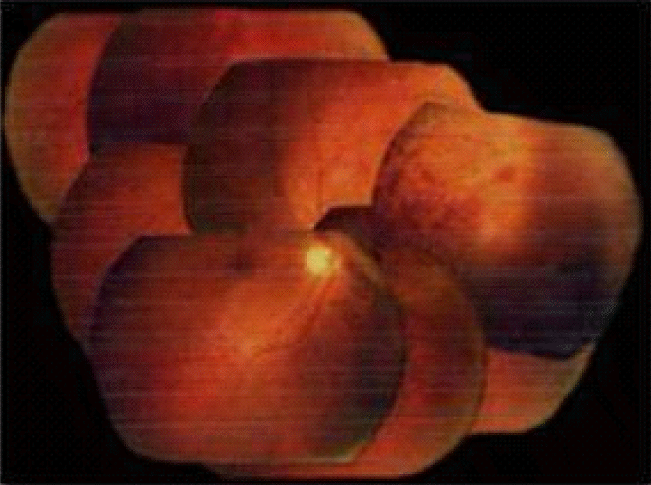
Figure 2.
Ultrasonography of the right eye revealed a highly echogenic choroidal mass with moderate to high internal reflectivity.
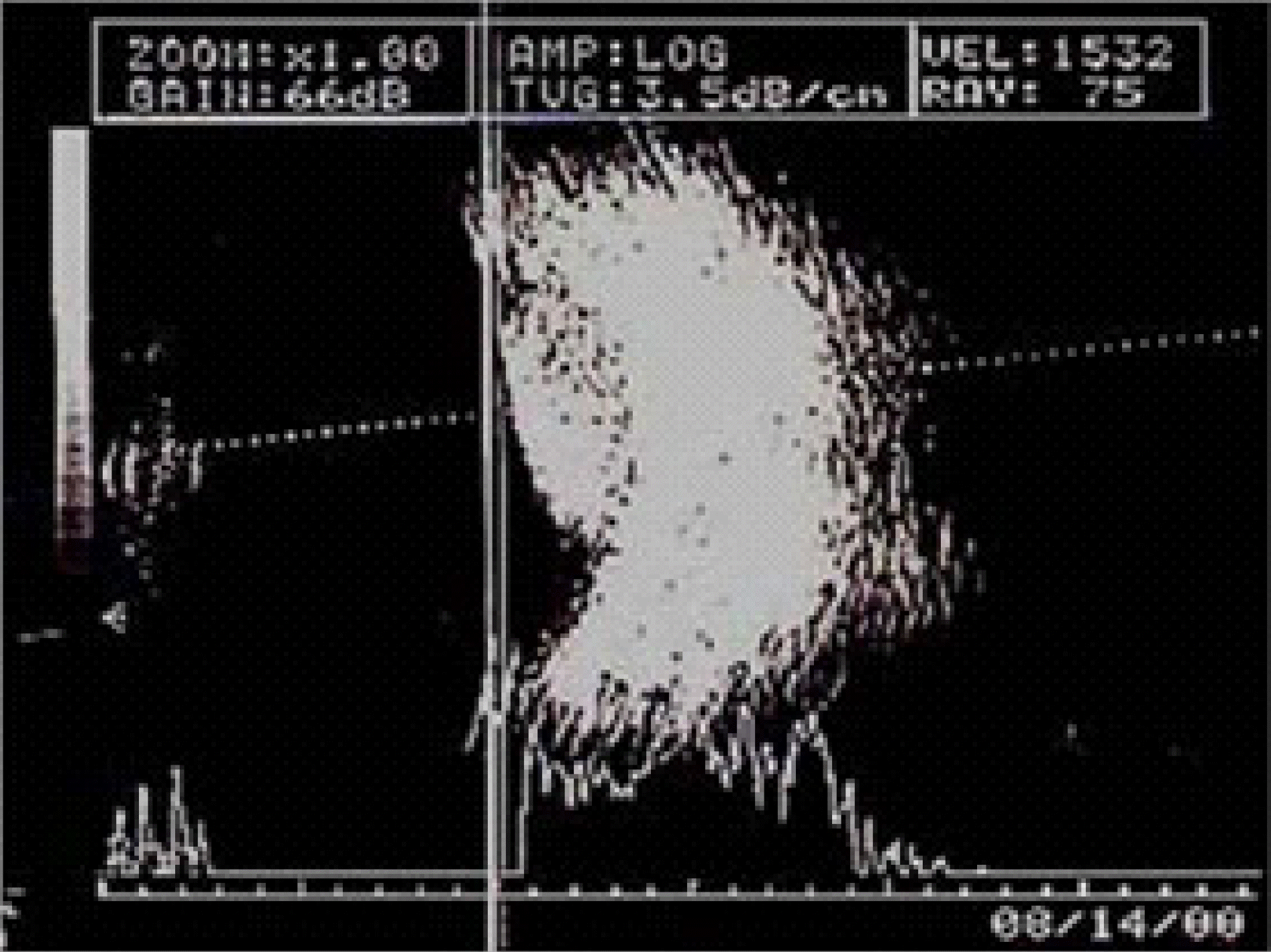
Figure 3.
Fluorescein angiography shows generalized hypofluorescence with pinpoint hyperfluorescence during the prearterial and arteriovenous phase (Top) and hyperfluorescence during the venous phase (Bottom) due to leakage.
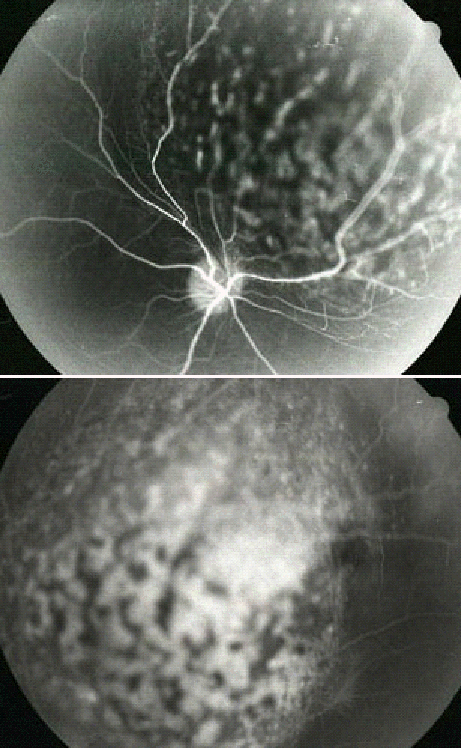
Figure 4.
The 99m Tc bone scan revealed multiple active bone lesions in the thoracic spine and the ribs on both sides.
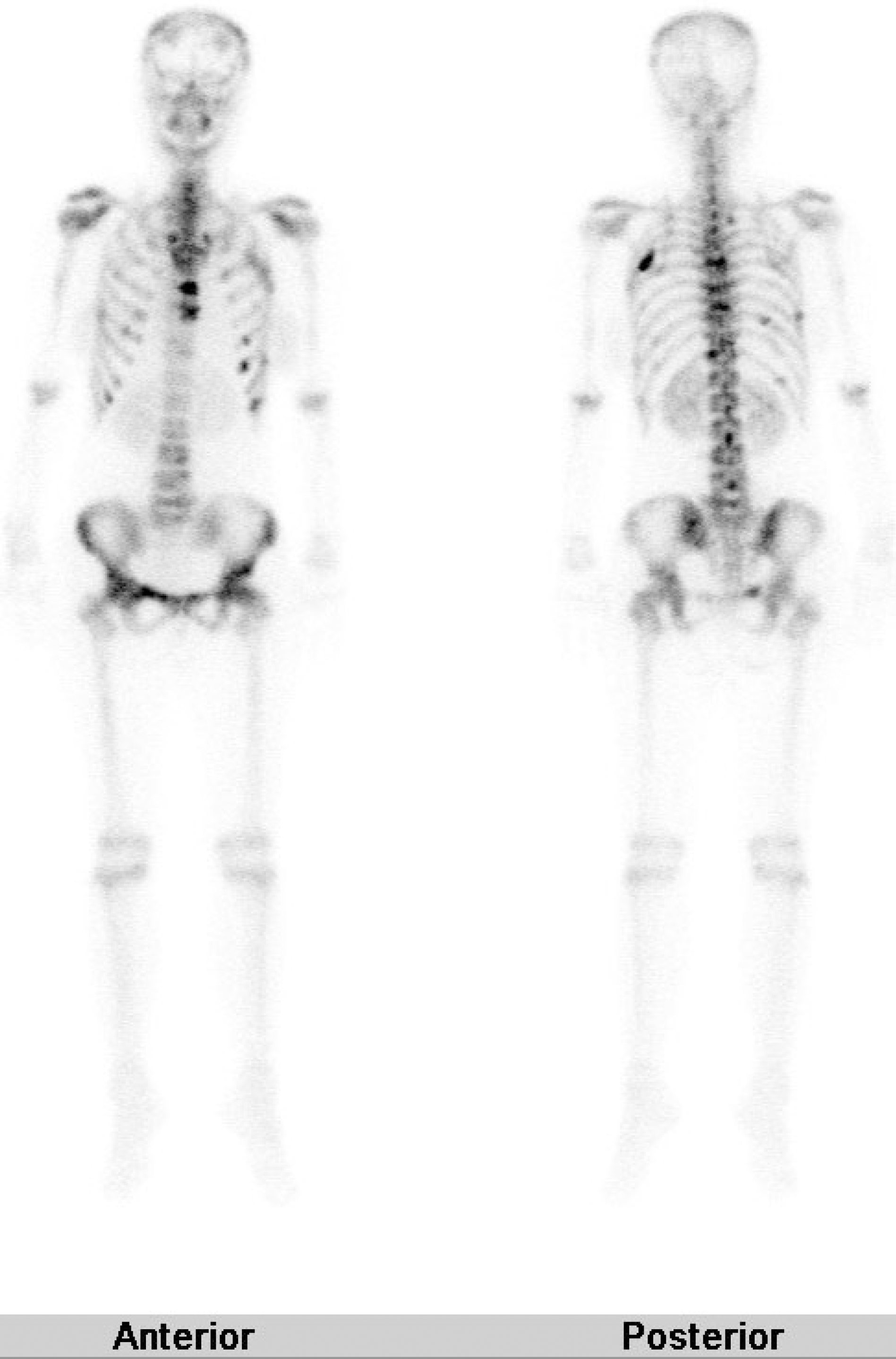
Figure 5.
Pathology of primary breast cancer. The invasive ductal carcinoma infiltrating into the adjacent stromal tissue.
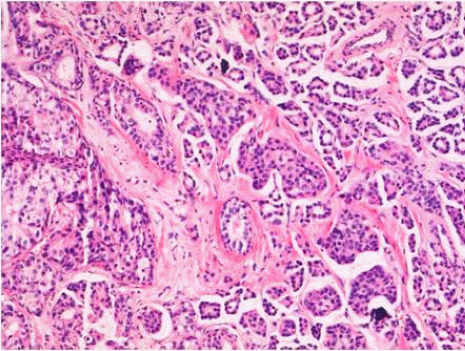
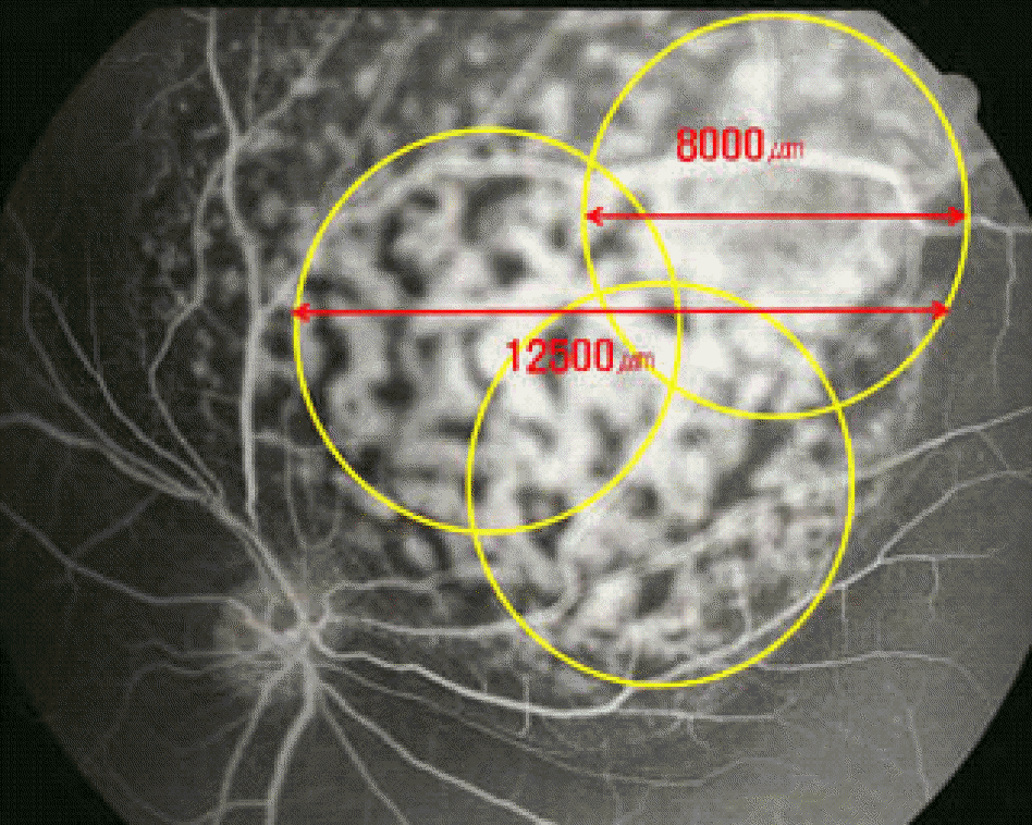
Figure 6.
The diameter of the treatment spot is calculated based on the lesion size measured on the pretreatment fluorescein angiogram. Fundus drawing demonstrating photodynamic therapy of the choroidal tumor using three overlapping spot (yellow circles). The greatest linear dimension of the retinal lesion is 12,500 μm. And each treatment spot size is 8,000 μm.


 PDF
PDF ePub
ePub Citation
 Print
Print


 XML Download