Abstract
Purpose
Corneal bee sting is a relatively rare injury. The authors report the significant endothelial changes despite complete resolution of corneal injury after a corneal bee sting.
Case summary
Two males, ages 55 and 30, presented to our clinic for unilateral decreased visual acuity and eyeball pain after bee sting injuries. At the first visit, localized corneal stromal edema and epithelial defect were detected. One week after medical treatments, both patients achieved restoration of vision and resolution of corneal injury. In one patient, specular microscopy of the traumatized eye at one and five months showed a significant decrease in endothelial densities as compared with the contralateral eye.
References
1. Arcieri ES, Franca ET, de Oliveria HB, et al. Ocular lesions arising after stings by hymenopteran insects. Cornea. 2002; 21:328–30.

2. Singh G. Bee sting of the cornea. Ann Ophthalmol. 1984; 16:320–2.
4. Song HS, Wray SH. Bee sting optic neuritis. A case report with visual evoked potentials. J Clin Neuroophthalmol. 1991; 11:45–9.
6. Smolin G, Wong I. Bee sting of the cornea: case report. Ann Ophthalmol. 1982; 14:342–3.
7. Chen CJ, Richardson CD. Bee sting-induced ocular changes. Ann Ophthalmol. 1986; 18:285–6.
9. King TP, Spangfort MD. Structure and biology of stinging insect venom allergens. Int Arch Allergy Immunol. 2000; 123:99–106.

10. Yildirim N, Erol N, Basmak H. Bee sting of the cornea: a case report. Cornea. 1998; 17:333–4.
12. Gilboa M, Gdal-On M, Zonis S. Bee and wasp stings of the eye. Retained intralenticular wasp sting: A case report. Br J Ophthalmol. 1977; 61:662–4.

14. Jain V, Shome D, Natarajan S. Corneal bee sting misdiagnosed as viral keratitis. Cornea. 2007; 26:1277–8.

15. Yuen KS, Lai JS, Law RW, Lam DS. Confocal microscopy in bee sting corneal injury. Eye. 2003; 17:845–7.

17. Gurlu VP, Erda N. Corneal bee sting-induced endothelial changes. Cornea. 2006; 25:981–3.
Figure 1.
Case 1. Slit-lamp biomicroscopic findings. (A&B) At the first visit, oval-shaped stromal edema and Descemet's foldings was detected in the inferotemporal paracentral cornea. (C) One week later, target-shaped stromal opacity remained with complete resolution of stromal edema.

Figure 2.
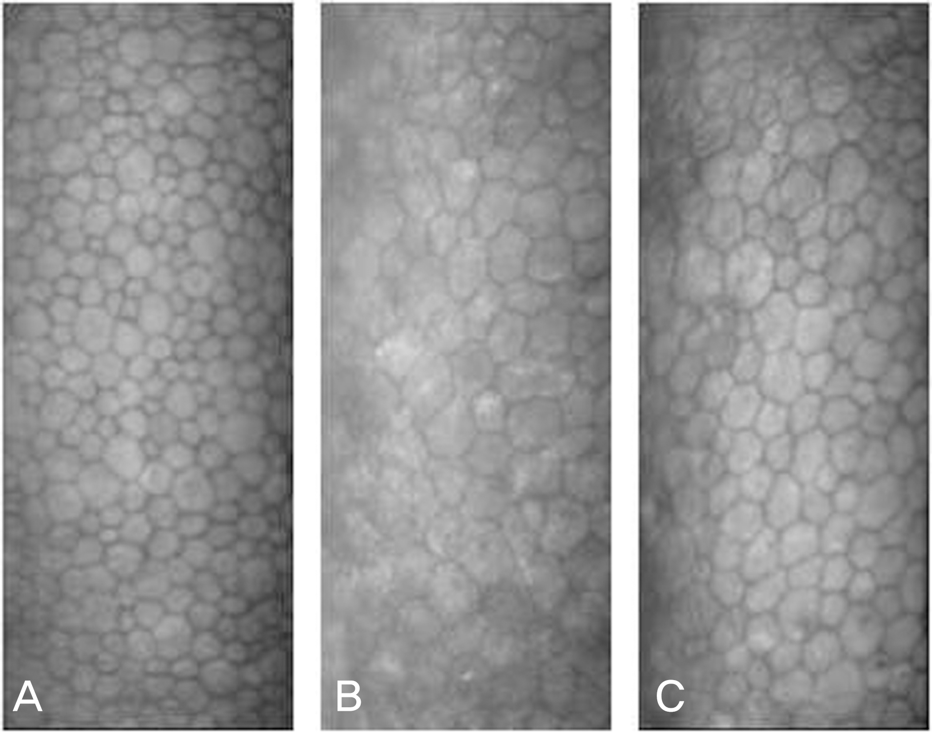
Case 1. Specular microscopic findings (ECD-CoV-Hex) at the center of the cornea. (A) Normal left eye (2621-36-48), (B) Right eye at 1 month after the initial visit (1386-45-57), (C) Right eye at 5 months after the initial visit (1429–44–55). Specular microscopy of the right eye at one and five months showed a significant decrease in endothelial densities compared with the left eye. ECD=endothelial cell densities (/mm2); CoV=coefficient of variation; Hex=hexagonaility (%).

Figure 3.
Case 2. Slit-lamp biomicroscopic findings. (A&B) At the first visit, localized round stromal edema and epithelial defect were detected at the nasal paracentral cornea. (C) One week later, only target-shaped stromal opacity remained with complete resolution of stromal edema.

Figure 4.
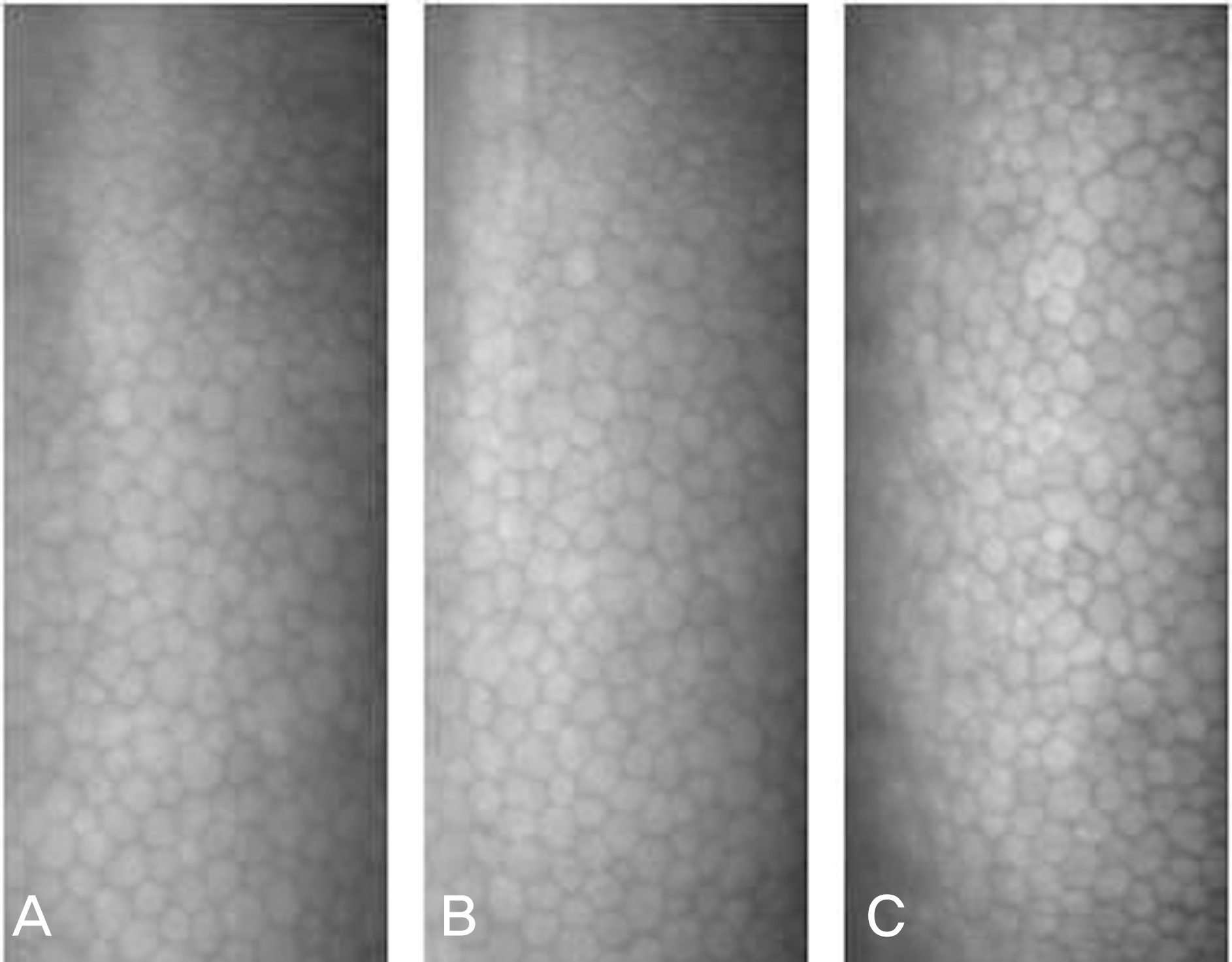
Case 2. Specular microscopic findings (ECD-CoV-Hex) at the center of the cornea. (A) Normal left eye (2802-38-57), (B) Right eye at 1 month after the initial visit (2445-30-44), (C) Right eye at 5 months after the initial visit (2568–29–51). Unlike case 1, specular microscopy of the right eye at one and five months showed a slight decrease in endothelial densities compared with the left eye. ECD=endothelial cell densities (/mm2); CoV=coefficient of variation; Hex=hexagonaility (%).


 PDF
PDF ePub
ePub Citation
 Print
Print


 XML Download