

 PDF
PDF ePub
ePub Citation
 Print
Print


Abstract
Purpose
To evaluate changes in muscle length when retracting the extraocular rectus muscle with muscle hooks during strabismus surgery.
Methods
Forty-four rectus muscles of 42 patients consecutively resected in two hospitals (A, B) were included in this study. After isolation of the rectus muscle, the length of the muscle was recorded and the portion to be resected was marked using gentian violet stain on the tip of calipers. After the rectus muscle was retracted with two muscle hooks in either direction, its length was measured again with the calipers.
Results
The length of the rectus muscle was not changed by retraction in 25 of 44 muscles (56.8%). The length of the muscle was changed by 0.5 mm in 13 muscles (29.5%) and by 1mm in six muscles (13.6%). Changes of rectus muscle length over 0.5 mm were observed in 15 of 27 muscles of patients treated at hospital A (55.5%) and four of 17 muscles of patients treated at hospital B (23.5%). The results for the two hospitals were significantly different (p=0.037).
References
1. von Noorden GK. Binocular vision and ocular motility. Theory and management of strabismus. 6th ed.St. Louis: Mosby;2002. p. 588–90.
2. Krieger F, Cvintal T, Bicas H. Applied force and elongation in the medial rectus in esotropic patients with and without movement restriction. Strabismus. 2004; 12:247–56.

3. Simonsz HJ, Kolling GH, Kaufmann H, van Dijk B. Intraoperative length and tension curves of human eye muscles: Including stiffness in passive horizontal eye movement in awake volunteers. Arch Ophthalmol. 1986; 104:1495–500.
4. Simonsz HJ, Kolling GH, van Dijk B, Kaufmann H. Length-tension curves of human eye muscles during succinylcholine-induced contraction. Invest Ophthalmol Vis Sci. 1988; 29:1320–30.
5. Simonsz HJ. Force-length recording of eye muscles during lo-cal-anesthesia surgery in 32 strabismus patients. Strabismus. 1994; 2:197–218.
6. Collins CC, Jampolsky A, Alden AB, et al. Length-tension abdominal system for strabismus surgery. IEEE Trans Biomed Eng. 1991; 38:230–7.
7. von Noorden GK. Binocular vision and ocular motility. Theory and management of strabismus. 6th ed.St. Louis: Mosby;2002. p. 101–7.
8. Pratt-Johnson JA, Tillson G. Management of strabismus and abdominal: A practical guide. New York: Thieme;1994. p. 227.
9. Kushner BJ, Fisher MR, Lucchese NJ, et al. Factors influencing abdominal to strabismus surgery. Arch Ophthalmol. 1993; 111:75–9.
10. Helveston EM. Reoperations in strabismus. Ophthalmology. 1979; 86:1379–88.
11. Kushner BJ, Preslan MW, Vrabec M. Artifacts of measuring abdominal strabismus surgery. J Pediatr Ophthalmol Strabismus. 1987; 24:159–64.
12. Scott WE, Martin-Casal A, Braverman DE. Curved ruler for measurement along the surface of the globe. Arch Ophthalmol. 1978; 96:1084.
13. Kushner BJ, Morton GV. A randomized comparison of surgical procedures for infantile esotropia. Am J Ophthalmol. 1984; 98:50–61.
14. France NK, France TD, Woodburn JD Jr, Burbank DP. Succinylcholine alteration of the forced duction test. Ophthalmology. 1980; 87:1282–7.
15. Brooks SE, Yu JC, Preston D, Johnson M. Quantitative forced ductions in an animal model-characterization of passive forces. J AAPOS. 1998; 2:239–45.
Figure 1.
(A) Medial rectus muscle was retracted with Jameson muscle hook. The resection amount was marked with Gentian violet (white arrow). (B) This picture shows the discrepancy between the resection amount (caliper) and the elongated rectus muscle length (white arrow) after retraction second Jameson muscle hook (white arrow head).
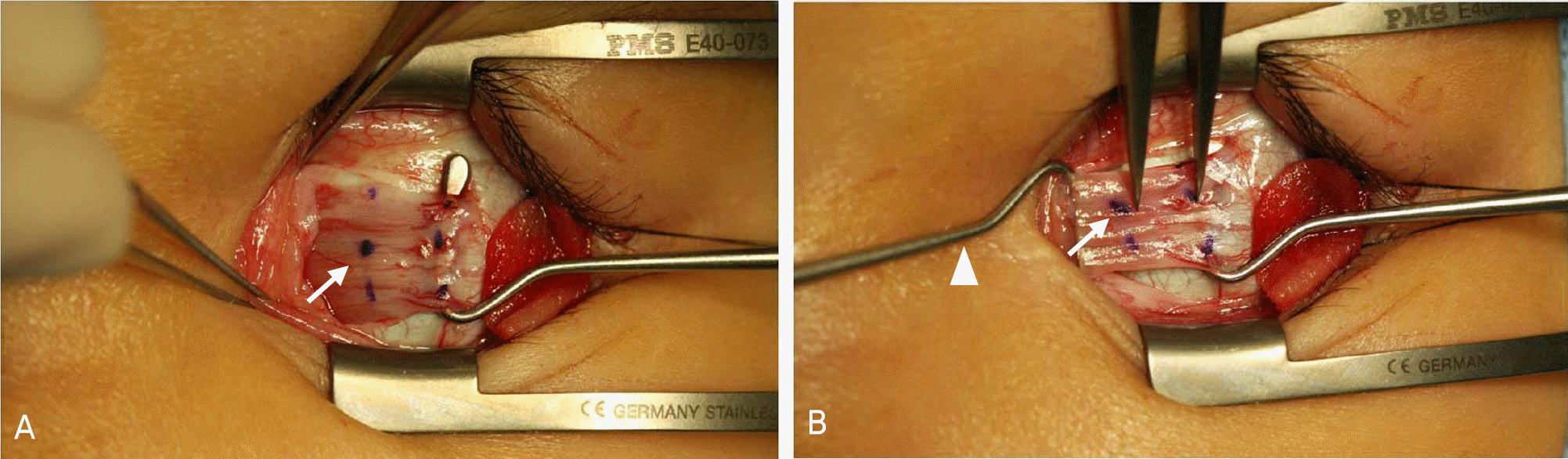
Table 1.
Demographic data of patients
| Number of patients | 42 |
| Sex (No. Male/Female) | 19/23 |
| Mean age (Mean ± SD, year) | 13.12±13.17 |
| Distant deviation angle (Mean± SD, PD*) | 29.24±13.60 |
| Near deviation angle (Mean± SD, PD*) | 31.37±14.23 |
| Diagnosis (No. XT†/ET‡/Vertical strabismus) | 37/4/1 |
| Muscle (No. MR§/LR∏, /SR∏) | 39/4/1 |
Table 2.
Statistical analysis of clinical factors between groups
| Factors | Group 1 | Group 2 | P-value |
|---|---|---|---|
| A Hospital (%)/B Hospital (%) | 73%/27% | 31%/69% | 0.037* |
| Sex (No. Male/Female) | 7/11 | 12/14 | 0.900* |
| Age (Mean± SD, year) | 13.44±13.32 | 12.87±13.30 | 0.890† |
| Diagnosis (No. XT‡/ET§/Vertical strabismus) | 17/1 / 0 | 22/3/1 | 0.487* |
| Muscle (No. MR∏/LR#/SR**) | 17/1 / 0 | 22/3/1 | 0.487* |
| Planned resection amount (Mean± SD, mm) | 5.08±1.11 | 5.36±0.87 | 0.190† |
 XML Download