

 PDF
PDF ePub
ePub Citation
 Print
Print


Abstract
Purpose
To evaluate the repeatability of macular thickness measurements using time domain (TD) OCT and spectral domain (SD) OCT in diabetic macular edema.
Methods
In 42 eyes of 42 patients with diabetic macular edema, three consecutive macular measurements were performed with TD OCT and SD OCT, and measurements for macular thickness and total macular volume obtained by the two OCTs were compared. The within-subject standard deviation (Sw), coefficient of variation (CVw), and intraclass correlation coefficient (ICC) were calculated to assess repeatability, with agreement between measurements assessed with Bland Altman plots. The correlations were also evaluated via the Pearson's correlation coefficient.
Results
The Sw of TD OCT and SD OCT for foveal thickness, total macular volume were 29.67 μ m/16.44 μ m, 1.26 mm3/0.23 mm3, respectively, and were significantly lower in SD OCT. The ranges of the respective CVw and ICC values were 1.10–2.78%, 0.78∼0.96 for TD OCT, and 0.29∼0.94%, 0.92∼0.99 for SD OCT. The SD OCT showed better repeatability for macular thickness measurements (all p≤0.001). The 95% limits of agreement for foveal and total macular volume were 88.9 μ m, 2.4 mm3, respectively. The Pearson's correlation coefficients of macular thickness and total macular volume between the two OCT methods were statistically significant (p=0.88–0.99).
Conclusions
Although both OCTs proved reliable for macular thickness measurements in diabetic macular edema, SD OCT shows better repeatability than TD OCT. Although macular thickness measurements obtained from the two OCTs cannot be used interchangeably, there were statistically significant correlations between measurements obtained using the two OCTs.
Go to : 
References
1. Zeimer RC, Mori MT, Khoobehi B. Feasibility test of a new method to measure retinal thickness noninvasively. Invest Ophthalmol Vis Sci. 1989; 30:2099–105.
2. Gieser JP, Rusin MM, Mori M, et al. Clinical assessment of the macular by retinal topography and thickness mapping. Am J Ophthalmol. 1997; 124:648–60.
3. Polito A, Shah SM, Haller JA, et al. Comparision between retinal thickness analyzer and optical coherence tomography for abdominal of foveal thickness in eyes with macular disease. Am J Ophthalmol. 2002; 134:240–51.
4. Puliafito CA, Hee MR, Schuman JS, et al. Optical coherence abdominal of ocular disease. Thorofare, NJ: Slack;1996. p. 369–74.
5. Muscat S, Parks S, Kemp E, Keating D. Repeatability and abdominal of macular thickness measurements with the Humphrey system. Invest Ophthalmol Vis Sci. 2002; 43:490–5.
6. Gurses-Ozden R, Teng C, Vessani R, et al. Macular and retinal nerve fiber layer thickness measurement reproducibility using abdominalal coherence tomography (OCT-3). J Glaucoma. 2004; 13:238–44.
7. Paunescu LA, Schuman JS, Price LL, et al. Reproducibility of nerve fiber thickness, macular thickness, and optic nerve head measurements using Stratus OCT. Invest Ophthalmol Vis Sci. 2004; 45:1716–24.
8. Polito A, Del Borrello M, Isola M, et al. Repeatability and abdominal of fast macular thickness mapping with Stratus abdominal coherence tomography. Arch Ophthalmol. 2005; 123:1330–7.
9. Hangai M, Ojima Y, Gotoh N, et al. Three-dimensional imaging of macular holes with high-speed optical coherence tomography. Ophthalmology. 2007; 114:763–73.

10. Ahlers C, Michels S, Beckendorf A, et al. Three-dimensional imaging of pigment epithelial detachment in age-related macular degeneration using optical coherence tomography, retinal thickness analysis and topographic angiography. Graefes Arch Clin Exp Ophthalmol. 2006; 244:1233–9.
11. Leung CK, Cheung CY, Weinreb RN, et al. Comparison of abdominal measurements between time domain and specral domain abdominal coherence tomography. Invest Ophthalmol Vis Sci. 2008; 49:4893–7.
12. Oh SB, Cho WB, Moon JW, Kim HC, et al. Repeatability and agreement of macular thickness measurement using time domain OCT and spectral domain OCT in normal subjects. J Korean Ophthalmol Soc. 2009; 50:710–6.
13. Bland JM, Altman DG. Statistical methods for assessing abdominal between two methods of clinical measurement. Lancet. 1986; 1:307–10.
14. Bland JM. Comparing within-subject variances in a study to compare two methods of measurement. Available at. http://www-users.york.ac.uk/∼mb55/meas/compsd.pdf.
15. Massin P, Vicaut E, Haouchine B, et al. Reproducibility of retinal mapping using optical coherence tomography. Arch Ophthalmol. 2001; 119:1135–42.
16. Shrout PE, Fleiss JL. Intraclass correlations: uses in assessing rater reliability. Pshychol Bull. 1979; 86:420–8.
17. Hendrickson A, Drucker D. The development of parafoveal and midperipheral human retina. Behav Brain Res. 1992; 49:21–31.
18. Spraul CW, Lang GE, Grossniklaus HE. Morphometric analysis of the choroid, Bruch's membrane, and retinal pigment abdominal in eyes with age-related macular degeneration. Invest Ophthalmol Vis Sci. 1996; 37:2724–35.
Go to :
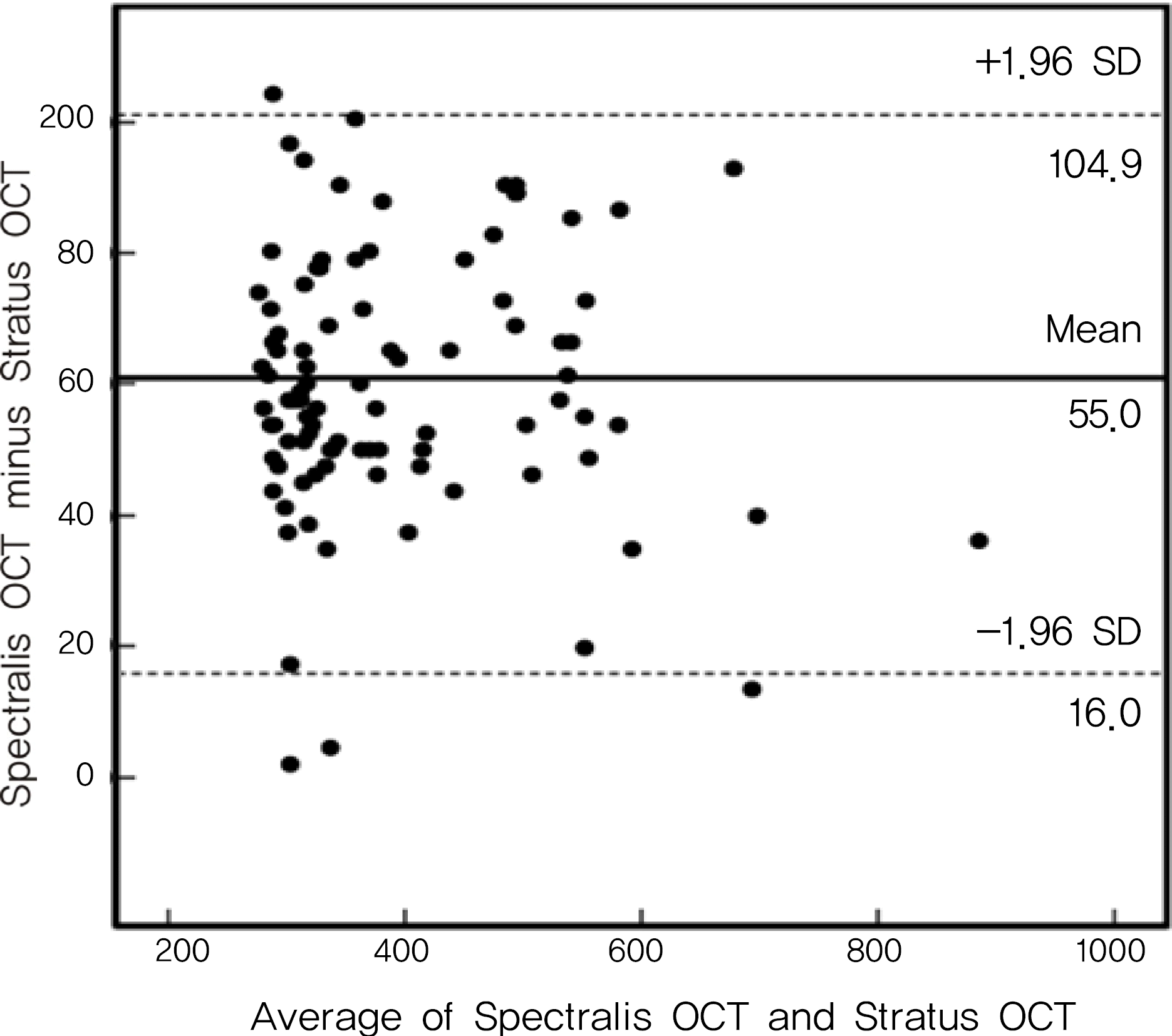 | Figure 1.Bland-Altman plots of foveal thickness measurements obtained by Stratus OCT and Spectralis OCT. Solid line indicates the average mean difference, while dotted lines delineates the 95% confidence limits of agreement. |
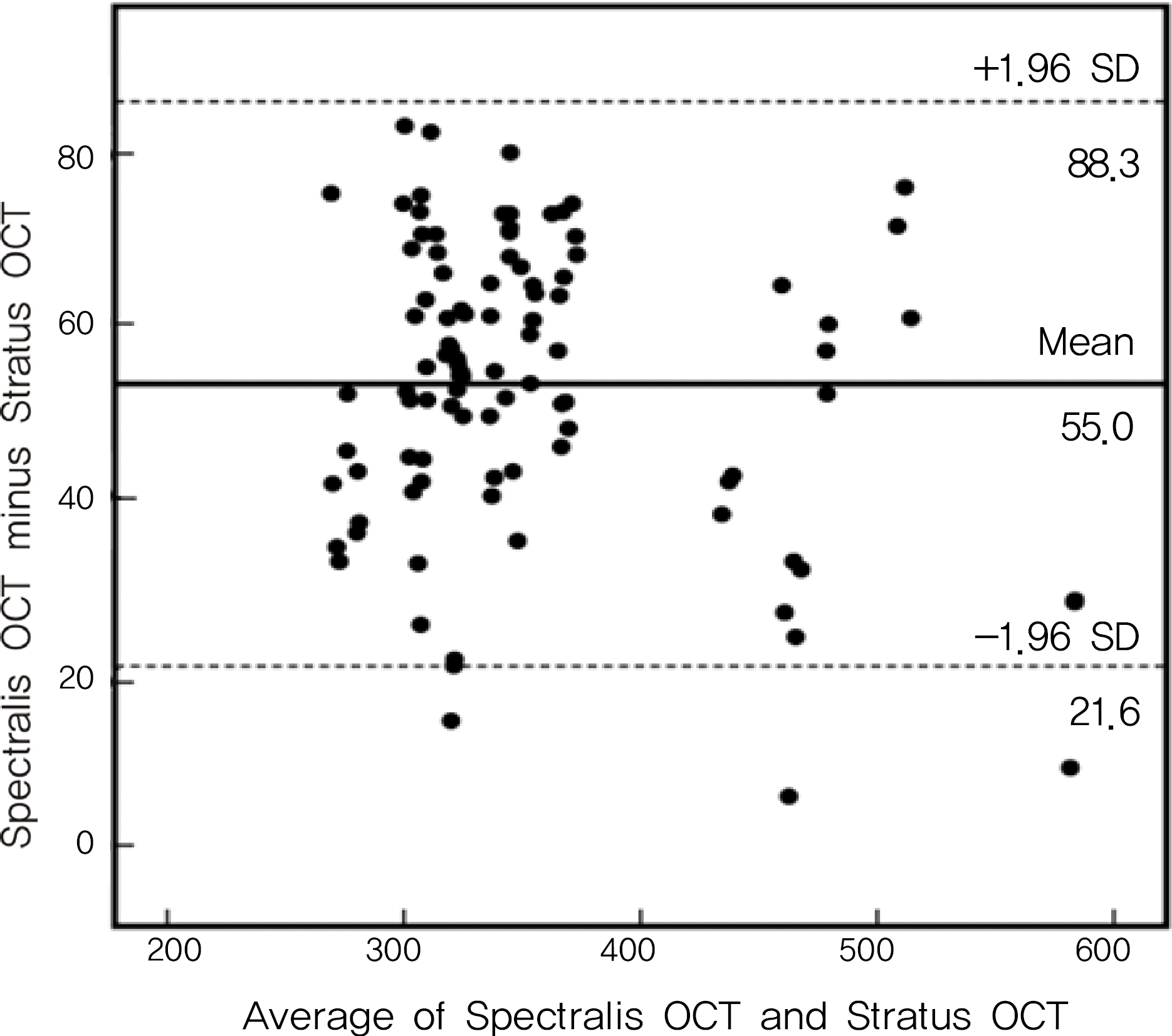 | Figure 2.Bland-Altman plots of total macular thickness obtained by Stratus OCT and Spectralis OCT. Solid line indicates the average mean difference, while dotted lines delineates the 95% confidence limits of agreement. |
Table 1.
Baseline characteristics
| Number of Eyes | 42 |
| Mean age ± SD* (years) | 58.6 ± 8.2 |
| Type of DME† | |
| Diffuse retinal thickness | 16 |
| CME‡ | 23 |
| Serous RD§ | 3 |
| Signal Strength | |
| Stratus OCT∏ | 6.5 ± 0.5 |
| Spectralis OCT (dB) | 24.0 ± 3.7 |
Table 2.
Comparison of regional and total macular thicknesses measured by spectral domain and time domain optical coherence tomography
Table 3.
Within-subject standard deviation (Sw), coefficient of variation (CVw), and intraclass correlation coefficient (ICC) of total and regional macular thicknesses obtained with time domain OCT and spectral domain OCT
| Foveal |
Inner subfield thickness |
Outer subfiled thicknes |
Total | Total | ||||||||
|---|---|---|---|---|---|---|---|---|---|---|---|---|
| Thickness | Temporal | Superior | Nasal | Inferior | Temporal | Superior | Nasal | Inferior | Thickness | Volume | ||
| Sw | TD OCT | 29.67 T | 23.71 | 31.84 | 20.94 | 34.73 | 13.81 | 17.31 | 14.27 | 31.38 | 8.56 | 1.26 |
| SD OCT | 16.44 T | 24.54 | 16.15 | 14.93 | 13.10 | 8.88 | 8.97 | 12.54 | 9.50 | 5.08 | 0.23 | |
| CVw | TD OCT | 2.92 T | 2.43 | 3.21 | 2.11 | 3.61 | 2.82 | 2.18 | 2.68 | 4.12 | 2.99 | 2.10 |
| SD OCT | 1.39 T | 2.14 | 1.39 | 1.28 | 1.17 | 1.97 | 1.45 | 1.25 | 2.07 | 1.50 | 1.97 | |
| ICC | TD OCT | 0.921 T | 0.934 | 0.937 | 0.932 | 0.924 | 0.917 | 0.893 | 0.784 | 0.843 | 0.912 | 0.801 |
| SD OCT | 0.952 T | 0.963 | 0.962 | 0.956 | 0.942 | 0.931 | 0.919 | 0.903 | 0.924 | 0.897 | 0.932 | |
| p-value* | 0.014 | 0.233 | 0.004 | 0.128 | <0.001 | 0.005 | 0.001 | 0.018 | <0.001 | <0.001 | 0.002 | |
Table 4.
Within-subject standard deviation (Sw), coefficient of variation (CVw), and intraclass correlation coefficient (ICC) of total and regional macular thicknesses obtained with time domain OCT and spectral domain OCT between below 400 μm and over 400 μm
|
FT< 400 μm (26 eyes) |
FT≥400 μm (16 eyes) |
|||||||
|---|---|---|---|---|---|---|---|---|
| Fovea | Total | Total | Fovea | Total | Total | |||
| Thickness | Thickness | Volume | Thickness | Thickness | Volume | |||
| Sw | TD OCT | 21.64 | 8.31 | 0.26 | 44.03 | 10.52 | 0.32 | |
| SD OCT | 14.25 | 3.77 | 0.16 | 27.15 | 7.42 | 0.45 | ||
| p=0. | .012 | |||||||
| CVw | TD OCT | 2.48 | 2.03 | 1.85 | 3.07 | 3.04 | 2.38 | |
| SD OCT | 1.19 | 1.38 | 1.60 | 1.72 | 2.65 | 2.48 | ||
| p=0. | .043 | |||||||
| ICC | TD OCT | 0.930 | 0.922 | 0.825 | 0.909 | 0.899 | 0.782 | |
| SD OCT | 0.961 | 0.925 | 0.941 | 0.937 | 0.882 | 0.920 | ||
| p-value* | 0.004 | 0.002 | <0.001 | 0.269 | 0.052 | 0.015 | ||
Table 5.
Pearson correlation coefficient of macular thicknesses and total macular volume obtained with time domain OCT and spectral domain OCT
| Foveal |
Inner subfielld thickness |
Outer subfiled thicknes | Total | Total | |||||||
|---|---|---|---|---|---|---|---|---|---|---|---|
| Thickness | Temporal | Superior | Nasal | Inferior | Temporal | Superior | Nasal | Inferior | Thickness | Volume | |
| Pearson | |||||||||||
| correlation coeffcient | 0.98 | 0.94 | 0.97 | 0.97 | 0.94 | 0.91 | 0.97 | 0.93 | 0.88 | 0.99 | 0.95 |
| p-value* | <0.001 | <0.001 | <0.001 | <0.001 | <0.001 | <0.001 | <0.001 | <0.001 | <0.001 | <0.001 | <0.001 |
 XML Download