Abstract
Purpose
To compare the outcomes of scleral buckling (SB) and primary pars plana vitrectomy (PPV) for treatment of simple rhegmatogenous retinal detachment (RD).
Methods
One hundred and fifteen Eyes undergoing SB or PPV for the treatment of simple rhegmatogenous RD were prospectively identified and followed up at least six months. Anatomic results, functional outcomes and complications were compared between eyes treated with SB and eyes treated with PPV.
Results
We detected no significant differences in overall success rates or final vision quality between the two treatment groups. However, the final vision quality of pseudophakic patients treated with PPV was significantly better than the final vision quality of pseudophakic patients treated with SB. Two treatment goups experienced similar rates of intraocular pressure elevation and proliferative vitreoretinopathy development. In phakic patients, the rates of cataract progression were higher in patients treated with PPV than in patients treated with SB.
Go to : 
References
1. Gonin J. Treatment of detached retina by searing the retinal tears. Arch Ophthalmol. 1930; 4:621–5.
2. Machemer R, Buettner H, Norton EW, Parel JM. Vitrectomy. A pars plana approach. Trans Am Acad Ophthalmol Otolaryngol. 1971; 75:813–6.
3. Escoffery RF, Olk RJ, Grand MG, Boniuk I. Vitrectomy without scleral buckling for primary rhegmatogenous retinal detachment. Am J Ophthalmol. 1985; 99:275–81.

4. Heimann H, Zou X, Jandeck C, et al. Primary vitrectomy for rhegmatogenous retinal detachment: an analysis of 512 cases. Graefes Arch Clin Exp Ophthalmol. 2006; 244:69–78.

5. Gartry DS, Chignell AH, Franks WA, Wong D. Pars plana abdominal for the treatment of rhegmatogenous retinal abdominal uncomplicated by advanced proliferative vitreoretinopathy. Br J Ophthalmol. 1993; 77:199–203.
6. Kang SW, Min JP. Vitrectomy without scleral buckling for the treatment of primary rhegmatogenous retinal detachment. J Korean Ophthalmol Soc. 1997; 38:227–35.
7. Brazitikos PD, Androudi S, Christen WG, Stangos NT. Primary pars plana vitrectomy versus scleral buckle surgery for the abdominal of pseudophakic retinal detachment: a randomized clinical trial. Retina. 2005; 25:957–64.
8. Lee M, Moon CS, Yang HS, Lew HM. Factor influencing abdominal failure of simple rhegmatogenous retinal detachment. J Korean Ophthalmol Soc. 2006; 47:407–14.
9. Park HJ, Seo MS. Clinical analysis according to treatment abdominal in simple retinal detachment. J Korean Ophthalmol Soc. 2001; 42:1277–83.
10. Heimann H, Bartz-Schmidt KU, Bornfeld N. et al. Scleral Buckling versus Primary Vitrectomy in Rhegmatogenous Retinal Detachment. Ophthalmology. 2007; 114:2142–54.
11. Woon WH, Burdon MA, Green WT, Chignell AH. Comparison of pars plana vitrectomy and scleral buckling for uncomplicated rhegmatogenous retinal detachment. Curr Opin Ophthalmol. 1995; 6:76–9.
12. Bartz-Schmidt KU, Kirchhof B, Heimann K. Primary vitrectomy for pseudophakic retinal detachment. Br J Ophthalmol. 1996; 80:346–9.

13. Newman DK, Burton RL. Primary vitrectomy for pseudophakic and aphakic retinal detachments. Eye. 1999; 13:635–9.

14. Ku M, Sohn HJ, Lee DY, Nam DH. Foveal reattachment after scleral buckling vs vitrectomy for macula-off retinal detachment. J Korean Ophthalmol Soc. 2009; 50:399–404.

15. Mendrinos E, Dang-Burgener NP, Stangos AN, et al. Primary abdominal without scleral buckling for pseudophakic abdominal retinal detachment. Am J Ophthalmol. 2008; 145:1063–70.
16. Ahmadieh H, Entezari M, Soheilian M, et al. Factors influencing anatomic and visual results in primary scleral buckling. Eur J Ophthalmol. 2000; 10:153–9.

17. Sharma YR, Karunanithi S, Azad RV, et al. Functional and abdominal outcome of scleral buckling versus primary vitrectomy in pseudophakic retinal detachment. Acta Ophthalmol Scand. 2005; 83:293–7.
18. Azad RV, Chanana B, Sharma YR, Vohra R. Primary vitrectomy versus conventional retinal detachment surgery in phakic abdominal retinal detachment. Acta Ophthalmol Scand. 2007; 85:540–5.
19. Hoeing C, Heidenkummer HP, Kampik A. Primary vitrectomy in rhegmatogenous retinal detachment. Ophthalmologe. 1995; 92:668–71.
20. Heimann H, Bornfeld N, Friedrichs W, et al. Primary vitrectomy without sclera buckling for rhegmatogenous retinal detachment. Graefes Arch Clin Exp Ophthalmol. 1996; 234:561–8.
21. Miki D, Hida T, Hotta K, et al. Comparison of sclera buckling and vitrectomy for superior retinal detachment caused by flap tears. Nippon Ganka Gakkai Zasshi. 2000; 104:24–8.
22. Oshima Y, Yamanishi S, Sawa M, et al. Two-year follow-up study comparing primary vitrectomy with scleral buckling for mac-ula-off rhegmatogenous retinal detachment. Jpn J Ophthalmol. 2000; 44:538–49.

23. Girard P, Mimoun G, Karpouzas I, Montefiore G. Clinical risk factors for proliferative vitreoretinopathy after retinal abdominal surgery. Retina. 1994; 14:417–24.
24. Hakin KM, Lavin MJ, Leaver PK. Primary vitrectomy for abdominal retinal detachment. Graefes Arch Clin Exp Ophthalmol. 1993; 231:344–6.
Go to : 
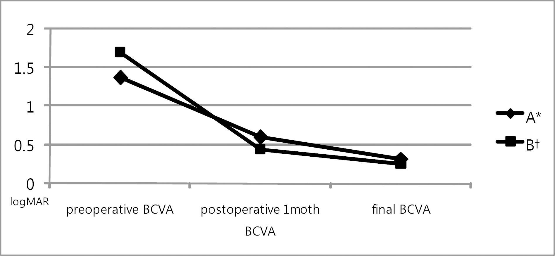
 | Figure 1.BCVA§ change of macula off patients showed primary success. § BCVA=best corrected visual acuity; * A=foveal reattached group after postoperative 1–2month by OCT (=optical coherence tomography); †B=foveal detached group after postoperative 1–2month by OCT. |
Table 1.
Demographics and clinical datas of the patients
| Vitrectomy (N=55) | SB† (N=60) | p | |
|---|---|---|---|
| Age (years) | 53.9±10.2 | 49.1±16.6 | 0.063 |
| Sex (Male/Female) | 27/28 | 32/28 | 0.711 |
| Mean F/U period (months) | 11.5±3.3 | 10.3±3.6 | 0.078 |
| Phakic/pseudophakic | 45/10 | 53/7 | 0.432 |
| Preoperative BCVA* (logMAR) | 1.13±1.07 | 0.95±1.00 | 0.342 |
Table 2.
Clinical aspects of breaks and detachment
|
No. of eyes (%) |
p | ||
|---|---|---|---|
| Vitrectomy(n=55) | Scleral buckling(n=60) | ||
| Large tear (>3DD*) | 17(30.9) | 10(16.7) | 0.082 |
| Multiple tear | 32(58.2) | 42(70.0) | 0.243 |
| Macula off detachment | 34(61.8) | 38(63.3) | 1.000 |
Table 3.
Primary operation procedure
| Methods |
No. of eyes (%) |
|
|---|---|---|
| Vitrectomy | Scleral buckling | |
| (N=55) | (N=60) | |
| With cataract operation | 6/45(13.3) | 1/53(1.9) |
| 23G vitrectomy | 15(27.3) | – |
| Tamponade | ||
| SF6 | 7(12.7) | – |
| C3 F8 | 48(87.3) | – |
| Buckle material | ||
| 506 sponge | – | 59(98.3) |
| 507 sponge | – | 1(1.7) |
| External SRFD* | – | 6(10) |
| Retinotomy | 9(16.4) | – |
Table 4.
Overall success rate
| Success rate | Vitrectomy | Scleral buckling | p |
|---|---|---|---|
| (N=55) | (N=60) | ||
| Primary | 92.7%(51/55) | 81.7%(49/60) | 0.099 |
| Final | 98.2%(54/55) | 96.7%(58/60) | 1.000 |
Table 5.
Success rate according to lens status of vitrectomy and scleral buckling
Table 6.
Best corrected visual acuity and number of eyes showed visual acuity improvement more than 0.3
|
BCVA* (logMAR) |
p |
BCVA improvement, No. of eyes(%) |
p | |||
|---|---|---|---|---|---|---|
| Vitrectomy | SB† | Vitrectomy | SB | |||
| Preoperative | 1.14±1.06 | 1.02±1.00 | 0.516 | – | – | – |
| Final | 0.20±0.24 | 0.28±0.29 | 0.114 | 36(65.5) | 35(58.3) | 0.450 |
Table 7.
BCVA change in phakic group and pseudophakic group
| Vitrectomy (logMAR) | SB (logMAR) | p | |
|---|---|---|---|
| (mean± SD) | (mean± SD) | ||
| Phakic group | |||
| Preoperative BCVA* | 1.17±1.08 | 1.02±0.99 | 0.463 |
| Final BCVA | 0.22±0.24 | 0.27±0.28 | 0.322 |
| Pseudophakic group | |||
| Preoperative BCVA | 1.00±1.02 | 1.00±1.12 | 1.00 |
| Final BCVA | 0.14±0.22 | 0.56±0.14 | 0.042 |


 PDF
PDF ePub
ePub Citation
 Print
Print


 XML Download