Abstract
Purpose
To evaluate changes in anterior chamber depth (ACD) and angle after phacoemulsification and intraocular lens implantation using anterior segment optical coherence tomography (AS-OCT).
Methods
Seventy-eight eyes of 69 patients had uneventful phacoemulsification and IOL implantation using a clear corneal incision. Anterior segment OCT images of nasal and temporal angle quadrants were obtained before and at one month after surgery. The angle-referenced (ACD1), pupil-referenced (ACD2), lens-referenced (ACD3) ACDs, crystalline lens rise (CLR), nasal and temporal iridocorneal angles, angle opening distance at 500 μ m (AOD500), and trabecular iris surface area at 750 μ m (TISA750) were measured. Preoperative and postoperative measurements were compared using paired sample t-tests.
Results
The mean ACD1 was 3.19±0.24 mm preoperatively and 3.22±0.21 mm at one month postoperatively (P=0.21); ACD2 was 2.99±0.40 mm preoperatively and 3.56±0.28 mm at one month postoperatively (P<0.05); ACD3 was 2.75±0.41 mm preoperatively and 4.00±0.27 mm at one month postoperatively (P<0.05). The mean iridocorneal angles, AOD500, and TISA750 for both nasal and temporal sides increased significantly at the postoperative examinations (P<0.05).
Go to : 
References
1. Kucumen RB, Yenerel NM, Gorgun E, et al. Anterior segment optical coherence tomography measurement of anterior chamber depth and angle changes after phacoemulsification and intraocular lens implantation. J Cataract Refract Surg. 2008; 34:1694–8.

2. Nolan WP, See JL, Aung T, et al. Changes in angle configuration after phacoemulsification measured by anterior segment optical coherence tomography. J Glaucoma. 2008; 17:455–9.

3. Memarzadeh F, Tang M, Li Y, et al. Optical coherence tomography assessment of angle anatomy changes after cataract surgery. Am J Ophthalmol. 2007; 144:464–5.

4. Baikoff G, Lutun E, Ferraz C, Wei J. Static and dynamic analysis of the anterior segment with optical coherence tomography. J Cataract Refract Surg. 2004; 30:1843–50.

5. Baikoff G, Jitsuo Jodai H, Bourgeon G. Measurement of the internal diameter and depth of the anterior chamber: IOLMaster versus anterior chamber optical coherence tomographer. J Cataract Refract Surg. 2005; 31:1722–8.

6. Radhakrishnan S, Goldsmith J, Huang D, et al. Comparison of optical coherence tomography and ultrasound biomicroscopy for detection of narrow anterior chamber angles. Arch Ophthalmol. 2005; 123:1053–9.

7. Kurimoto Y, Park M, Sakaue H, Kondo T. Changes in the anterior chamber configuration after small-incision cataract surgery with posterior chamber intraocular lens implantation. Am J Ophthalmol. 1997; 124:775–80.

8. Goldsmith JA, Li Y, Chalita MR, et al. Anterior chamber width measurement by high-speed optical coherence tomography. Ophthalmology. 2005; 112:238–44.

9. Leung CK, Chan W-M, Ko CY, et al. Visualization of anterior chamber angle dynamics using optical coherence tomography. Ophthalmology. 2005; 112:980–4.

10. Hayashi K, Hayashi H, Nakao F, Hayashi F. Changes in anterior chamber angle width and depth after intraocular lens implantation in eyes with glaucoma. Ophthalmology. 2006; 107:698–703.

11. Rabsilber TM, Khoramnia R, Auffarth GU. Anterior chamber measurements using Pentacam rotating Scheimpflug camera. J Cataract Refract Surg. 2006; 32:456–9.

12. Nonaka A, Kondo T, Kikuchi M, et al. Angle widening and alteration of ciliary process configuration after cataract surgery for primary angle closure. Ophthalmology. 2006; 113:437–41.

13. Pereira FA, Cronemberger S. Ultrasound biomicroscopic study of anterior segment changes after phacoemulsification and foldable intraocular lens implantation. Ophthalmology. 2003; 110:1799–806.

14. Pavlin CJ, Harasiewicz K, Foster FS. Ultrasound biomicroscopy of anterior segment structures in normal and glaucomatous eyes. Am J Ophthalmol. 1992; 113:381–9.

15. Kohnen T, Thomala MC, Cichocki M, Strenger A. Internal anterior chamber diameter using optical coherence tomography compared with white-to-white distances using automated measurements. J Cataract Refract Surg. 2006; 32:1809–13.

16. Dada T, Sihota R, Gadia R, et al. Comparison of anterior segment optical coherence tomography and ultrasound biomicroscopy for assessment of the anterior segment. J Cataract Refract Surg. 2007; 33:837–40.

17. Radhakrishnan S, Rollins AM, Roth JE, et al. Real-time optical coherence tomography of the anterior segment at 1310 nm. Arch Ophthalmol. 2001; 119:1179–85.

18. Nolan WP, See JL, Chew PT, et al. Detection of primary angle closure using anterior segment optical coherence tomography in Asian eyes. Ophthalmology. 2007; 114:33–9.

19. Radhakrishnan S, See JL, Smith SD, et al. Reproducibility of anterior chamber angle measurements obtained with anterior segment optical coherence tomography. Invest Ophthalmol Vis Sci. 2007; 48:3683–8.

20. Spaeth GL. The normal development of the human anterior chamber angle: a new system of descriptive grading. Trans Ophthalmol Soc U K. 1971; 91:709–39.
Go to : 
 | Figure 1.(A) Anterior segment OCT image of the left eye of an 55-year-old man before phacoemulsification and IOL implantation; ACD1 is 3.02 mm (2.47+0.55), ACD3 is 2.47 mm, and CLR is 0.55 mm. (B) Postoperative image of the same eye; ACD1 is 3.06 mm (3.86–0.80). and ACD3 is 3.86 mm (ACD1=angle-referenced anterior chamber depth; ACD3=lens-referenced anterior chamber depth; CLR=crystalline lens rise). |
 | Figure 2.(A) Anterior segment OCT image of the right eye of an 51-year-old man before phacoemulsification and IOL implantation; ACD2 is 3.48 mm. (B) Postoperative image of the same eye; ACD2 is 3.81 mm (ACD2=pupil-referenced anterior chamber depth). |
 | Figure 3.(A) Anterior segment OCT image of the left eye of an 70-year-old man before phacoemulsification and IOL implantation; the nasal iridocorneal angle is 29.5 degrees and the temporal iridocorneal angle is 26.0 degrees, the nasal AOD500 is 0.390 mm and the temporal AOD500 is 0.435 mm, the nasal TISA750 is 0.274 mm2 and the temporal TISA750 is 0.306 mm2. (B) Postoperative image of the same eye; the nasal iridocorneal angle is 34.2 degrees, and the temporal iridocorneal angle is 36.5 degrees, the nasal AOD500 is 0.698 mm and the temporal AOD500 is 0.460 mm, the nasal TISA750 is 0.458 mm2 and the temporal TISA750 is 0.319 mm2. (AOD500=angle opening distance at 500 um; TISA750=trabecular-iris space at 750 μm). |
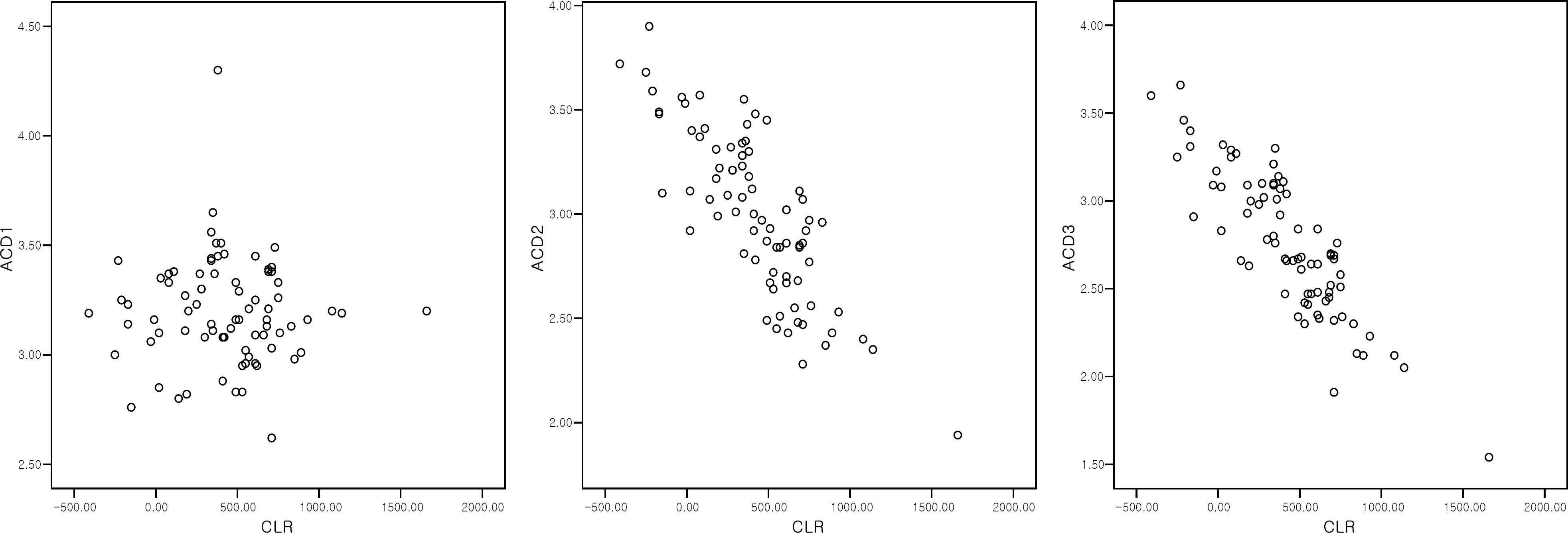
 | Figure 4.Correlation between CLR and preoperative ACD.
* Pearson correlation test (CLR=crystalline lens rise; ACD=anterior chamber depth; ACD1=angle-referenced anterior chamber depth; ACD2= pupil-referenced anterior chamber depth; ACD3=lens-referenced anterior chamber depth).
|
Table 1.
Changes in anterior chamber parameters before and after cataract surgery
| Parameter |
Mean± SD† |
Increase | P-value* | |
|---|---|---|---|---|
| preoperative | postoperative | |||
| ACD1‡ (mm) | 3.19±0.24 | 3.22±0.21 | 0.90% | P=0.215 |
| ACD2§ (mm) | 2.99±0.40 | 3.56±0.28 | 20.70% | P<0.001 |
| ACD3∏ (mm) | 2.75±0.41 | 4.00±0.27 | 45.50% | P<0.001 |
| Nasal angle (degrees) | 19.87±12.53 | 32.79±10.52 | 65% | P<0.001 |
| Temporal angle (degrees) | 21.54±12.10 | 33.02±8.89 | 53.30% | P<0.001 |
| AOD500# nasal (mm) | 0.322±0.199 | 0.546±0.202 | 69.60% | P<0.001 |
| AOD500# temporal (mm2) | 0.366±0.239 | 0.559±0.234 | 52.70% | P<0.001 |
| TISA750** nasal (mm2) | 0.218±0.127 | 0.362±0.127 | 66.10% | P<0.001 |
| TISA750** temporal (mm2) | 0.248±0.145 | 0.371±0.149 | 49.60% | P<0.001 |


 PDF
PDF ePub
ePub Citation
 Print
Print


 XML Download