

 PDF
PDF ePub
ePub Citation
 Print
Print


Abstract
Purpose
To report three cases of ophthalmoplegic migraine which is a rare condition characterized by the association of headaches with an oculomotor nerve palsy.
Case summary:
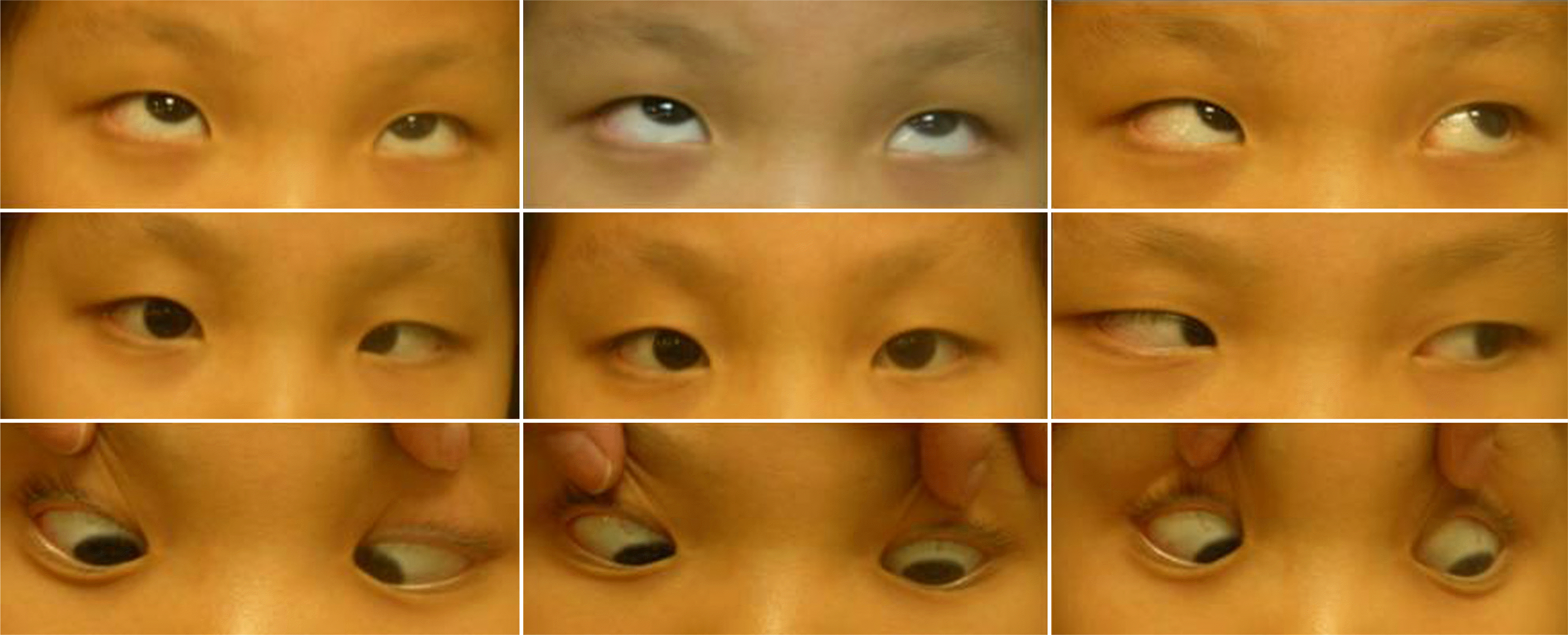
A 44-year-old male and two eight-year-old females were presented with diplopia developed after headaches. All of the three patients showed abnormal eye movement and they had past episodes of transient diplopia with headaches. Visual evoked potential (VEP), cerebrospinal fluid (CSF) examination, laboratory findings, and other neurologic tests were all normal, also there was no tumor or cerebrovascular disease on brain MRA & MRI. The symptoms of all patients improved gradually within several weeks from first the visit, with complete recovery seen in all three.
References
1. Headache ClassiIfication SubCommittee of the International Headache society. The International Classification of Headache Disorders, 2nd edtion. Cephalalgia. 2004; 24:1–160.
2. Hansen SL, Borelli-Moller L, Strange P, et al. Ophthalmoplegic migraine: diagnostic criteria, incidence of hospitalization, and possible etiology. Acta Neurol Scand. 1990; 81:54–60.

3. Lance JW, Zagami AS. Ophthalmoplegic migraine: a recurrent demyelinating neuropathy? Cephalalgia. 2001; 21:84–9.
4. Straube A, Bandmann O, Büttner U, Schmidt H. Contrast aberrations lesion of the III nerve on MR of a patient with ophthalmoplegic migraine as evidence for a Tolosa-Hunt syndrome. Headache. 1993; 33:446–8.
5. Mark AS, Casswlman J, Brown D, et al. Opthalmoplegic migraine: reversible enhancement and thickening of the cisternal segment of the oculomotor nerve on contrastenhanced MRI images. AJNR Am J Neuroradiol. 1998; 19:1887–91.
6. Wong , Wong WC. Enhancement of oculomotor nerve:a diagnostic cisterion for ophthalmic migraine? Pediatric Neurology. 1997; 17:70–73.
7. Galdstone JP. An approach to the patient with painful aberrations. with a focus on Tolosa-Hunt syndrome Curr Pain aberrations Rep. 2007; 11:317–25.
8. Lee TG, Choi WS, Chung KC. Ophthalmoplegic migraine with reversible enhancement of intraparenchymal abducens nerve on MRI. Headache. 2002; 42:140–1.
9. Verhagen WI, Prick MJ, van Dijk Azn R. Onset of ophthalmoplegic migraine with abduens palsy at middle age? Headache. 2003; 43:798–800.
10. Celebisoy N, Sirin H, Gökçay F. Ophthalmoplegic migraine: two patients, one at middle age with abducens palsy. Cephalalgia. 2004; 25:151–3.
11. Carlow TJ. Oculomotor ophthalmoplegic migraine: is it really migrane? J Neuroophthalmol. 2002; 22:215–21.
12. Troost BT. The Headaches. Philadelphia: lippincott Williams & Wilkins;2000. p. 511–6.
13. Lance JW, Zagami AS. Ophthalmoplegic migraine: a recurrent demylination. Cephalagia. 2001; 21:84–9.
14. Peck R. Ophthalmoplegic migraine presenting in infancy: a aberrations case Cephalagia. 2006; 26:1242–4.
15. O'Halloran HS, Lee WB, Baker RS, Pearson PA. aberrations Migraine With Unusual Feature. Headache. 1999; 39:670–3.


 XML Download