Abstract
Purpose
To evaluate intrableb morphology and function after trabeculectomy using anterior segment optical coherence tomography (AS-OCT)
Methods
Twenty-eight post-trabeculectomy eyes from patients with primary open-angle glaucoma were examined using AS-OCT. Intrableb structures, including bleb wall thickness, subconjunctival fluid, suprascleral fluid and the route under the scleral flap, were measured at the center of the bleb using AS-OCT and were classified according to their slit-lamp appearance. Blebs were classified as successful (IOP≤18 mmHg without ocular hypotensive medication) and failed (IOP>18 mmHg or use of ocular hypertensive medication.) in order to compare parameters between the two groups.
Results
The blebs were classified as either diffuse filtering (n=17), cystic (n=5), encapsulated (n=3) or flattened (n=3) according to slit lamp appearance and were correlated with AS-OCT images. Blebs were classified as successful and failed in order to compare parameters between the two groups. Significant differences were observed between the two groups in regard to sub-conjunctival fluid space (p=0.0004). No significant differences were observed in bleb wall thickness (p=0.098), suprascleral fluid space (p=0.87) or the route under the scleral flap (p=0.196).
References
1. Cairns JE. Trabeculectomy. Preliminary report of a new method. Am J Ophthalmol. 1968; 66:673–9.
2. Picht G, Grehn F. Classification of filtering blebs in aberrations: biomicroscopy and functionality. Curr Opin Ophthalmol. 1998; 9:2–8.
3. Hu CY, Matsuo H, Tomita G, et al. Clinical characteristics and leakage of functioning blebs after trabeculectomy with mitomycin-C in primary glaucoma patients. Ophthalmology. 2003; 110:345–52.

4. DeBry PW, Perkins TW, Heatly G, et al. Incidence of lateonset bleb-related complications following trabeculectomy with aberrations. Arch Ophthalmol. 2002; 120:297–300.
5. Soltau JB, Rothman RF, Budenz DL, et al. Risk factors for aberrations filtering bleb infections. Arch Ophthalmol. 2000; 118:338–42.
6. Kronfeld FC. The chemical demonstration of transconjunctival passage of aqueous after antiglaucomatous operations. Am J aberrations. 1952; 35:38–45.

7. Cantor LB, Mantravadi A, WuDunn D, et al. Morphologic aberrations of filtering blebs after glaucoma filtration surgery: the Indiana Bleb Appearance Grading Scale. J Glaucoma. 2003; 12:266–71.
8. Wells AP, Crowston JG, Marks J, et al. A pilot study of system for grading of drainage blebs after glaucoma surgery. J Glaucoma. 2004; 13:454–60.
9. Yamamoto T, Sakuma T, Kitazawa Y. An ultrasound aberrations study of filtering blebs after mitomycin C trabeculectomy. Ophthalmology. 1995; 102:1770–6.
10. Pavlin CJ, Harasiewicz K, Foster FS. Ultrasound biomicroscopy of anterior segment structures in normal and glaucomatous eyes. Am J Ophthalmol. 1992; 113:381–9.

11. Singh M, Chew PT, Friedman DS, et al. Imaging of aberrations blebs using anterior segment optical coherence tomography. Ophthalmology. 2007; 114:47–53.
12. Leung CK, Yick DW, Kwong YY, et al. Analysis of bleb aberrations after trabeculectomy with Visante anterior segment optical coherence. Br J Ophthalmol. 2007; 91:340–4.
13. Kronfeld PC. The mechanism of filtering operations. Trans Pac Coast Oto Ophthalmol Soc Annu Meet. 1949; 33:23–40.
14. Savini G, Zanini M, Barboni P. Filtering blebs imaging by optical coherence tomography. Clin Exp Ophthalmol. 2005; 33:483–9.

15. Zhang Y, Wu Q, Zhang M, et al. Evaluating subconjunctival bleb function after trabeculectomy using slit-lamp optical coherence tomography and ultrasound biomicroscopy. Chin Med J. 2008; 121:1274–9.

16. Theelen T, Wesseling P, Keunen JE, Klevering BJ. A plot study on slit lamp-adapted optical coherence tomography imaging of aberrations filtering blebs. Graefes Arch Clin Exp Ophthalmol. 2007; 245:877–82.
17. Kawana K, Kiuchi T, Yasuno Y, Oshika T. Evaluation of aberrations blebs using 3-dimensional cornea and anterior segment optical coherence tomography. Ophthalmology. 2009; 116:848–55.
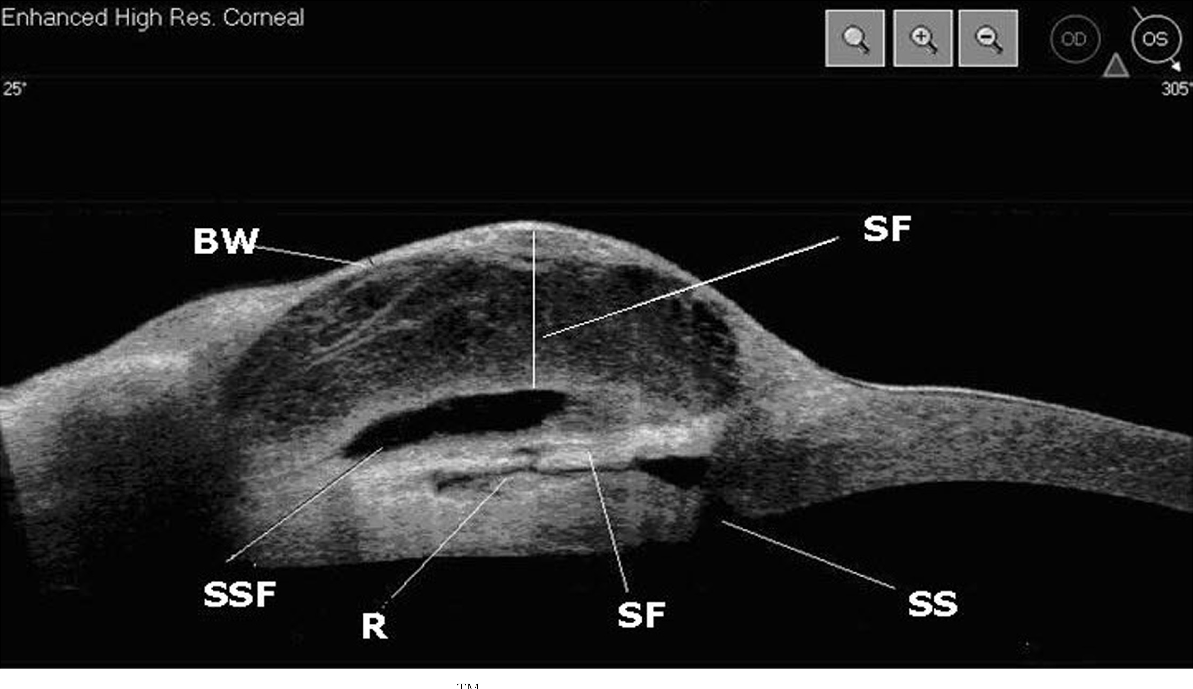
Figure 1.
Morphology of bleb using Visante TM OCT. BW=bleb wall thickness; SF=subconjunctival fluid; SS=scle-rotomy site; SF=scleral flap; R=route under scleral flap; SSF=suprascleral fluid.

Figure 2.
AS-OCT image and corresponding slit-lamp appearance. (A) Diffuse functioning bleb. (B) Cystic bleb. (C) Encapsulated bleb. (D) Flattened bleb

Table 1.
Comparison of clinical characteristics and intrableb parameters between 4 different bleb type
| | Diffuse functioning bleb (n=17) | Cystic bleb (n=5) | Encapsulated bleb (n=3) | Flattened bleb (n=3) |
|---|---|---|---|---|
| Age (y) | 50.76±13.04 | 59.00±16.29 | 52.33±14.15 | 65.00±19.97 |
| Sex (M/F) | 14/3 | 2/3 | 3/0 | 2/1 |
| Postop (months) | 44.82±34.17 | 8.4±2.70 | 35±42.79 | 103±63.24 |
| IOP (mmHg) | 11.29±4.62 | 11.60±2.70 | 22.00±7.00 | 25.00±7.00 |
| Successful/Failed* (n) | 8/9 | 3/2 | 0/3 | 0/3 |
| Bleb wall thickness(mm) | 0.14±0.04 | 0.11±0.05 | 0.43±0.06 | 0.25±0.09 |
| Subconjunctival fluid (mm) | 0.48±0.34 | 0.73±0.39 | 0 | 0.02±0.02 |
| Suprascleral space (mm) | 0.21±0.13 | 0.14±0.05 | 2.20±0.36 | 0.08±0.02 |
| Route under scleral flap (+/-) | 17/0 | 3/2 | 2/1 | 0/3 |
Table 2.
Comparison of clinical characteristics and intrableb parameters between successful and failed bleb groups
| | Successful (n=12) | Failed (n=16) | p-value |
|---|---|---|---|
| Age (y) | 51.75±17.04 | 55.50±12.57 | 0.51 |
| Sex (M/F) | 7/5 | 14/2 | 0.1 |
| Postop (months) | 23.41±23.94 | 58.56±46.64 | 0.115 |
| IOP (mmHg) | 9.42±3.73 | 17.37±6.77 | 0.001* |
| Bleb wall thickness (mm) | 0.13±0.05 | 0.22±0.13 | 0.09 |
| Subconjunctival fluid (mm) | 0.70±0.37 | 0.21±0.24 | 0.004* |
| Suprascleral space (mm) | 0.24±0.16 | 0.53±0.84 | 0.87 |
| Route under scleral flap (+/-) | 11/1 | 11/5 | 0.2 |
Table 3.
Comparison of characteristics and intrableb parameters between successful and failed bleb groups in diffuse functioning bleb and cystic bleb
| | Successful (n=12) | Failed (n=10) | p-value |
|---|---|---|---|
| Bleb type (n) | | | |
| Diffuse functioning bleb | 8 | 9 | |
| Cystic bleb | 4 | 1 | |
| Age (y) | 51.75±17.05 | 53.60±9.63 | 0.76 |
| Sex (M/F) | 7/5 | 9/1 | 0.09 |
| Postop (months) | 23.42±23.94 | 52.30±37.98 | 0.06 |
| IOP (mmHg) | 9.42±3.73 | 13.70±3.62 | 0.01* |
| Bleb wall thickness (mm) | 0.13±0.05 | 0.14±0.04 | 0.73 |
| Subconjunctival fluid (mm) | 0.70±0.37 | 0.33±0.22 | 0.01* |
| Suprascleral space (mm) | 0.24±0.16 | 0.16±0.04 | 0.12 |
| Route under scleral flap (+/-) | 11/1 | 9/1 | 0.89 |


 PDF
PDF ePub
ePub Citation
 Print
Print


 XML Download