

 PDF
PDF ePub
ePub Citation
 Print
Print


Abstract
Purpose
To describe a transconjunctival sutureless technique for pars plana vitrectomy using conventional 20-gauge instruments.
Methods
We performed transconjunctival sutureless pars plana vitrectomy (TSV) using conventional 20-gauge instruments in 36 eyes of 35 patients. We made 20-gauge transconjunctival beveled sclerotomies using microvitreoretinal (MVR) blades and used traditional 20-gauge instruments for the operations.
Results
Eighty-three (81.4%) of 102 sclerotomies self-sealed without the need for sutures. The sutureless rate was even higher in the last one-third of the patients: 32 (94.1%) of 34 sclerotomy sites were sutureless. No serious complications were observed in our series, including postoperative hypotony, wound leakage, or endophthalmitis.
Go to : 
References
1. Fujii GY, De Juan E Jr, Humayun MS, et al. A new 25-gauge instrument system for transconjunctival sutureless vitrectomy surgery. Ophthalmology. 2002; 109:1807–12.
2. Eckardt C. Transconjunctival sutureless 23-gauge vitrectomy. Retina. 2005; 25:208–11.

3. Lakhanpal RR, Humayun MS, de Juan E Jr, et al. Outcomes of 140 consecutive cases of 25-gauge transconjunctival surgery for posterior segment disease. Ophthalmology. 2005; 112:817–24.
4. Shimada H, Nakashizuka H, Mori R, et al. 25-gauge scleral tunnel transconjunctival vitrectomy. Am J Ophthalmol. 2006; 142:871–3.
5. Shimada H, Nakashizuka H, Nakajima M, et al. Twenty-gauge Transconjunctival Vitrectomy. Jpn J Ophthalmol. 2005; 49:257–60.
6. Jorge R, Gomes AV, Siqueira RC, et al. 20-gauge transconjunctival pars plana vitrectomy. Ophthalmic Surg Lasers Imaging. 2007; 38:342–4.
7. Gotzaridis EV. Sutureless Transconjunctival 20 gauge pars plana Vitrectomy. Semin Ophthalmol. 2007; 22:179–83.
8. Inoue M, Shinoda K, Shinoda H, et al. Two-step oblique incision during 25-gauge vitrectomy reduces incidence of postoperative hypotony. Clin Experiment Ophthalmol. 2007; 35:693–6.
9. Parolini B, Prigione G, Romanelli F, et al. Postoperative aberrations and intraocular pressure in 943 consecutive cases of 23-gauge transconjunctival pars plana vitrectomy with 1-year follow-up. Retina. 2009. Oct 7.[Epub ahead of print].
10. Ku M, Sohn HJ, Lee DY, Nam DH. Partial fluid-air-exchange at the end of 23 gauge sutureless vitrectomy to prevent postoperative hypotony. J Korean Ophthalmol Soc. 2009; 50:359–64.
11. Lafeta AP, Claes C. Twenty-gauge transconjunctival sutureless vitrectomy trocar system. Retina. 2007; 27:1136–41.
Go to :
 | Figure 1.(A) A traditional 20-gauge MVR blade is bent at the middle of the shaft before the operation to facilitate a transconjunctival sclera tunnel incision. (B) Diathermy is applied on the conjunctiva 3.5 mm from the limbus using pressure, while the conjunctiva is displaced laterally with a cotton-tipped applicator to misalign the conjunctival and scleral openings. (C) Temporary thinning and adhesion of the conjunctiva to the sclera is achieved. Three plane incision can make scleral tunnel more stable (D), but 1 plane incision is also acceptable (E). Direction of the tunnel can be either away from the operator (F) or toward the operator (G). (H) A 6 mm infusion cannula and endoilluminator, both of which are used in conventional 20-gauge pars plana vitrectomy, are placed after phacoemulsification has been performed. (I) Appearance at the end of the operation. Note self-sealing sclerotomies without leakage. |
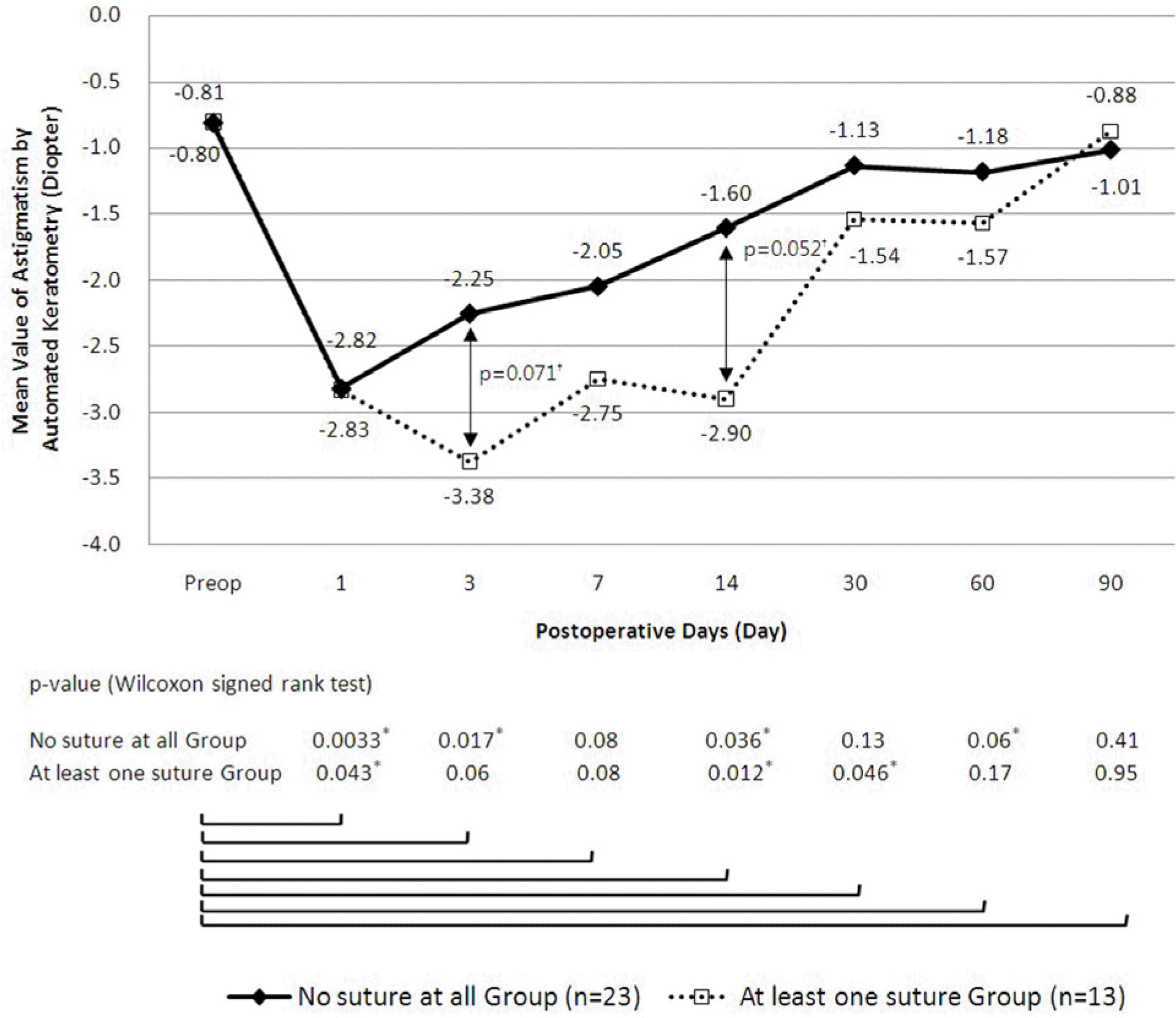 | Figure 3.Postoperative astigmatism change. *Statistically significant values †p-value by Mann-Whitney U test. |
 | Figure 5.(A) Severe conjunctival chemosis enough to interfere with preceding the operation occurred in two patients. By applying small incision on the conjunctiva and Tenon's capsule to drain the fluid (B), the remainder of the operation could be completed without difficulty (C). |
 | Figure 6.Postoperative appearance at 1 day (A), 2 weeks (B), and 4 weeks (C) postoperatively. |
Table 1.
Surgical indications
Table 2.
Sutureless rate showing a steep learning curve
| A series of consecutive cases |
No. of sclerotomies |
Sutureless rate | p-value* | ||
|---|---|---|---|---|---|
| Sutured | Sutureless | Total | |||
| 1st 12 Cases | 14 | 20 | 34 | 58.8% | |
| 2nd 12 Cases | 3 | 31 | 34 | 91.2% | p<0.001 |
| Last 12 Cases | 2 | 32 | 34 | 94.1% | |
| Total | 19 | 83 | 102 | 81.4% | |
Table 3.
Sutureless rate by location of sclerotomy sites
| Sclerotomy location |
No. of sclerotomies |
Sutureless rate | p-value* | ||
|---|---|---|---|---|---|
| Sutured | Sutureless | Total | |||
| 2 o'clock (usually used for an endoilluminator) | 4 | 27 | 31 | 87.1% | |
| 10 o'clock (usually used for intraocular instruments) | 6 | 29 | 35 | 82.9% | 0.431 |
| 4 or 8 o'clock (used for an infusion cannula) | 9 | 27 | 36 | 75.0% | |
| Total | 19 | 83 | 102 | 81.4% | |
 XML Download