

 PDF
PDF ePub
ePub Citation
 Print
Print


Abstract
Purpose
To estimate the horizontal and vertical white-to-white diameters (WTW) and anterior chamber depths (ACD) with a dual Scheimpflug camera (GALILEI™, Ziemer, Switzerland) and to compare the estimates measured by a measuring caliper and ultrasound biomicroscopy (UBM PlusTM, Paradigm Inc., Utah, USA) in normal subjects.
Methods
Forty-four eyes of 23 subjects were evaluated. Corneal diameter as measured by GALILEI was directly compared with the white-to-white diameter (WTW) measured by a caliper and the correlation with ciliary sulcus diameter (STS) by UBM was evaluated. The anterior chamber depth (ACD) as measured by GALILEI™ was compared with the estimates measured by UBM.
Results
The horizontal and vertical diameters of WTW by GALILEI were not significantly different from the measurements taken by calipers (p>0.05, p>0.05, respectively), and there were no correlations between the measurements (r=-0.074, p>0.05 at 180 o r=0.297, p>0.05 at 90 o, respectively). The estimates by GALILEI did not correlate with those measured with UBM (r=-0.006, p>0.05 at 180 o r=-0.222, p>0.05 at 90 o, respectively). However, the mean ACD by GALILEI was deeper than thatby UBM (p<0.01), and the measurements correlated strongly with each other (r=0.760; p<0.01).
Conclusions
The mean WTW measured by GALILEI was not significantly different from the measurements taken by calipers and the measurements did not correlate with each other. There was also no correlation with the measurementsby GALILEI and UBM. ACD by GALILEI was measured to be deeper those that by UBM.
References
1. Choi KH, Chung SE, Chung TY, Chung ES. Ultrasound aberrations for determining visian implantable contact lens length in phakic IOL implantation. J Refract Surg. 2007; 23:362–7.
2. Koranyi G, Lydahl E, Norrby S, Taube M. Anterior chamber depth measurement: a-scan versus optical methods. J Cataract Refract Surg. 2002; 28:243–7.

3. Holladay JT, Prager TC, Chandler TY, et al. A three-part system for refining intraocular lens power calculations. J Cataract Refract Surg. 1988; 14:17–24.
4. Binder PS. Ectasia after laser in situ keratomileusis. J Cataract Refract Surg. 2003; 29:2419–29.
5. Jiménez-Alfaro I, Gómez-Tellería G, Bueno JL, Puy P. Contrast sensitivity after posterior chamber phakic intraocular lens aberrations for high myopia. J Cataract Refract Surg. 2001; 17:641–5.
6. Kohnen T, Kasper T, Terzi E. Intraocular lenses for the aberrations of refraction errors. Part II. Phakic posterior chamber lenses and refractive lens exchange with posterior chamber lens implantation. Ophthalmologe. 2005; 102:1105–17.
7. Kohnen T, Baumeister M, Cichocki M. Intraocular lenses for the correction of refraction errors. Part I. Phakic anterior chamber lenses. Ophthalmologe. 2005; 102:1003–7.
8. Gonvers M, Bornet C, Othenin-Girard P. Implantable contact lens for moderate to high myopia: relationship of vaulting to cataract formation. J Cataract Refract Surg. 2003; 29:918–24.
9. Jiménez AI, Benítez JM, García FJ, et al. Safety of posterior chamber phakic intraocular lenses for the correction of high myopia: anterior segment changes after posterior chamber phakic intraocular lens implantation. Ophthalmology. 2001; 108:90–9.
10. Oh J, Shin HH, Kim JH, et al. Direct measurement of the ciliary sulcus diameter by 35-megahertz ultrasound biomicroscopy. Ophthalmology. 2007; 114:1685–8.
11. Pop M, Payette Y, Mansour M. Predicting sulcus size using ocular measurements. J Cataract Refract Surg. 2001; 27:1033–8.
12. Lee SC, Jin KH. Ciliary sulcus size according to refractive error using ultrasound biomicroscopy. J Korean Ophthalmol Soc. 2004; 45:2093–8.
13. Rosen E, Gore C. Staar Collamer posterior chamber phakic aberrations lens to correct myopia and hyperopia. J Cataract Refract Surg. 1998; 24:596–606.
14. Jea SY, Jung SC, Oum BS. Quantified values of anterior chamber depth and angle measurements using ultrasound biomicroscopy and topography. J Korean Ophthalmol Soc. 2006; 47:97–104.
15. Kim HJ, Kim HJ, Joo CK. Comparison of IOL Master, a-scan and Orbscan 2 for measurement of axial length and anterior chamber depth. J Korean Ophthalmol Soc. 2003; 44:1519–27.
16. Ryu HW, Kim KR, Chung SK. Comparison of A-scan, aberrations camera, and Orbscan for measurement of anterior chamber depth. J Korean Ophthalmol Soc. 2006; 47:1287–91.
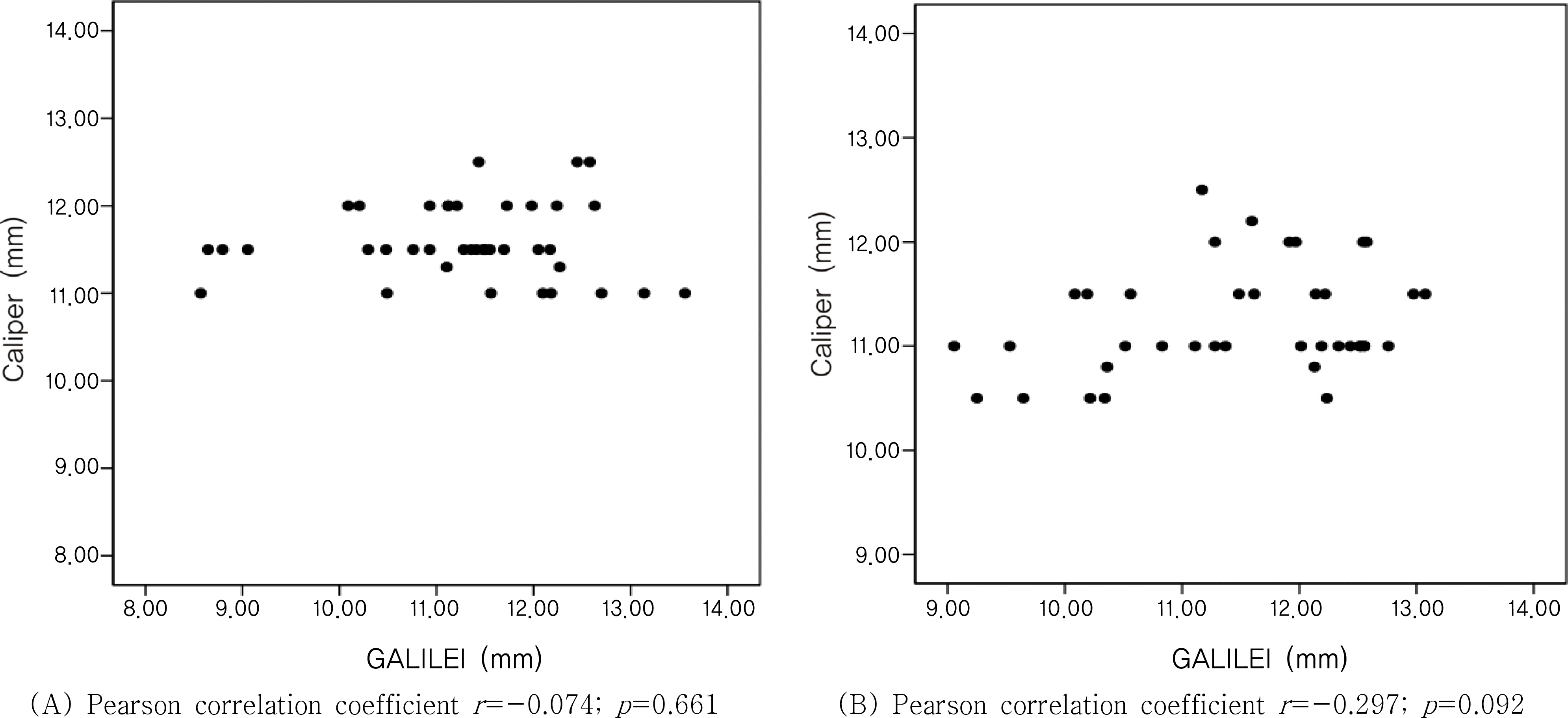
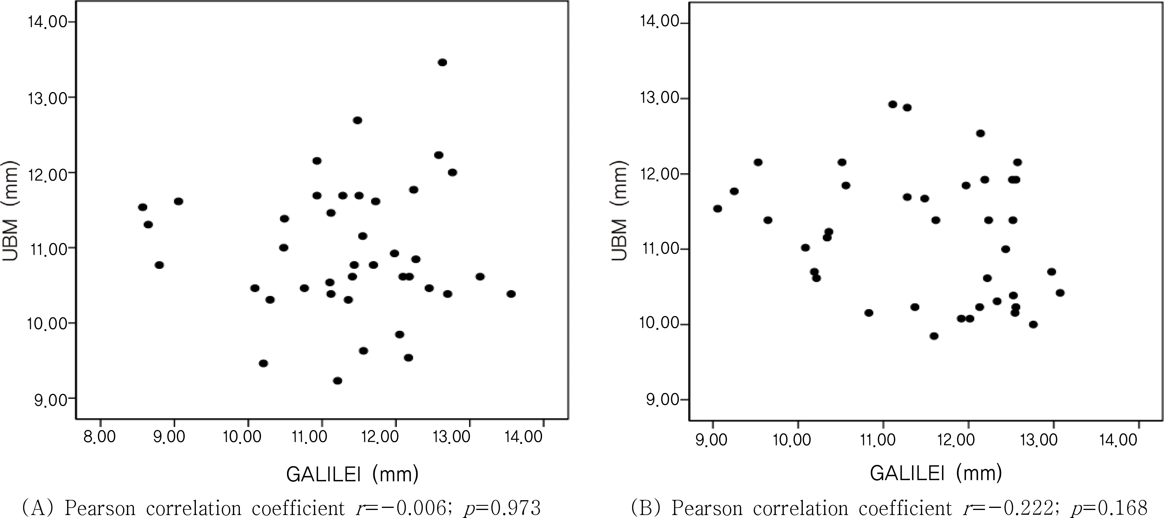
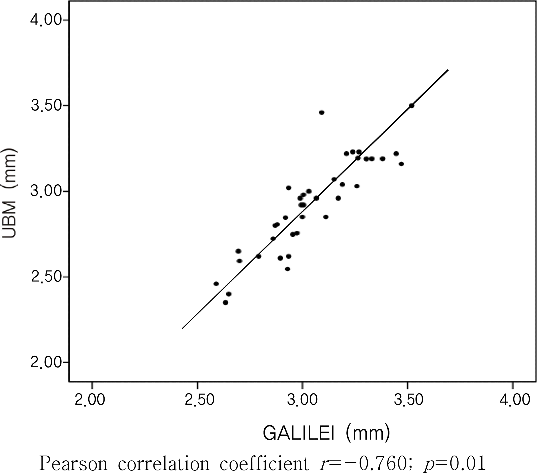
Table 1.
Demographics of patients (Mean± SD)
| Characteristics | Values |
|---|---|
| No. of patients (eyes) | 23 (44) |
| Sex (M:F) | 4:19 |
| Ages (years) | 30.2±7.44 |
| Spherical equivalent (diopter) | −2.74±1.94 |
Table 2.
Comparison of estimates by calipers, GALILEI TM and UBM (Mean± SD)
| | Caliper (WTW†) | GALILEI (WTW) | UBM (STS‡) | p-value* |
|---|---|---|---|---|
| Horizontal (mm) | 11.6±0.50 | 11.3±1.18 | 11.0±0.89 | 0.111 |
| Vertical (mm) | 11.2±0.51 | 11.5±1.10 | 11.1±0.92 | 0.138 |
Table 3.
Comparison of the anterior chamber depth (ACD) by GALILEI TM and UBM (Mean± SD)
| Measurement | GALILEI | UBM | p-value* |
|---|---|---|---|
| ACD (mm) | 3.1 ±0.29 | 2.9 ± 0.28 | <0.01 |
 XML Download