

 PDF
PDF ePub
ePub Citation
 Print
Print


Abstract
Case summary
A nine-year-old boy visited our clinic with conjunctival injection and decreased visual acuity in the left eye, which had developed two month previously. Slit lamp examination revealed a distorted pupil and an anteriorly protruding ciliary body displacing the peripheral iris toward the cornea on the inferonasal side. Funduscopic examination showed total retinal detachment accompanied by an inferior hypervascular mass. The examination of the right eye was unremarkable. On computer tomography imaging, a calcified mass was identified behind the iris of the left globe in the inferomedial aspect, and ultrasound biomicroscopy revealed a medium to high echogenic tumor with an uneven oval cystic cavity in the ciliary body. At the follow-up examination, the size of the mass was increased, so we performed enucleation of the left eye. Pathology demonstrated that the retrolental mass abutting the lens had arisen from the retina. Histological examination revealed that the tumor had originated from the retina and extended into the ciliary body, and most of the tumor was composed of hyaline cartilage with calicification. Tumor cells were identified in the periphery, forming elongated tubules and cord-like structures that were immunohistochemically positive for vimentin, neuron-specific enolase, and CD56 compatible with a teratoid medulloepithelioma. The patient was followed up for eight months without any metastasis in the orbit or elsewhere.
References
1. Broughton WL, Zimmerman LE. A clinicopathologic study of 56 cases of intraocular medulloepitheliomas. Am J Ophthalmol. 1978; 85:407–18.

2. Shields JA, Eagle RC Jr, Shields CL, Potter PD. Congenital neoplasms of the nonpigmented ciliary epithelium. Ophthalmology. 1996; 103:1998–2006.
3. Steinkuller PG, Font RL. Congenital malignant teratoid neoplasm of the eye and orbit: a case report and review of the literature. Ophthalmology. 1997; 104:38–42.
4. Kim JH, Park ES, Yang HN. A case of benign nonteratoid medulloepithelioma of the optic nerve head. J Korean Ophthalmol Soc. 1999; 40:1727–31.
5. Park YH, Lee HJ, Chung SM, et al. Medulloepithelioma of the ciliary body. J Korean Ophthalmol Soc. 1999; 40:2942–7.
6. Zimmerman LE. Verhoeff's “terato-neuroma”. A critical reappraisal in light of new observations and current concepts of embryonic tumors. Am J Ophthalmol. 1971; 72:1039–57.
7. Sosi ska-Mielcarek K, Senkus-Konefka E, Jaskiewicz K, et al. Intraocular malignant teratoid medulloepithelioma in an adult: clinicopathological case report and review of the literature. Acta Ophthalmol Scand. 2006; 84:259–62.
8. Font RL, Rishi K. Diffuse retinal involvement in malignant non-teratoid medulloepithelioma of ciliary body in an adult. Arch Ophthalmol. 2005; 123:1136–8.
9. Chavez M, Mafee MF, Castillo B, et al. Medulloepithelioma of the optic nerve. J Pediatr Ophthalmol Strabismus. 2004; 41:48–52.
10. Chung EM, Specht CS, Schroeder JW. From the archives of the AFIP: pediatric orbit tumors and tumorlike lesions: neuroepithelial lesions of the ocular globe and optic nerve. Radiographics. 2007; 27:1159–86.
11. Zhou M, Xu G, Bojanowski CM, et al. Differential diagnosis of anterior chamber cysts with ultrasound biomicroscopy: ciliary body medulloepithelioma. Acta Ophthalmol Scand. 2006; 84:137–9.
12. Ayres B, Brasil OM, Klejnberg C, et al. Ciliary body medulloepithelioma: clinical, ultrasound biomicroscopic and histopathologic correlation. Clin Experiment Ophthalmol. 2006; 34:695–8.
13. Al-Salam S, Algawi K, Alashari M. Malignant non-teratoid medulloepithelioma of ciliary body with retinoblastic differentiation: a case report and review of literature. Neuropathology. 2008; 28:551–6.
Figure 1.
A.B. Anterior segment photographs at the first visit. Pupil is dragged inferonasally and band keratopathy is observed (A). On slit-lamp view, bulged iris touches the cornea (B). (C) Funduscopic finidng of the intraocular mass. Total retinal detachment is accompanied with inferior hypervascular mass.

Figure 2.
Ultrasound biomicroscopy of mass. The ultrasound biomicroscopy reveals the medium to high echogenic tumor in the retrolental space.
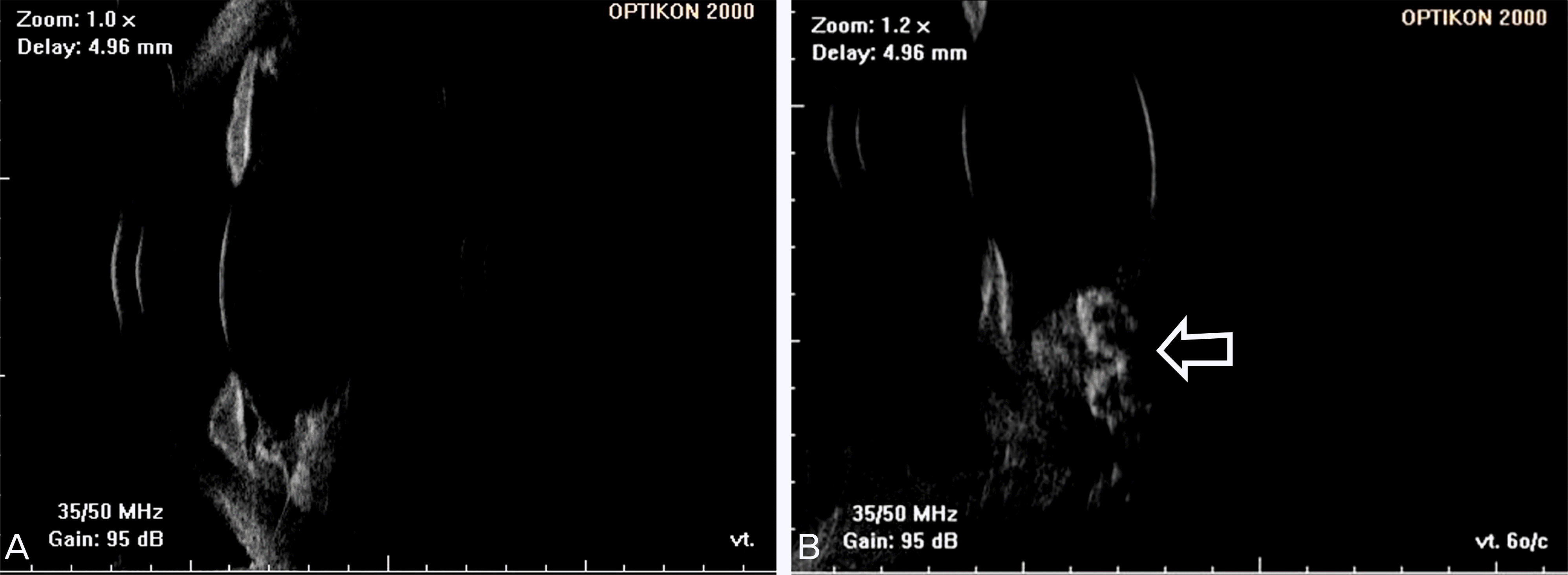
Figure 3.
Preoperative CT and MRI images (A,B). Preoperative CT images; Calcified mass is detected behind of iris in inferomedial aspect of left globe. Left globe looks slightly smaller. (C,D) MRI images; T1-weighted image showing marked enhancement of the ocular mass (C) and T2-weighted image showing ‘V’ shaped subretinal high attenuation (D).

Figure 4.
Microscopic findings of the specimen. (A) a 1.0 × 0.8 × 0.5 cm sized mass is confined to the posterior chamber of the left orbit. (B) The islands of cartilage with calcification are observed. (C)Tumor cells are present at the periphery of the ciliary body showing elongated tubules and cord-like structures. (D) Tumor cells (arrow head) and teratoid cartilage (asterisk) are located at the inner surface of the retinal pigment epithelium (arrow). (H&E, A: ×10, B: ×40, C: ×100, D: ×100)


 XML Download