Abstract
Purpose
Posterior scleritis is known to be a rare disease. The authors of the present study herein report a case of posterior scleritis, which occurred in a patient's eye, accompanied by hyperthyroidism and recurring in the other eye one year later.
Case summary
A 39-year-old female patient visited the hospital for ocular pain in the left eye and a headache. The patient was diagnosed with posterior scleritis through fundus examination, ultrasonography, CT and MRI, and an effective outcome of treatment was obtained by oral administration of methylprednisolone. Four months after discharge, the patient received left subtotal thyroidectomy for thyroid papillary cancer. Seven months after surgery she visited again, due to ocular pain that started 1 week earlier in the left eye, as well as a headache, and was diagnosed with posterior scleritis upon fundus examination, ultrasonography and MRI. Methylprednisolone was administered orally and an effective treatment result was obtained. After discharge, the patient was followed up for 5 months and did not show any signs of recurrence.
Go to : 
References
1. Kim SW, Lee KH, Lee EK. A case of posterior scleritis associated with ciliochoroidal detachment and anterior uveitis in background diabetic retinopathy patient. J Korean Ophthalmol Soc. 1995; 36:1234–8.
2. Rosenbaum JT, Robertson JE. Recognition of posterior scleritis and its treatment with indomethacin. Retina. 1993; 13:17–21.

3. Kim MW, Chung YT. Retinal pigment epithelial detachment in posterior scleritis. J Korean Ophthalmol Soc. 1989; 30:823–7.
5. Joo SH, Choi JK. A case of posterior scleritis associated with relapsing polychondritis. J Korean Ophthalmol Soc. 1989; 30:665–70.
6. Benson WE, Shields IA, Tasman W, et al. Posterior scleritis: a cause of diagnostic confusion. Arch Ophthalmol. 1979; 97:1482–6.

7. McCluskey P, Wakefield D. Intrascleritis. Arch Ophthalmol. 1987; 105:793–7.
8. McGavin DD, Williamson J, Forrester JV, et al. Episcleritis and scleritis. A study of their clinical manifestations and association with rheumatoid arthritis. Br J Ophthalmol. 1976; 60:192–226.

9. Anderson B Sr. Ocular lesions in relapsing polychondritis and other rheumatoid syndromes. Am J Ophthalmol. 1967; 64:35–50.

10. Foster GS. Immunosuppressive therapy for external ocular inflammatory disease. Ophthalmology. 1980; 87:140–50.

11. Watson PG, Hazleman BL. The sclera and systemic disorders. London: Saunders;1976.
12. Vitale AT, Maza MS. Scleral inflammatory disease. Stephen J Ryan, editor. Retina. 4th revised ed.Los Angeles: Elsevier Mosby;2006. 2:chap.p. 98.

13. Hedges TR Jr, Leopold IH. Parallel retinal folds; Their significance in orbital space-taking lesions. Arch Ophthalmol. 1959; 62:353–5.
14. Nettleship E. Peculiar lines in the choroid in a case of postpapillitic atrophy. Trans Ophthalmol. 1981; 88:565–74.
15. Jellinek EH. The orbital pseudotumor syndrome and its differentiation from endocrine exophthalmos. Brain. 1969; 92:35–8.
16. Berger B, Reeser F. Retinal pigment epithelial detachment in posterior scleritis. Am J Ophthalmol. 1980; 90:604–7.
17. Cappaert WE, Purnell EW, Frank KE. Use of B-sector scan ultrasound in the diagnosis of benign choroidal folds. Am J Ophthalmol. 1977; 84:375–9.

18. McCluskey P, Wakefield D. Current concept in the management of scleritis: a study of their clinical manifestations and association with rheumatoid arthritis. Br J Ophthalmol. 1988; 16:169–76.
Go to : 
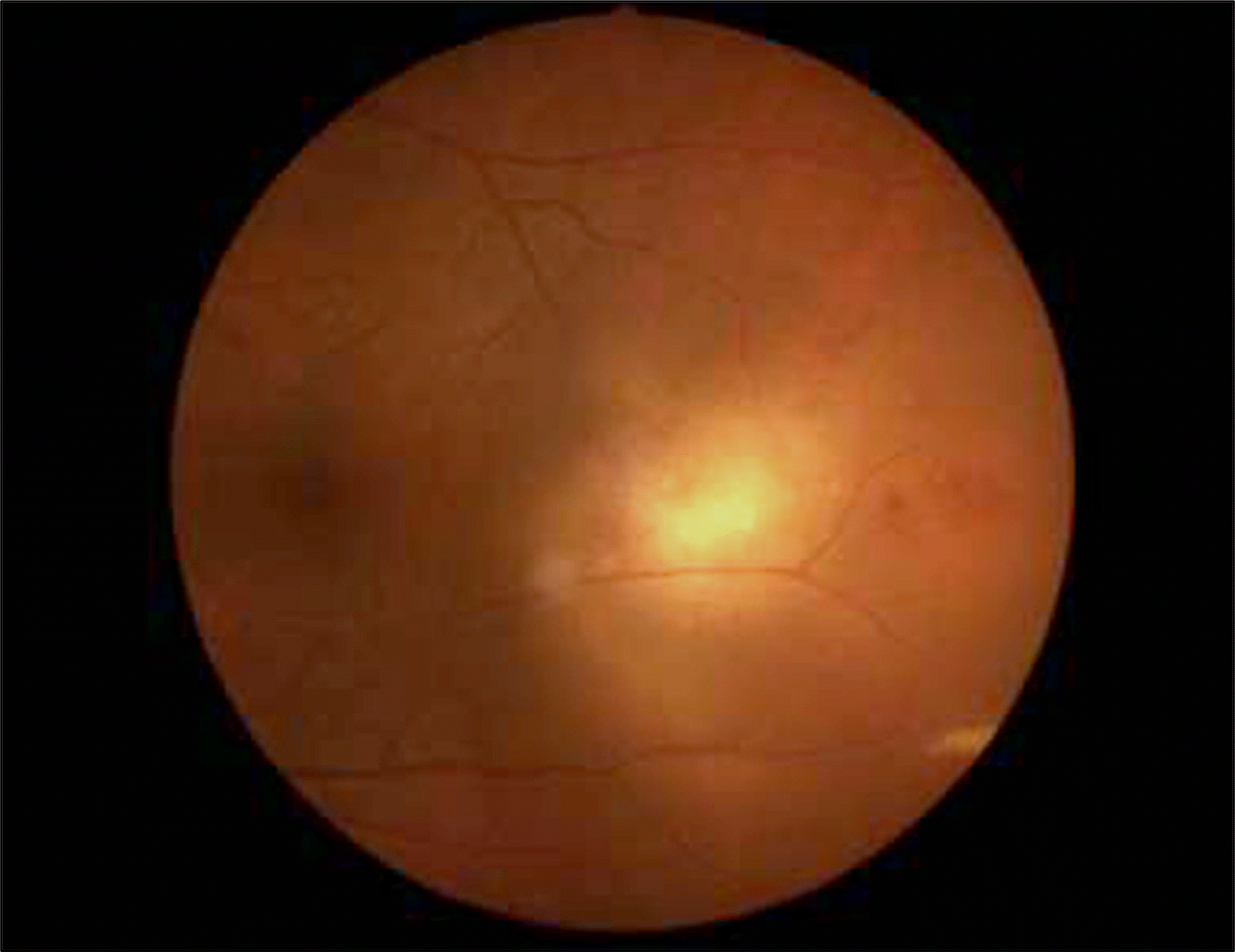 | Figure 1.Fundus Photograghy shows exudative retinal detachment, retinal hemorrhage, and whitish subretinal whitish lesion at 3 o'clock. |
 | Figure 2.(A) B-scan ultrasonogram shows scleral thickening and “T-sign” in the left eye. The squaring off of the normally rounded optic nerve shadow with extension of the edema along the back of the eye is called the “T-sign.” (B) Orbit CT shows lining pattern contrast enhancement of the sclera in the left eye. (C) Orbit MRI shows soft tissue density of the bulbar area and lining pattern contrast enhancement of the sclera in the left eye. |
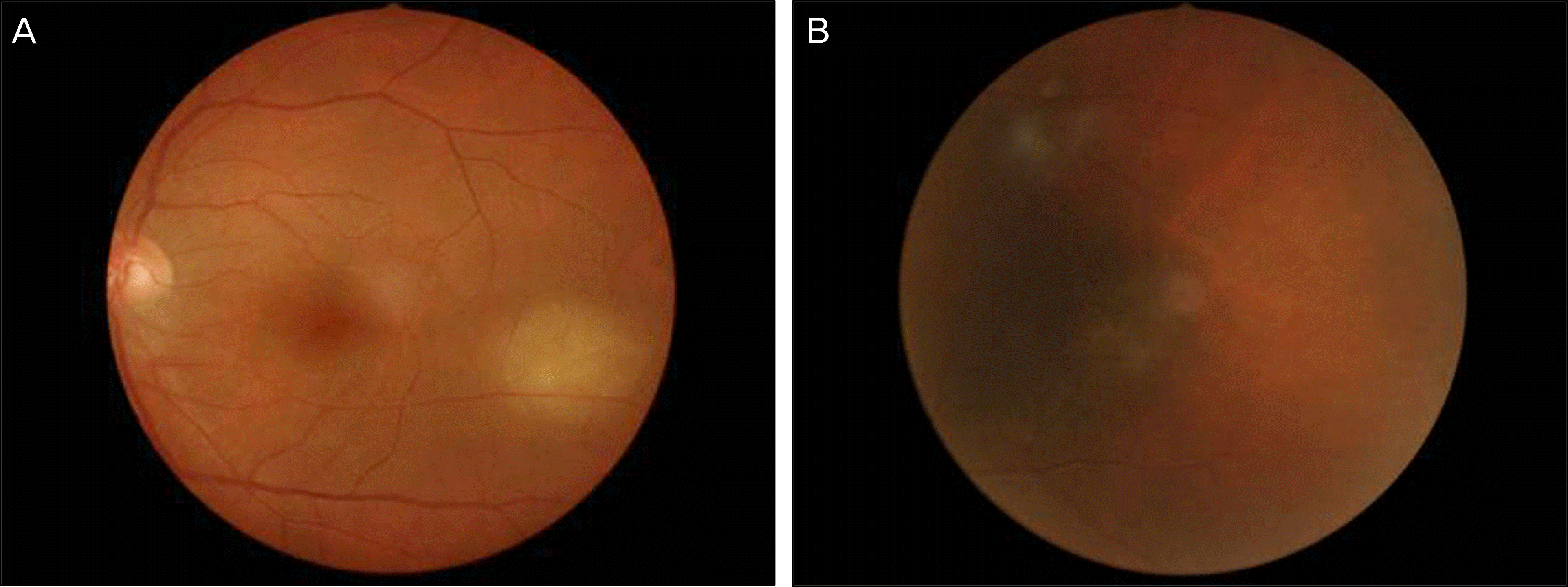 | Figure 3.(A) Exudative retinal detachment, retinal hemorrhage, and whitish subretinal lesion are decreased in the fundus photograph 7 days later. (B) Exudative retinal detachment, retinal hemorrhage, and whitish subretinal lesion are resolved in the fundus photograph 2 weeks later. |
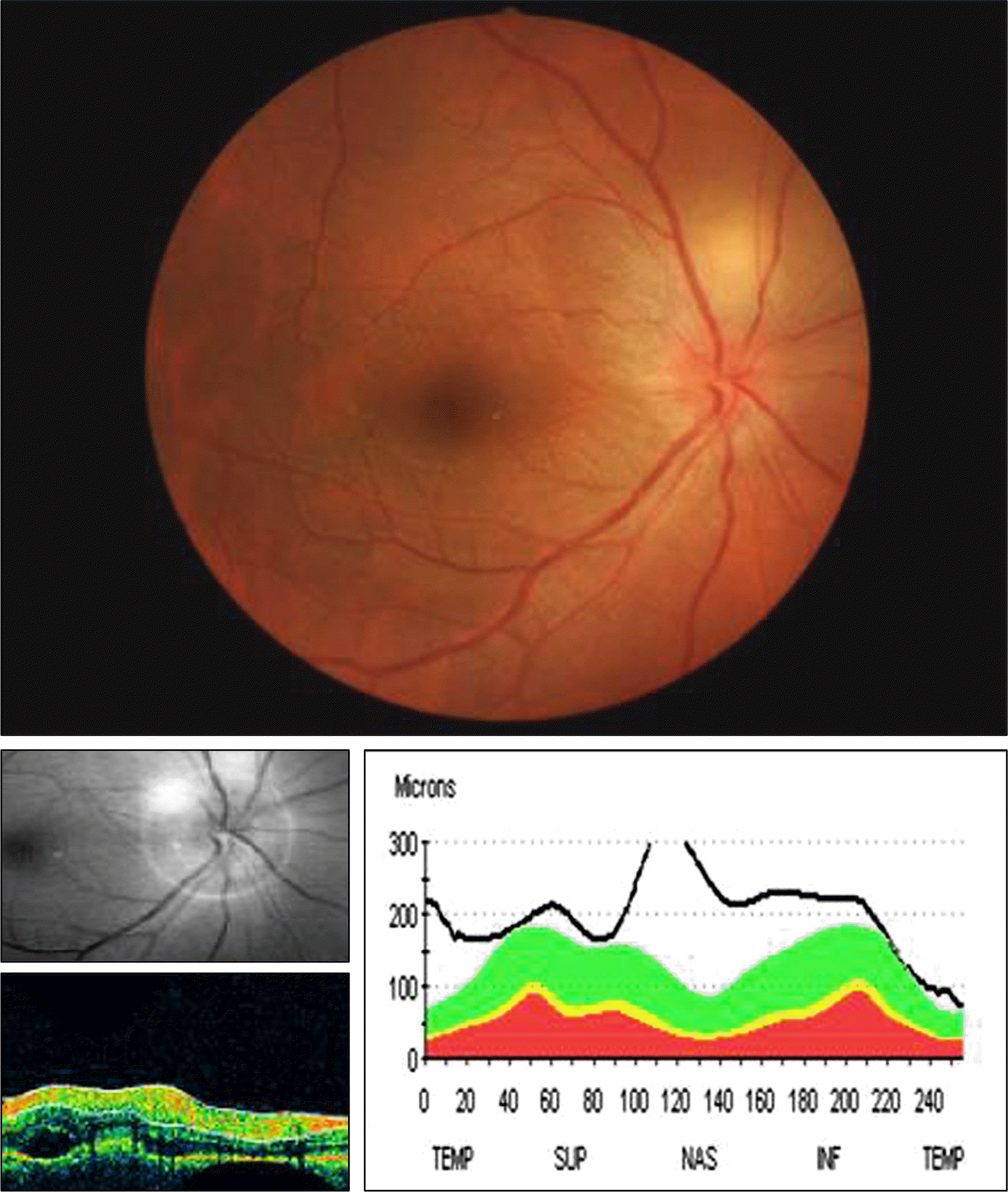 | Figure 4.Fundus photogragh shows serous retinal detachment and blurred margin at the optic disc. OCT images show edema of the optic disc. |
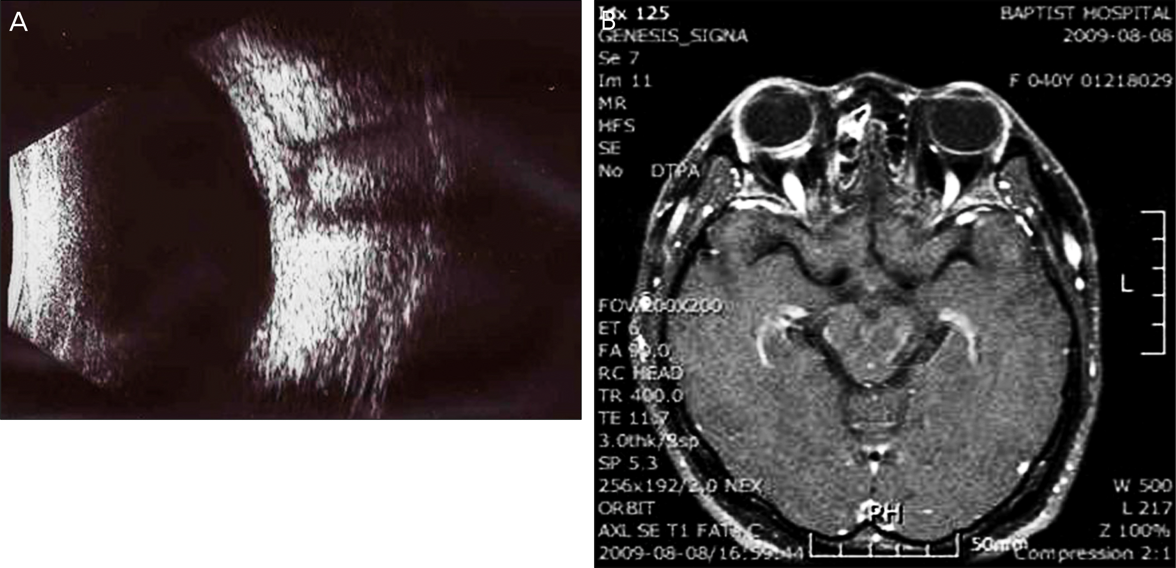 | Figure 5.(A) B-scan ultrasonogram showed scleral thickening and “T-sign” in the right eye. (B) Orbit MRI shows soft tissue density of the bulbar area and lining pattern contrast enhancement of the sclera in the right eye. |
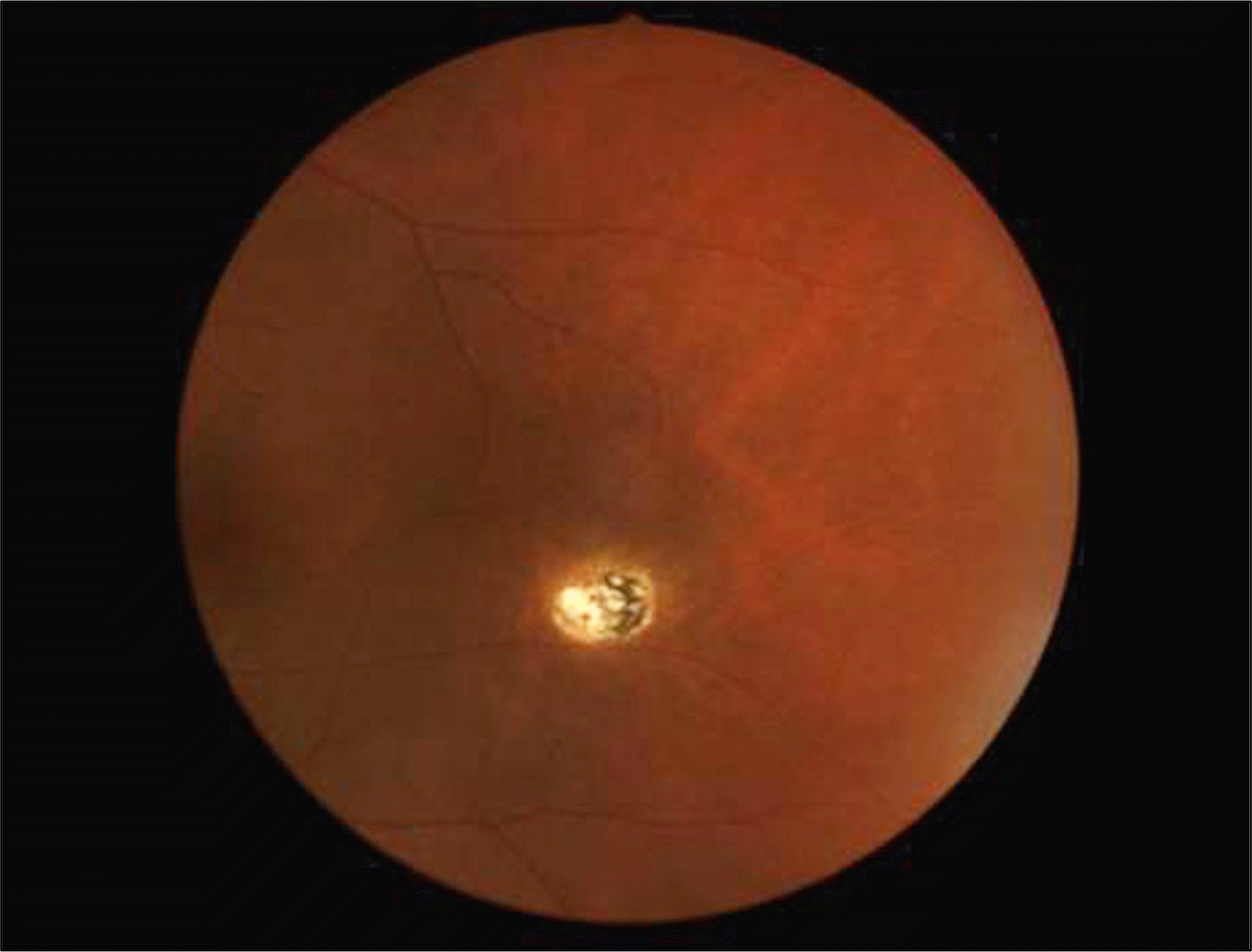 | Figure 6.Fundus Photogragh shows degeneration that exudative retinal detachment resolved 11 months later. |
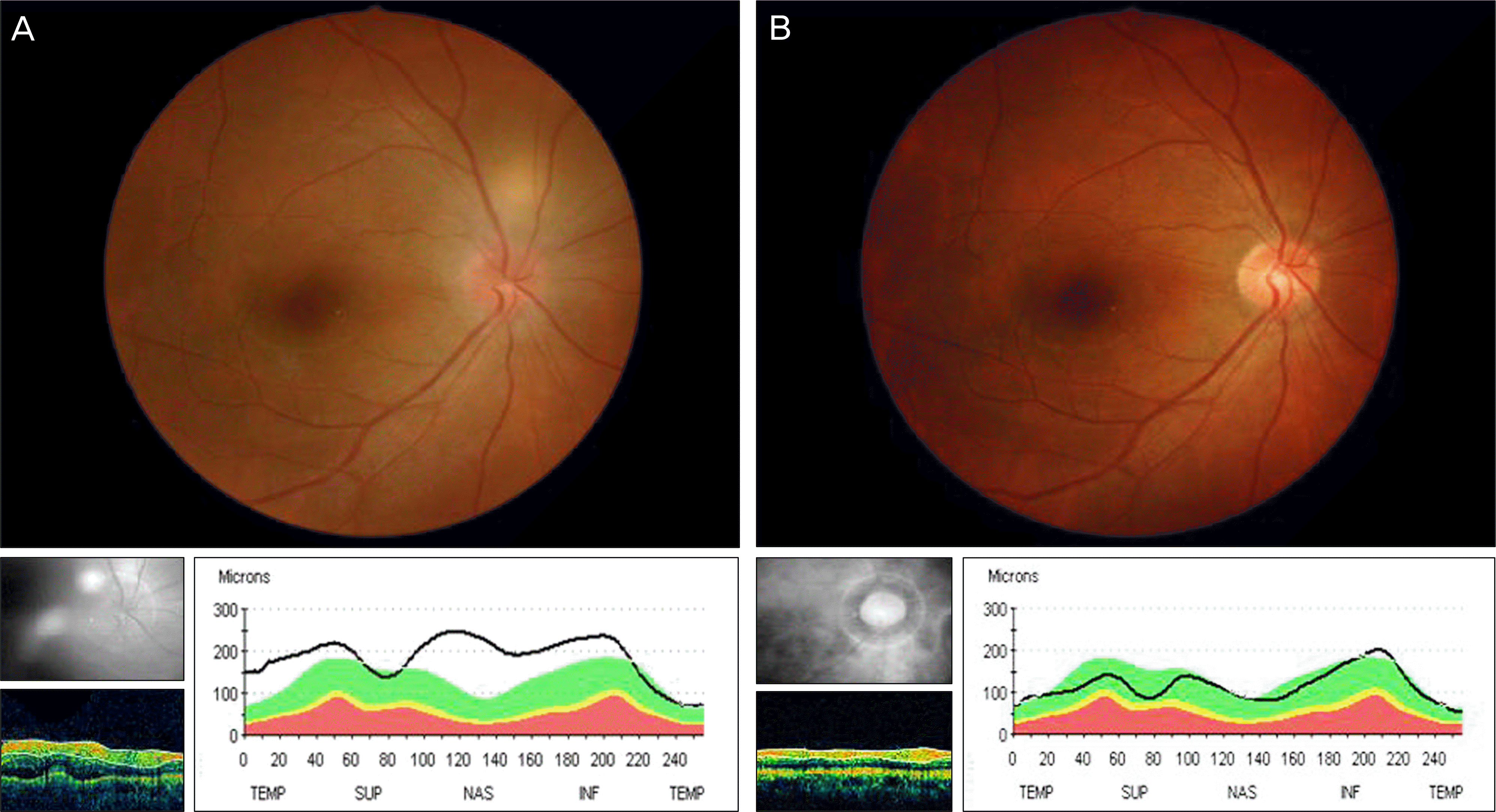 | Figure 7.(A) Serous retinal detachment and blurr the margin at the optic disc is decreased in the fundus photograph 3 days later. OCT images shows that edema of the optic disc is decreased. (B) Serous retinal detachment and blurred margin at the optic disc the s resolved in the fundus photograph 2 months later. OCT images show that edema of the optic disc is resolved. |


 PDF
PDF ePub
ePub Citation
 Print
Print


 XML Download