

 PDF
PDF ePub
ePub Citation
 Print
Print


Abstract
Purpose
To compare the therapeutic effects and complications after panretinal photocoagulation (PRP) and discomforts of patients using patterned PRP versus conventional PRP for diabetic retinopathy.
Methods
Eighty patients who required PRP due to diabetic retinopathy were enrolled in a prospective randomized controlled study. The patients were randomly divided into two groups: a patterned PRP group in which PRP was performed with a short laser exposure time (0.02 seconds) and a conventional PRP group with a long exposure time (0.2 seconds). At the 1-year follow-up visit, the progressions of diabetic retinopathy, best-corrected visual acuity, and central macular thickness were evaluated. All patients were questioned about the grade of pain during PRP. In addition, the complications after PRP were investigated.
Results
There were no statistical differences in clinical characteristics between both groups. The progression of diabetic retinopathy was not different in both groups at the 1-year follow-up visit. The best-corrected visual acuities at 1, 2, 4, and 8 weeks after PRP were decreased in both groups and, in the conventional PRP group, the decrements of visual acuity were greater than in the patterned PRP group. The increments of central macular thickness were also greater in the conventional PRP group than the patterned PRP group.
Go to : 
References
1. Wessing A, Meyer-Schwickerath G. Treatment of diabetic retinopathy by light-coagulation. Diabetologia. 1969; 5:312–7.
2. Beetham WP, Aiello LM, Balodimos MC, Koncz L. Ruby laser photocoagulation of early diabetic neovascular retinopathy: preliminary report of long-term controlled study. Arch Ophthalmol. 1970; 83:261–72.
3. Diabetic Retinopathy Study Research Group. Photocoagulation treatment of proliferative diabetic retinopathy: clinical application of Diabetic Retinopathy Study (DRS) findings. DRS report no.8. Ophthalmology. 1981; 88:583–600.
4. Early Treatment Diabetic Retinopathy Study Research Group. Techniques for scatter and local photocoagulation treatment of diabetic retinopathy: Early Treatment Diabetic Retinopathy Study Report No. 3. Int Ophthalmol Clic. 1987; 27:254–64.
5. McDonald HR, Schatz H. Macular edema following panretinal photocoagulation. Retina. 1985; 5:5–10.

6. Khosla PK, Rao V, Tewari HK, Kumar A. Contrast sensitivity in diabetic retinopathy after panretinal photocoagulation. Ophthalmic Surg. 1994; 25:516–20.
7. Pahor D. Visual field loss after argon laser panretinal photocoagulation in diabetic retinopathy: full- versus mild-scatter coagulation. Int Ophthalmol. 1998; 22:313–9.
8. Yuki T, Kimura Y, Nanbu S, et al. Ciliary body and choroidal detachment after laser photocoagulation for diabetic retinopathy. A high-frequency ultrasound study. Ophthalmology. 1997; 104:1259–64.
9. Scott J, Huskisson EC. Graphic representation of pain. Pain. 1976; 2:175–84.
10. Patz A. Studies on retinal neovascularization. Invest Ophthalmol Vis Sci. 1980; 19:1133–8.
11. Molnar J, Oastry S, Tsacopuolos M. Effect of laser photocoagulation on oxygenation of the retina in miniature pigs. Invest Ophthalmol Vis Sci. 1985; 26:1410–4.
12. Yoshimura N, Matsumoto M, Shimizy H, et al. Photocoagulated human retinal pigment epithelial cells produce an inhibitor of vascular endothelial cell proliferation. Invest Ophthalmol Vis Sci. 1995; 36:1686–91.
13. Martidis A, Duker JS, Greenberg PB, et al. Intravitreal triamcinolone for refractory diabetic macular edema. Ophthalmology. 2002; 109:920–7.
14. Wu L, Martinez-Castellanos MA, Quiroz-Mercado H, et al. Twelve-month safety of intravitreal injections of bevacizumab (Avastin): results of the Pan-American Collaborative Retina Study Group (PACORES). Graefes Arch Clin Exp Ophthalmol. 2008; 246:81–7.
15. Chun DW, Heier JS, Topping TM, et al. A pilot study of multiple intravitreal injections of ranibizumab in patients with center-in-volving clinically significant diabetic macular edema. Ophthalmology. 2006; 113:1706–12.
16. Moorman CM, Hamilton AMP. Clinical applications of the micro-pulse diode laser. Eye. 1999; 13:145–50.
17. Bandello F, Brancoto R, Menchini U, et al. Light panretinal photocoagulation (LPRP) versus classic panretinal photocoagulation (CPRP) in proliferative diabetic retinopathy. Semin Ophthalmol. 2001; 16:12–8.
18. Wade EC, Blankenship GW. The effect of short versus long exposure times of argon laser panretinal photocoagulation on proliferative diabetic retinopathy. Graefes Arch Clin Exp Ophthalmol. 1990; 228:226–31.
19. Mainster MA. Decreasing retinal photocoagulation damage: principles and techniques. Semin Ophthalmol. 1999; 14:200–9.
20. Paulus YM, Jain A, Gariano RF, et al. Healing of retinal photocoagulation lesions. Invest Ophthalmol Vis Sci. 2008; 49:5540–5.
21. Cuthbertson FM, Newsom RS, Wainwright AC. Kinetic anesthesia for laser surgery. Eye. 2005; 19:1205–7.
22. Vaideanu D, Taylor P, McAndrew P, et al. Double masked randomized controlled trial to assess the effectiveness of paracetamol in reducing pain in panretinal photocoagulation. Br J Ophthalmol. 2006; 90:713–7.
23. Weinberger D, Ron Y, Lichter H, et al. Analgesic effect of topical sodium diclofenac 0.1% drops during retinal laser photocoagulation. Br J Ophthalmol. 2000; 84:135–7.
24. Al-Hussainy S, Dodson PM, Gibson JM. Pain response and follow-up of patients undergoing panretinal laser photocoagulation with reduced exposure times. Eye. 2008; 22:96–9.
25. Sanghvi C, McLauchlan R, Delgado C, et al. Initial experience with the Pascal photocoagulator: a pilot study of 75 procedures. Br J Ophthalmol. 2008; 92:1061–4.
26. Cho BJ, Kim TW, Woo SJ, et al. Short-term clinical outcome of patterned scanning laser photocoagulation with short exposure time in diabetic retinopathy. J Korean Ophthalmol Soc. 2009; 50:376–82.
Go to :
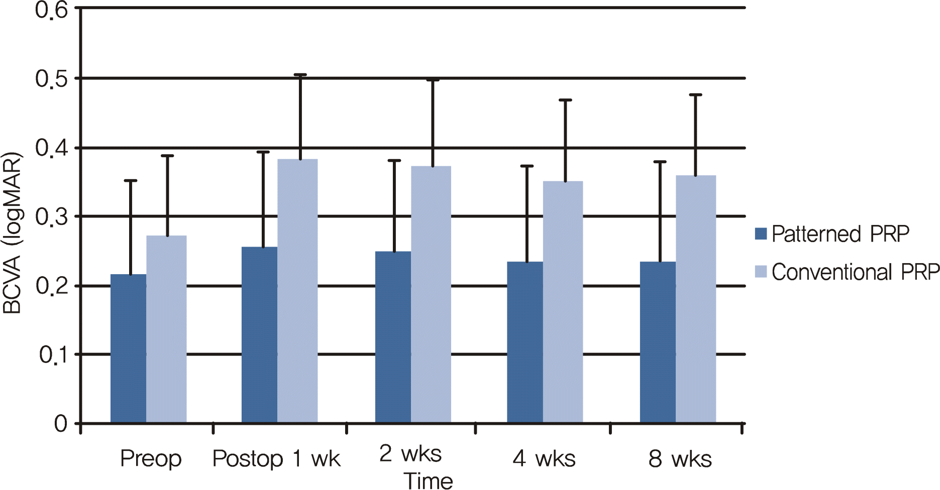 | Figure 1.Changes in best-corrected visual acuities (logarithm of the minimal angle of resolution) after panretinal photocoagulation. The best-corrected visual acuities at post-laser 1, 2, 4, and 8 weeks decreased in patterned PRP* group and conventional PRP† group, and in conventional PRP group the decrements of visual acuities were greater (p = 0.01, repeated measures ANOVA).* patterned PRP group = patients who received patterned panretinal photocoagulation using 0.02 second laser exposure time in a single treatment session; † conventional PRP group = patients who received panretinal photocoagulation using 0.2 second laser exposure time in three treatment sessions; BCVA = best-corrected visual acuity. |
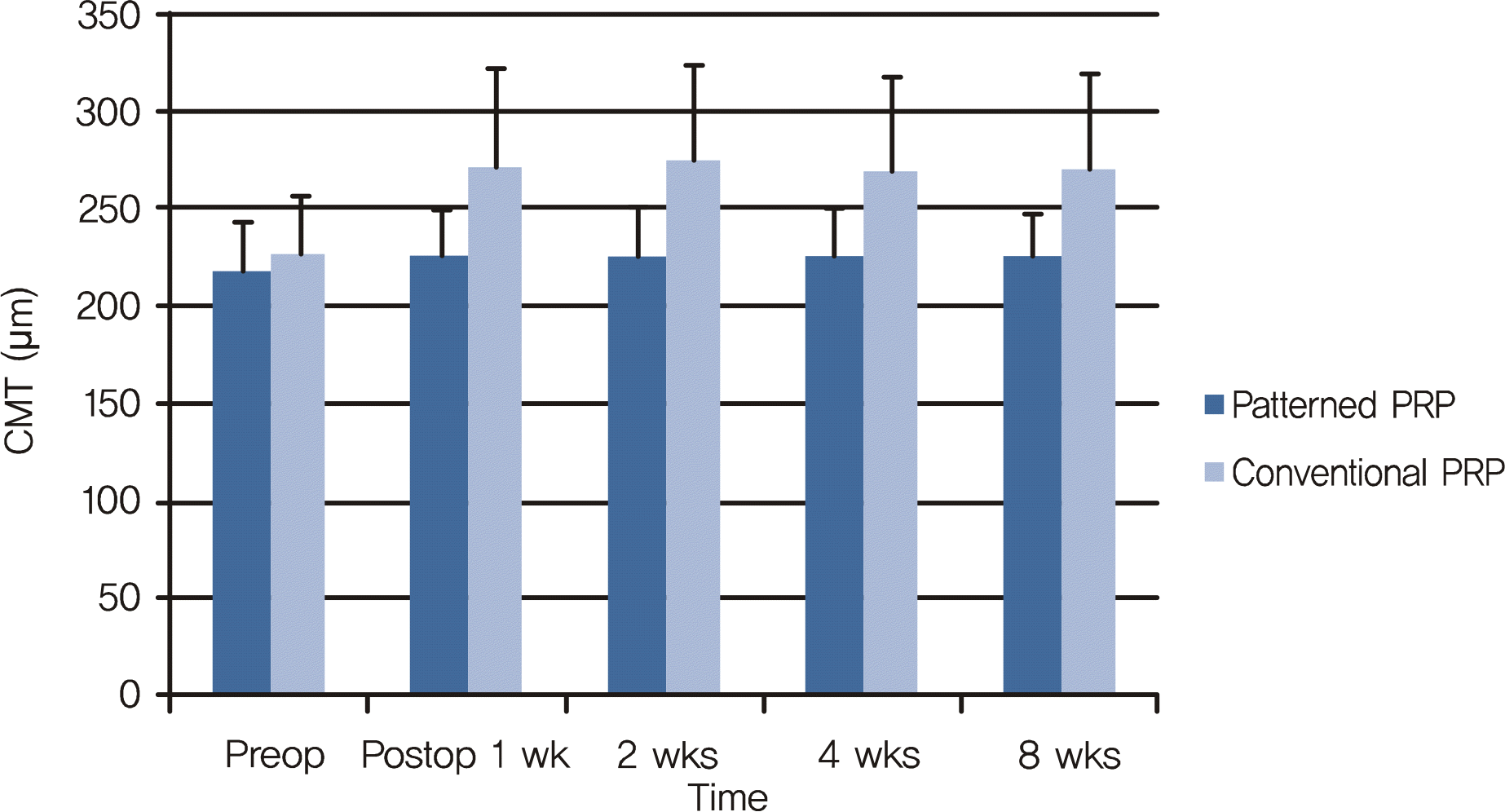 | Figure 2.Changes in central macular thickness after panretinal photocoagulation. The central macular thickness increased in patterned PRP group and conventional PRP group, and in conventional PRP group the increments of central macular thickness were greater (p = 0.01, repeated measures ANOVA). CMT = central macular thickness. |
Table 1.
Clinical characteristics of the patients in both groups
| | Patterned PRP Group | Conventional PRP Group | p value |
|---|---|---|---|
| Age (yr) | 60.03 ± 13.66 | 58.78 ± 11.30 | 0.657∏ |
| Sex | Male: Female = 20:20 | Male: Female = 19:21 | 0.826# |
| Duration of DM* (yr) | 9.28 ± 8.72 | 12.12 ± 6.83 | 0.216∏ |
| Severity of diabetic retinopathy | Severe NPDR†: 40/78 | 35/77 | 0.505# |
| | Very severe NPDR: 21/78 | 17/77 | 0.684# |
| | Early PDR‡: 11/78 | 15/77 | 0.583# |
| | High risk PDR: 6/78 | 10/77 | 0.648# |
| HBP§ | 55% (22/40) | 50% (20/40) | 0.508# |
| HbA1C | 8.75 ± 1.92 | 8.36 ± 1.53 | 0.422∏ |
| Blood urea nitrogen (mg/dL) | 18.42 ± 6.30 | 16.87 ± 4.95 | 0.308∏ |
| Creatinine (mg/dL) | 1.13 ± 0.83 | 1.07 ± 0.46 | 0.585∏ |
| Total cholesterol (mg/dL) | 191.11 ± 38.97 | 188.39 ± 47.47 | 0.835∏ |
Table 2.
Comparisons of laser parameters and pain in both groups
| | Patterned PRP Group | Conventional PRP Group | p value* |
|---|---|---|---|
| Laser power (mW) | 468.83 ± 106.08 | 307.10 ± 12.18 | 0.01 |
| Fluence (J/cm2) | 26.21 ± 7.29 | 189.40 ± 45.33 | 0.01 |
| Laser spot number | 1,728.90 ± 324.39 | 1,624.92 ± 511.92 | 0.205 |
| Pain | 2.18 ± 0.97 | 5.88 ± 1.06 | 0.02 |
Table 3.
Progression of diabetic retinopathy in one year follow-up after panretinal photocoagulation in both groups
| | Progression of DMR* |
Number of eyes (%) |
p value∏ | |
|---|---|---|---|---|
| Patterned PRP group | Conventional PRP Group | |||
| NPDR‡ | Number of progression to PDR§ | 5/61 (8.2%) | 7/52 (13.5%) | 0.274 |
| PDR | Number of complete NV# regression | 11/17 (65.7%) | 17/25 (68%) | |
| | Number of partial NV regression | 6/17 (35.3%) | 8/25 (32%) | 0.541 |
| | Number of no NV regression | 0/17 (0%) | 0/25 (0%) | |
| | Number of vitrectomy | 2/17 (11.8%) | 3/25 (12%) | 0.683 |
 XML Download