

 PDF
PDF ePub
ePub Citation
 Print
Print


Abstract
Methods
A retrospective chart review of the medical records from 94 patients (100 eyes), diagnosed to have a nevus of Ota from September 2005 to February 2010, was performed.
Results
The mean age of detection of a nevus of Ota was 8 months, and the mean extent of the nevus was 253 degrees, which covered more than 2 quadrants in most cases. The patients with a faint nevus tended to be younger. Pigmentation did not reach the fornix, but the limbus was mostly pigmented. Combined conjunctival pigmentation was observed in 61.7% of cases. The pigmentation was significantly associated with a greater extent of the nevus. Iris pigmentation was demonstrated in 98.7% of cases.
Conclusions
The clinical features of nevi of Ota were diverse regarding the location, extent, and color of the lesion. Conjunctival pigmentation was associated with the extent of the nevus. Iris pigmentation was revealed in almost all cases; therefore, this feature had the diagnostic value for a nevus of Ota. Young patients with a nevus of Ota may mimic osteo-genesis imperfecta, which necessitates careful consideration upon differential diagnosis.
Go to : 
References
1. Gupta GP, Gangwar DN. Naevus of Ota. Br J Ophthalmol. 1965; 49:364–8.

2. Cho KY, Eun HC, Lee YS. Clinical aspects of nevus of Ota and ex-tracutaneous pigmentation. Korean J Dermatol. 1986; 24:67–71.
3. Teekhasaenee C, Ritch R, Rutnin U, Leelawongs N. Ocular findings in oculodermal melanocytosis. Arch Ophthalmol. 1990; 108:1114–20.
4. Al-Sadhan Y, Shawaf S, Tabbara K. Oculodermal melanosis with choroidal melanoma in a black patient: a case report. Eye (Lond). 2006; 20:1437–8.
5. Qian Y, Zakov ZN, Schoenfield L. Iris melanoma arising in iris nevus in oculo (dermal) melanocytosis. Surv Ophthalmol. 2008; 53:411–5.
6. Teekhasaenee C, Ritch R, Rutnin U, et al. Glaucoma in oculodermal melanocytosis. Ophthalmology. 1990; 97:562–70.
7. Araie M. Oculodermal melanocytosis. J Glaucoma. 2002; 11:454–7.
8. Yoon JT, Tchah HW. The surgical treatment of nevus of Ota with ocular involvement. J Korean Ophthalmol Soc. 1999; 40:3229–33.
9. Cho BJ, Han YG, Kim JH, et al. Cosmetic repair of nevus of Ota. J Korean Ophthalmol Soc. 2006; 47:996–9.
Go to :
 | Figure 1.Manifestations of nevus of Ota. (A) Skin pigmentation with scleral pigmentation. (B) Iris pigmentation compared to contralateral eye without nevus of Ota (inside box). (C) Conjunctival pigmentation (black arrow), scleral (blue arrow) and deeper scleral (green arrow) pigmentation. (D, E) Anterior segment photograph of patients in dark group (D) and that of patient in faint group (E). |
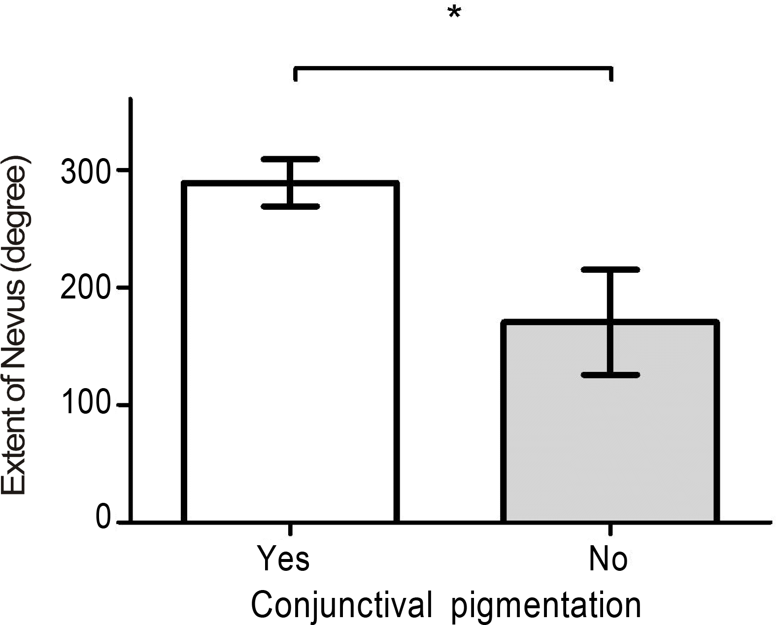 | Figure 2.The association between conjunctival pigmentation and the extent of nevus (Error bar indicates 95% confidence interval).* Statistically significant (p value <0.001) by Student's t test. |
Table 1.
Quadrant location and extent of Ota nevus
 XML Download