Abstract
Purpose
To report a case of prolonged bilateral inferior altitudinal visual field defect in a young migraine patient.
Case summary
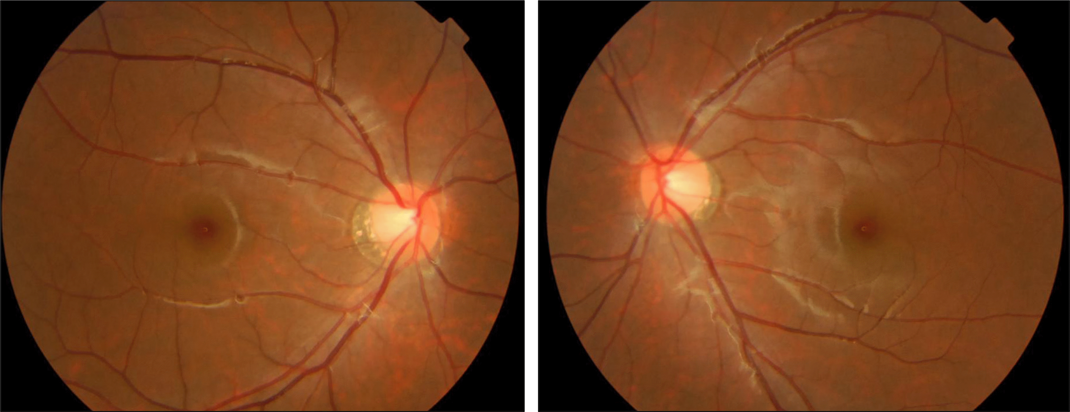
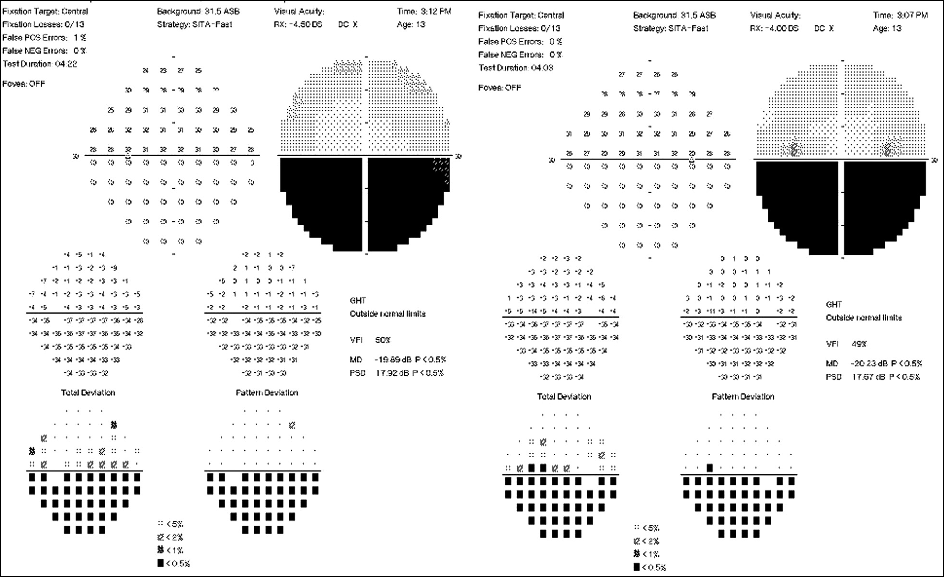
A 13-year-old female patient presented with bilateral disturbance of visual acuity and visual field, which had begun one month before. She complained of headache, with recently increasing frequency, that occurred 3 or 4 days a week for about 2∼3 hours duration, sometimes accompanied by nausea and located in the frontotemporal and retrobulbar area. Brain magnetic resonance imaging showed no abnormal finding in the brain and orbit. Her visual acuity was hand motion in both eyes and Humphrey visual field test showed bilateral inferior altitudinal visual field defect. Pupillary re-sonse was normal and extraocular muscle movement, anterior segment and fundus were also normal in ophthalmologic examination. Her best corrected visual acuity was 1.0 in both eyes by fogging method, but bilateral inferior altitudinal visual field defect persisted for 6 months follow-up.
References
1. Choi WJ, Chung J-Y, Lee DI, et al. Comparison of effectiveness of stellate ganglion block between chronic tension headache and chronic migraine patients. Korean J Anesthesiol. 2006; 51:201–6.

2. Lewis RA, Vijayan N, Watson C, et al. Visual field loss in migraine. Ophthalmology. 1989; 96:321–6.

3. Razeghinejad MR, Masoumpour M, Bagheri MH. Migrainous prolonged and reversible bilateral inferior altitudinal visual field defect. Headache. 2009; 49:773–6.

4. Relja G, Granato A, Ukmar M, et al. Persistent aura without infarction: description of the first case studied with both brain SPECT and perfusion MRI. Cephalalgia. 2005; 25:56–9.

5. Kang SJ, Kim JK, Kim MJ, et al. A case of recurrent and transient unilateral visual disturbance improved by beta-blocker: a variant of retinal migraine? Korean J Headache. 2007; 8:97–9.
6. Falavarjani KG, Sanjari MS, Modarres M, Aghamohammadi F. Clinical profile of patients with nonarteritic anterior ischemic optic neuropathy presented to a referral center from 2003 to 2008. Arch Iran Med. 2009; 12:472–7.
7. Mensah A, Vignal-Clermont C, Chadi M, et al. Dysthyroid optic neuropathy: Atypical initial presentation and persistent visual loss. Orbit. 2009; 28:354–62.

8. Lee AG, Brazis PW, Miller NR. Posterior ischemic optic neuropathy associated with migraine. Headache. 1996; 36:506–10.

9. González-Martín-Moro J, Pilo-de-la-Fuente B, Moreno-Martín P. Migraineous anterior optic ischemic neuropathy. Arch Soc Esp Oftalmol. 2009; 84:473–6.
10. Trobe JD. The neurology of vision. New York: Oxford University Press;2001. p. 295.
11. Tzourio C, Tehindrazanarivelo A, Iglésias S, et al. Case-control study of migraine and risk of ischaemic stroke in young women. BMJ. 1995; 310:830–3.

12. Agostoni E, Rigamonti A. Migraine and cerebrovascular disease. Neurol Sci. 2007; 28(Suppl 2):S156–60.

14. Streifler J-Y, Eliasziw M, Benavente OR, et al. North American Symptomatic Carotid Endarterectomy Trial. The risk of stroke with first-ever retinal vs hemispheric transient ischemic attacks and high-grade carotid stenosis. Arch Neurol. 1995; 52:246–9.
15. Lashley KS. Patterns of cerebral integration indicated by the scotomas of migraine. Arch Neurol Psychiatry. 1941; 46:331–9.

16. Dahlem MA, Engelmann R, Lowel S, Muller SC. Does the migraine aura reflect cortical organization? Eur J Neurosci. 2000; 12:767–70.

18. Wilkins A, Nimmo-Smith I, Tait A, et al. A neurological basis for visual discomfort. Brain. 1984; 107:989–1017.

19. Wakakura M, Ichibe Y. Permanent homonymous hemianopias following migraine. J Clin Neuroophthalmol. 1992; 12:198–202.
Figure 2.
(A) Humphrey visual fied test and (B) Goldmann visual field test showed bilateral inferior altitudinal visual field defect respecting the horizontal meridian.
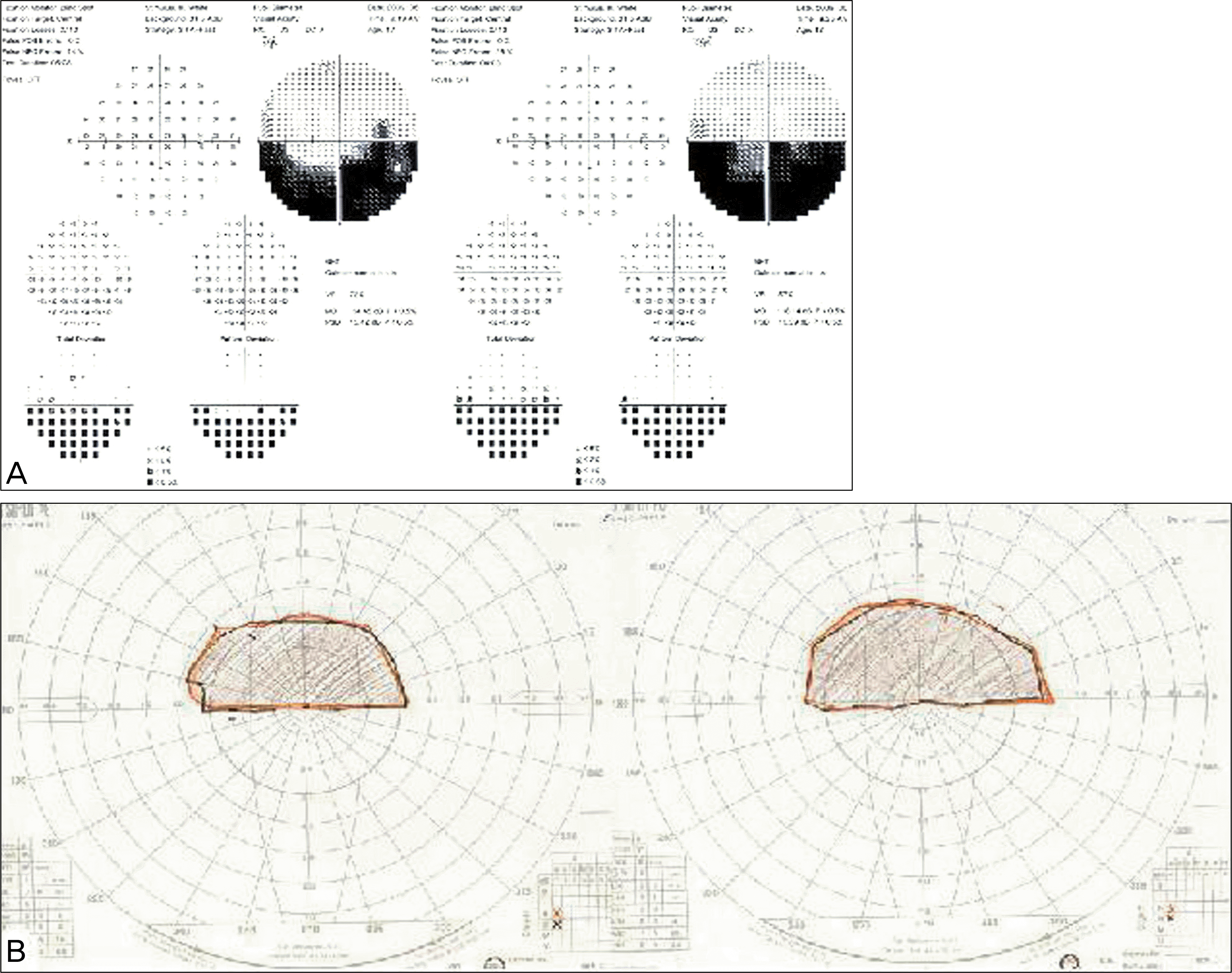
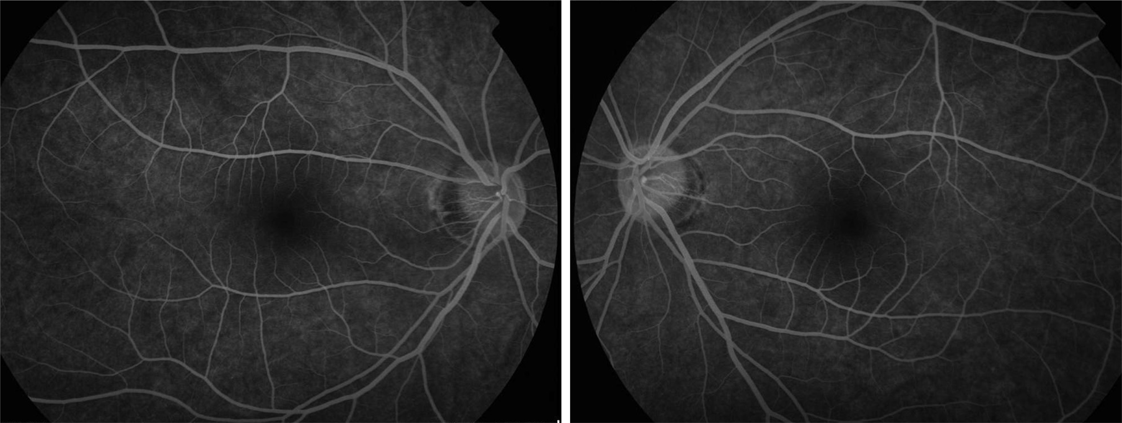
Figure 3.
Fluorescein angiography did not show any leakage or abnormal finding in optic disc and retina at recirculation phase.


 PDF
PDF ePub
ePub Citation
 Print
Print


 XML Download