Abstract
Purpose
To report a case of acyclovir-induced acute renal failure (ARF) suspected as acute retinal necrosis syndrome.
Case summary
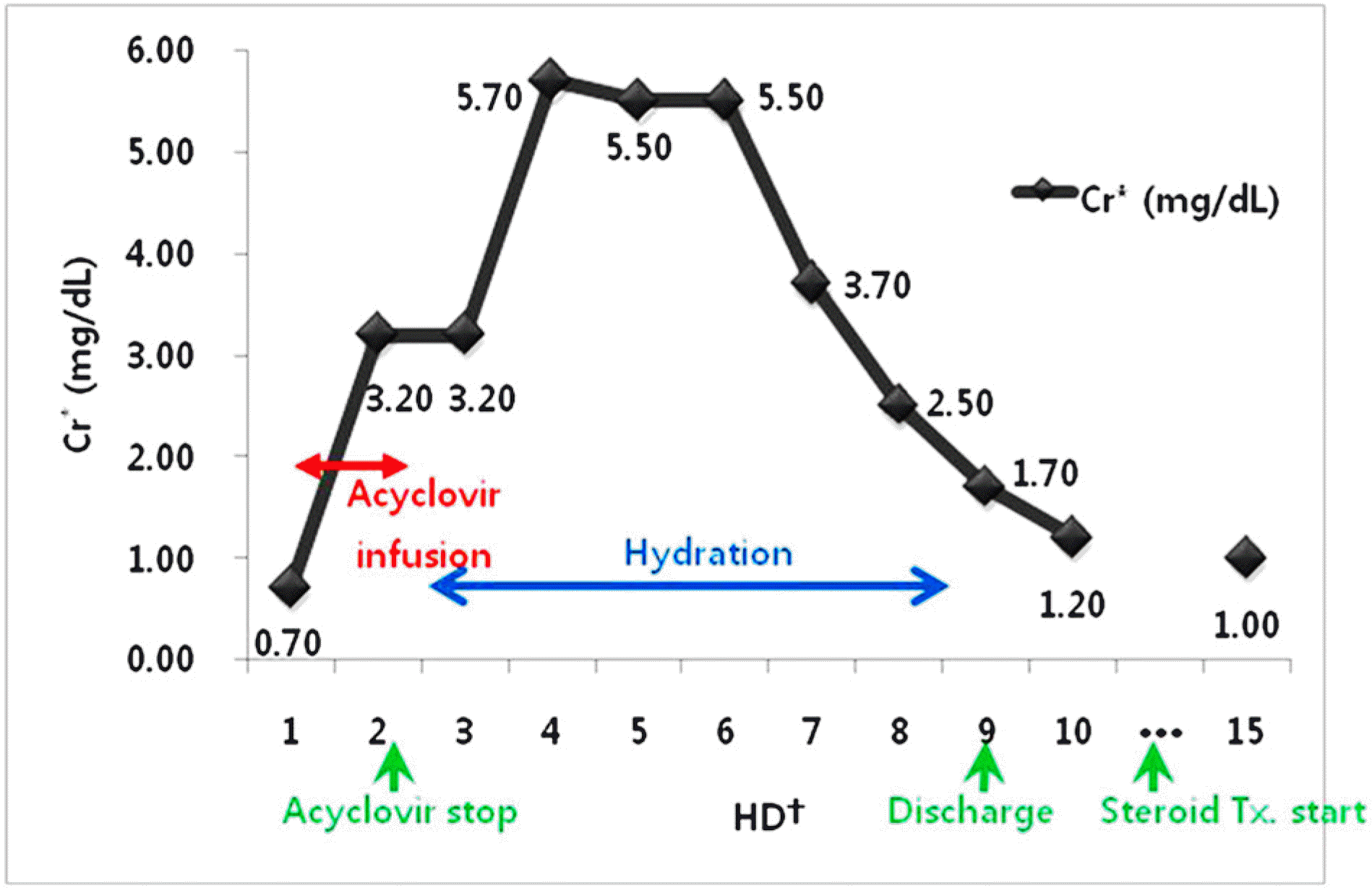
The authors report a 55-year-old male patient who presented with left eye visual disturbance due to suspected acute retinal necrosis syndrome. Non-oliguric ARF developed after the infusion of intravenous acyclovir (850 mg every 8 hours). The patient did not show any uremic symptoms or signs. The crystal was not discovered in the urine. After stopping the acyclovir infusion and hydration, acyclovir-induced ARF was reversed.
Go to : 
References
1. Clarkson JG, Blumenkranz MS, Culbertson WW, et al. Retinal detachment following the acute retinal necrosis syndrome. Ophthalmology. 1984; 91:1665–8.

2. Freeman WR, Thomas EL, Rao NA, et al. Demonstration of herpes group virus in acute retinal necrosis syndrome. Am J Ophthalmol. 1986; 102:701–9.

3. de Boer JH, Verhagen C, Bruinenberg M, et al. Serologic and polymerase chain reaction analysis of intraocular fluids in the diagnosis of infectious uveitis. Am J Ophthalmol. 1996; 121:650–8.

4. Usui Y, Goto H. Overview and diagnosis of acute retinal necrosis syndrome. Semin Ophthalmol. 2008; 23:275–83.

5. Matsuo T. Vitrectomy and silicone oil tamponade as an initial surgery for retinal detachment after acute retinal necrosis syndrome. Ocul Immunol Inflamm. 2005; 13:91–4.
6. McDonald HR, Lewis H, Kreiger AE, et al. Surgical management of retinal detachment associated with the acute retinal necrosis syndrome. Br J Ophthalmol. 1991; 75:455–8.

7. Sawyer MH, Webb DE, Balow JE, Straus SE. Acyclovir-induced renal failure. Clinical course and histology. Am J Med. 1988; 84:1067–71.
8. Holland GN. Standard diagnostic criteria for the acute retinal necrosis syndrome. Executive Committee of the American Uveitis Society. Am J Ophthalmol. 1994; 117:663–7.
9. Kim SJ, Yu HG. Bilateral acute retinal necrosis syndrome in the patient with acquired immunodeficiency syndrome. J Korean Ophthalmol Soc. 2003; 44:2445–50.
10. Duker JS, Blumenkranz MS. Diagnosis and management of the acute retinal necrosis (ARN) syndrome. Surv Ophthalmol. 1991; 35:327–43.

11. Tam PM, Hooper CY, Lightman S. Antiviral selection in the management of acute retinal necrosis. Clin Ophthalmol. 2010; 4:11–20.
13. Criteria for diagnosis of Behçet's disease. International Study Group for Behçet's Disease. Lancet. 1990; 335:1078–80.
14. Biswas J, Sharma T, Gopal L, et al. Eales disease an update. Surv Ophthalmol. 2002; 47:197–214.
15. Markowitz GS, Perazella MA. Drug-induced renal failure: a focus on tubulointerstitial disease. Clin Chim Acta. 2005; 351:31–47.

17. Tucker WE Jr, Macklin AW, Szot RJ, et al. Toxicology studies with acyclovir: acute and subchronic tests. Fundam Appl Toxicol. 1983; 3:573–8.
18. Bean B, Aeppli D. Adverse effects of highdose intravenous acyclovir in ambulatory patients with acute herpes zoster. J Infect Dis. 1985; 151:362–5.

19. Izzedine H, Launay-Vacher V, Deray G. Antiviral drug-induced nephrotoxicity. Am J Kidney Dis. 2005; 45:804–17.

Go to : 
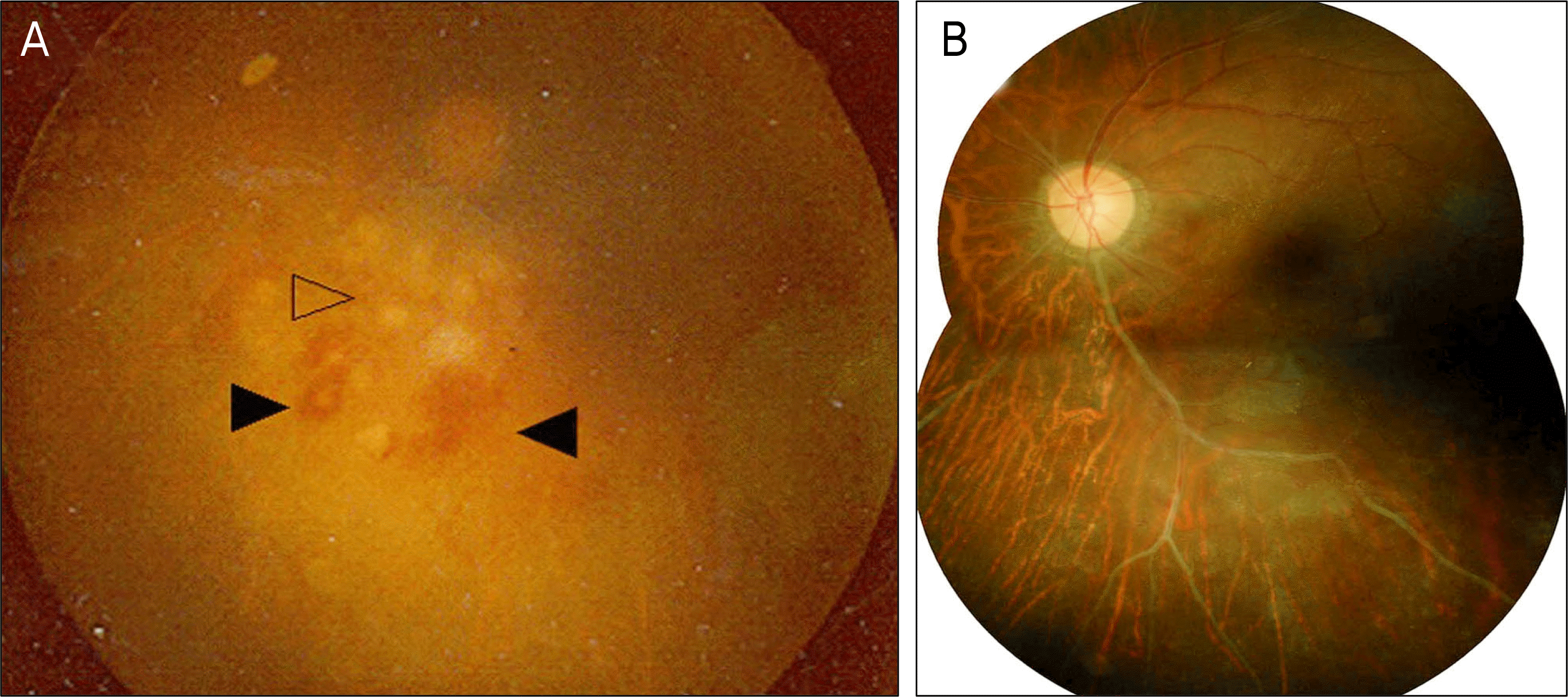
 | Figure 1.Initial fundus photograph of the left eye shows retinal necrosis (arrowhead) with retinal hemorrhages (arrowheads), obscured by dense vitreous opacity (A). After 3 months of the initial diagnosis, the vitreous inflammation and necrotic retinal lesions resolved almost completely, leaving behind ischemic retina with occluded retinal vessels inferotemporally (B). |


 PDF
PDF ePub
ePub Citation
 Print
Print


 XML Download