

 PDF
PDF ePub
ePub Citation
 Print
Print


Abstract
Purpose
To compare optical performances in eyes implanted with aspheric and spherical intraocular lenses (IOLs) after cataract surgery.
Methods
In 88 eyes of 69 patients, spherical (Sensar AR40; Acrysof natural SN60AT) or aspherical (Tecnis ZA9003; Acrysof IQ SN60WF) IOLs were implanted. After one month, contrast sensitivity, depth of focus, and spherical aberration were measured and compared between the groups.
Results
Contrast sensitivities of ZA9003 and SN60WF were significantly higher in 12,18 cycles per degree (CPD) under photopic conditions and were also higher in 6,12,18 CPD under mesopic conditions compared to those of respective spherical IOLs. Depths of focus were 1.31 D in ZA9003, 1.67 D in SN60WF, 1.52 D in AR40, and 1.49 in SN60AT, and the differences were not significant. Spherical aberration (Z40) with a 4 mm pupil was −0.032 μ m in ZA9003, 0.022 μ m in SN60WF, 0.076 μ m in AR40, and 0.072 μ m in SN60AT. Spherical aberration of SN60WF was significantly lower than spherical IOLs, and that of ZA9003 had the lowest among all IOL groups. Depth of focus significantly correlated with spherical aberration.
Go to : 
References
1. Miller D, Gurland JE, Isbey EK. Optics, Refraction and Contact Lenses. San Francisco, CA: American Academy of Ophthalmology;1992. p. 106.
2. Kim HS, Kim SW, Ha BJ, et al. Ocular aberrations and contrast sensitivity in eyes implanted with aspheric and spherical intraocular lenses. J Korean Ophthalmol Soc. 2008; 49:1256–62.

3. Wang L, Dai E, Koch DD, Nathoo A. Optical aberrations of the human anterior cornea. J Cataract Refract Surg. 2003; 29:1514–21.
4. Artal P, Berrio E, Guirao A, Piers P. Contribution of the cornea and internal surfaces to the change of ocular aberrations with age. J Opt Soc Am A Opt Image Sci Vis. 2002; 19:137–43.
5. Owsley C, Sekuler R, Siemsen D. Contrast sensitivity throughout adulthood. Vision Res. 1983; 23:689–99.
6. Michaels DD. Optics, refraction, and visual function. Curr Opin Ophthalmol. 1992; 3:69–70.
7. Packer M, Fine IH, Hoffman RS, Piers PA. Improved functional vision with a modified prolate intraocular lens. J Cataract Refract Surg. 2004; 30:986–92.
8. Charman WN. Ablation design in relation to spatial frequency, depth-of-focus, and age. J Refract Surg. 2004; 20:S542–9.
9. Johansson B, Sundelin S, Wikberg-Matsson A, et al. Visual and optical performance of the Akreos Adapt Advanced Optics and Tecnis Z9000 intraocular lenses: Swedish multicenter study. J Cataract Refract Surg. 2007; 33:1565–72.
10. Casprini F, Balestrazzi A, Tosi GM, et al. Glare disability and spherical aberration with five foldable intraocular lenses: a prospective randomized study. Acta Ophthalmol Scand. 2005; 83:20–5.
11. Yoon JU, Chung JL, Hong JP, et al. Comparison of wavefront analysis and visual function between monofocal and multifocal aspheric intraocular lenses. J Korean Ophthalmol Soc. 2009; 50:195–201.
12. Kang IS, You IC, Park YG, Yoon KC. Comparison of visual function among aspheric intraocular lenses. J Korean Ophthalmol Soc. 2009; 50:691–7.
13. Bae HW, Kim EK, Kim TI. Spherical Aberration, Contrast Sensitivity and Depth of Focus With Three Aspherical Intraocular Lenses. J Korean Ophthalmol Soc. 2009; 50:1639–44.
14. Weghaupt H, Pieh S, Skorpik C. Comparison of pseudoaccommodation and visual quality between a diffractive and refractive multifocal intraocular lens. J Cataract Refract Surg. 1998; 24:663–5.
15. Walkow L, Klemen UM. Patient satisfaction after implantation of diffractive designed multifocal intraocular lenses in dependence on objective parameters. Graefes Arch Clin Exp Ophthalmol. 2001; 239:683–7.
16. Ahn HS, Kim SW, Kim EK, Kim TI. Wavefront and visual function analysis after aspherical and spherical intraocular lenses implantation. J Korean Ophthalmol Soc. 2008; 49:1248–55.
17. Atchison DA. Design of aspheric intraocular lenses. Ophthalmic Physiol Opt. 1991; 11:137–46.
18. van Gaalen KW, Koopmans SA, Jansonius NM, Kooijman AC. Clinical comparison of the optical performance of aspheric and spherical intraocular lenses. J Cataract Refract Surg. 2010; 36:34–43.
19. Kamlesh , Dadeya S, Kaushik S. Contrast sensitivity and depth of focus with aspheric multifocal versus conventional monofocal intraocular lens. Can J Ophthalmol. 2001; 36:197–201.
20. Caporossi A, Martone G, Casprini F, Rapisarda L. Prospective randomized study of clinical performance of 3 aspheric and 2 spherical intraocular lenses in 250 eyes. J Refract Surg. 2007; 23:639–48.
21. Nio YK, Jansonius NM, Fidler V, et al. Age-related changes of de-focus-specific contrast sensitivity in healthy subjects. Ophthalmic Physiol Opt. 2000; 20:323–34.
22. Pepose JS, Qazi MA, Edwards KH, et al. Comparison of contrast sensitivity, depth of field and ocular wavefront aberrations in eyes with an IOL with zero versus positive spherical aberration. Graefes Arch Clin Exp Ophthalmol. 2009; 247:965–73.
23. Nochez Y, Majzoub S, Pisella PJ. Effects of spherical aberration on objective optical quality after microincision cataract surgery. J Fr Ophtalmol. 2010; 33:16–22.
24. Nanavaty MA, Spalton DJ, Boyce J, et al. Wavefront aberrations, depth of focus, and contrast sensitivity with aspheric and spherical intraocular lenses: fellow-eye study. J Cataract Refract Surg. 2009; 35:663–71.
25. Packer M, Fine IH, Hoffman RS. Aspheric intraocular lens selection based on corneal wavefront. J Refract Surg. 2009; 25:12–20.
Go to :
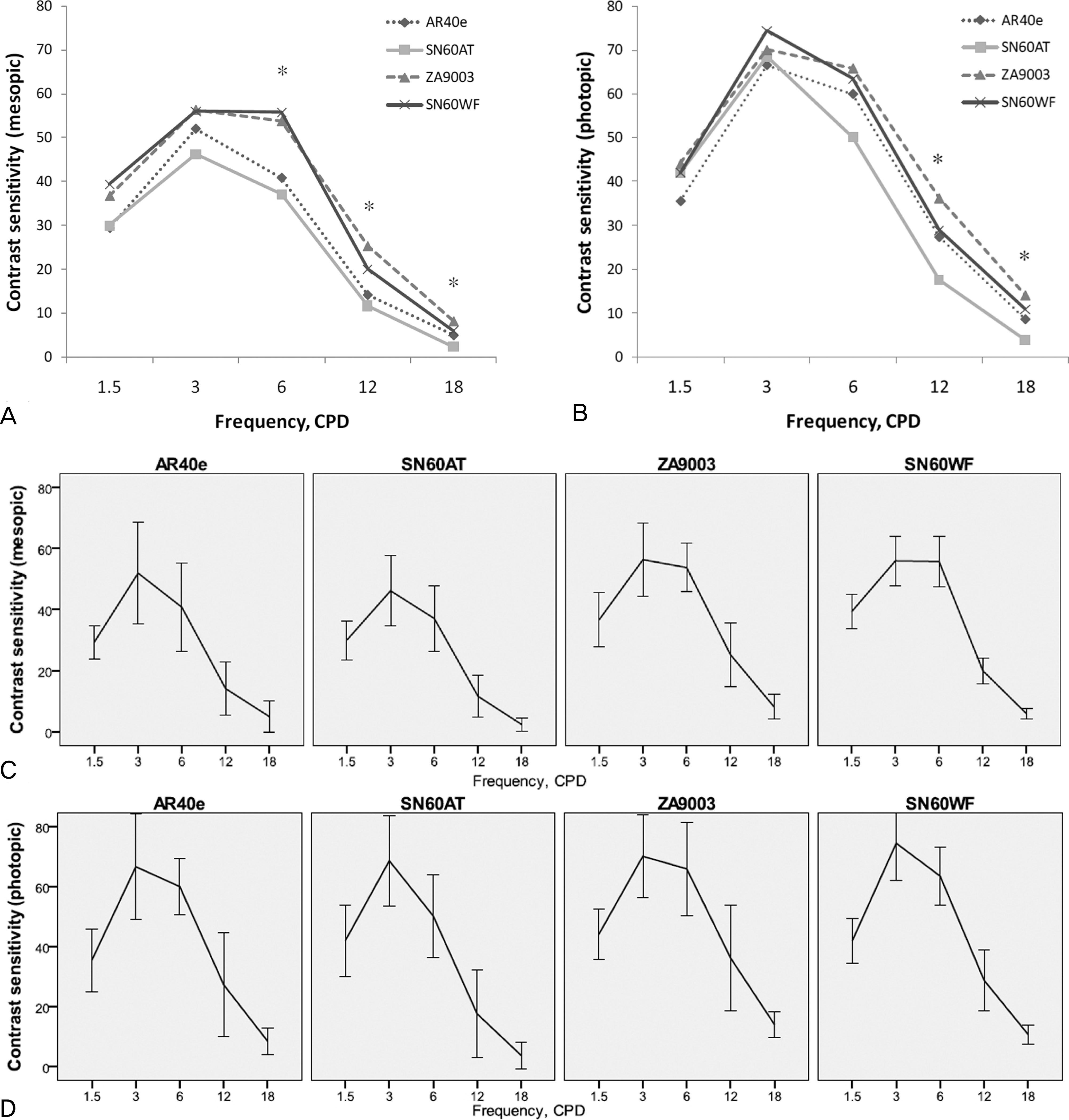 | Figure 1.Contrast sensitivity values under mesopic (3 cd/m2) (A,C) and photopic (85 cd/m2) (B,D) lighting conditions 1 month postoperatively. The asterisks(*) indicate the differences in values were significant (P<0.05) between the aspheric and spherical IOL groups. Error bars are indicated on each IOL's contrast sensitivity curve (C,D). |
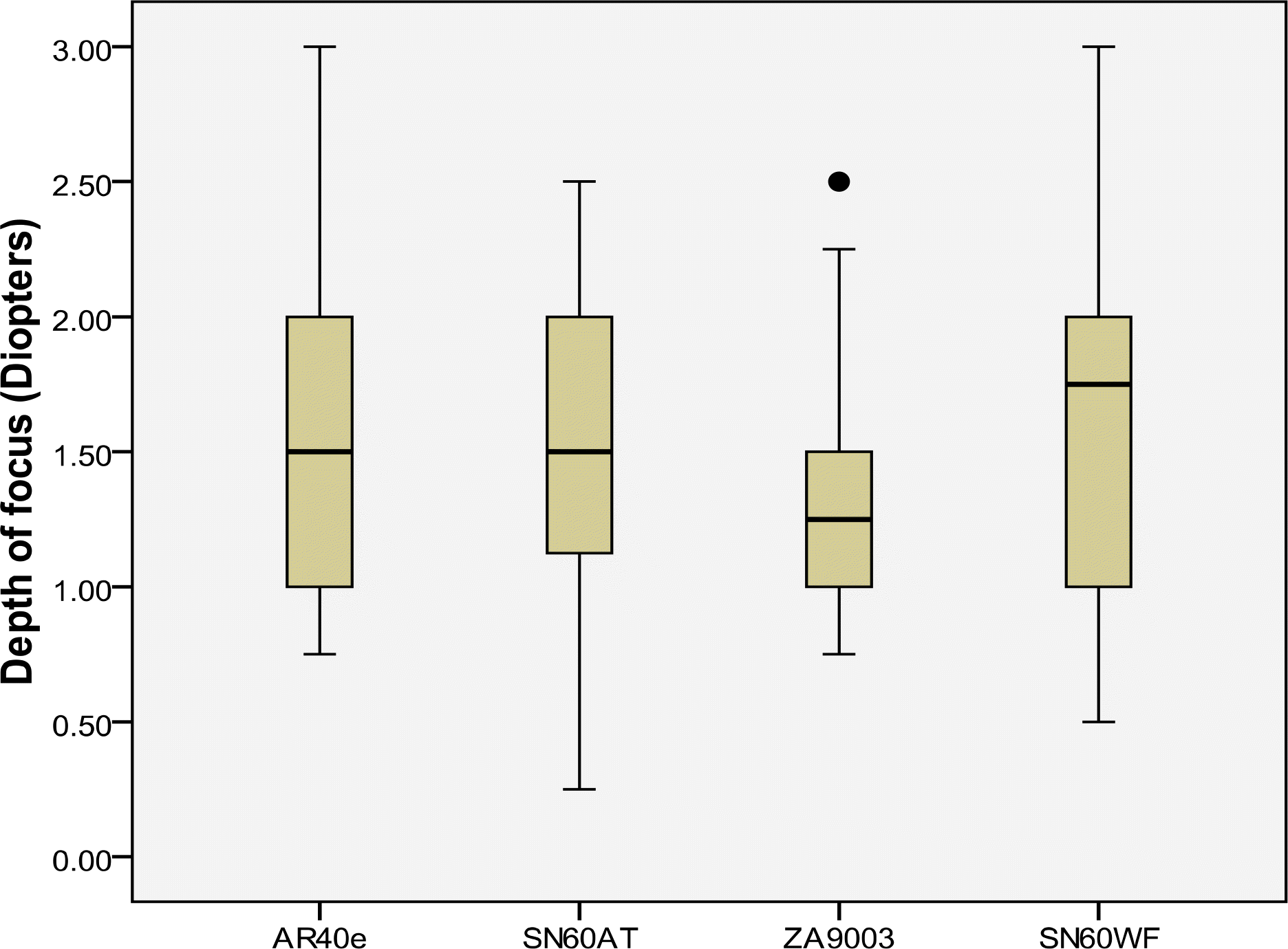 | Figure 2.Comparison of the depth of focus for the spherical and aspheric IOL groups. Data are expressed as mean (lower and upper quartiles, box) ± standard deviation (bars), extra-ordinary value (round dot). |
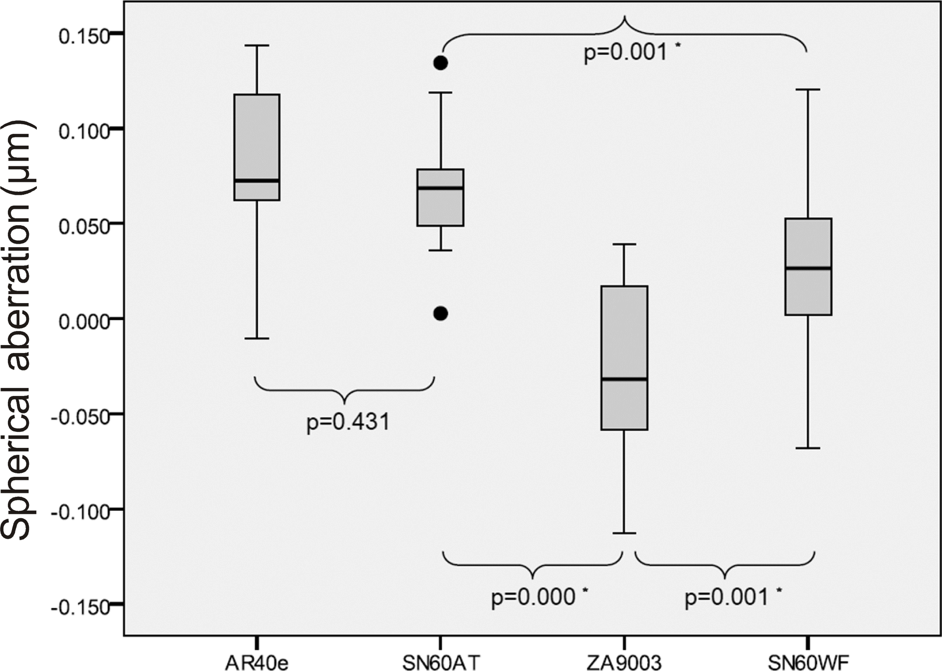 | Figure 3.Comparison of the postoperative ocular spherical aberrations for the spherical and aspheric IOL groups. Data are expressed as mean (lower and upper quartiles, box) ± standard deviation (bars), extraordinary value (round dot). * Mann-Whitney U test, statistically significant(p<0.05). |
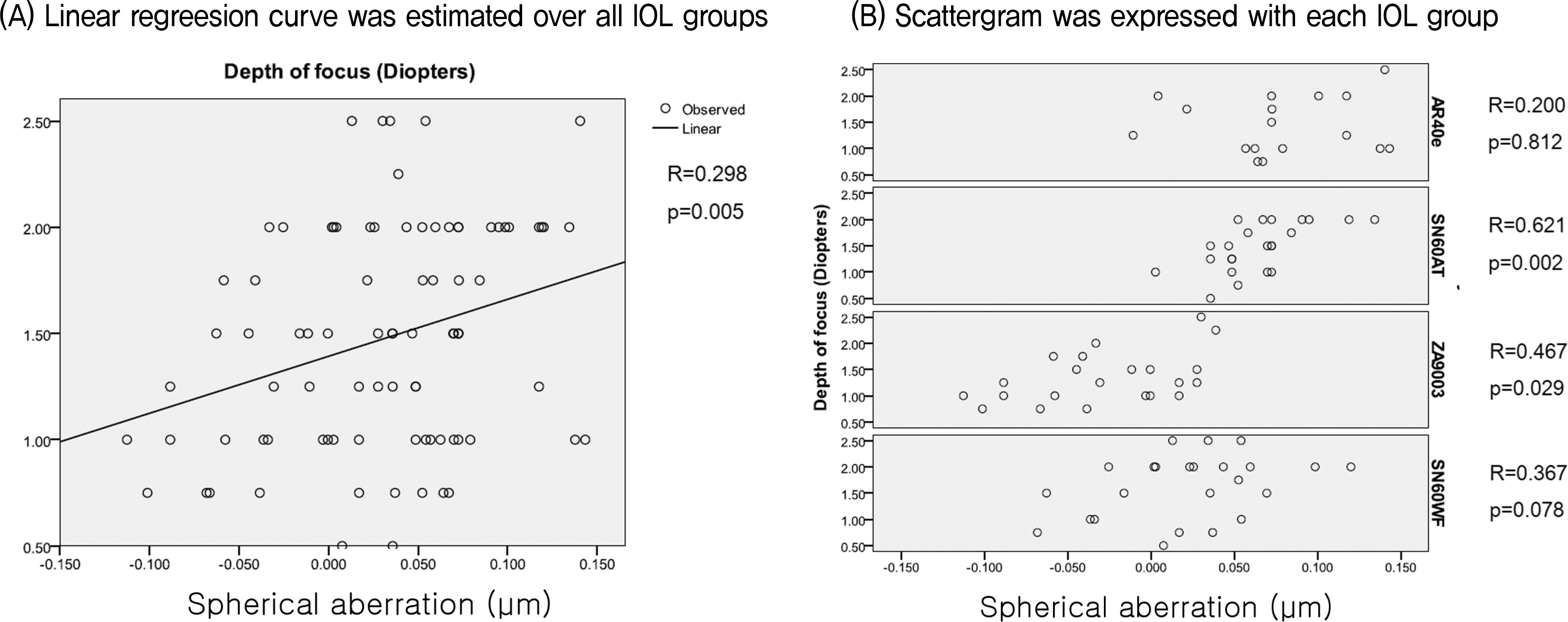 | Figure 4.Scattergrams of post-operative intraocular spherical aberrations and the depth of focus in the spherical and aspheric IOL groups. |
Table 1.
Optical characteristics of the spherical and aspheric IOLs used in the study
| Characteristic |
Spherical IOLs |
Aspheric IOLs |
||
|---|---|---|---|---|
| Sensar AR40e (AMO) | Acrysof SN60AT (Alcon) | Tecnis ZA9003 (AMO) | Acrysof IQ SN60WF (Alcon) | |
| Optic type | Monofocal | Monofocal | Monofocal | Monofocal |
| Lens | 3-piece | 1-piece | 3-piece | 1-piece |
| Optical material | Hydrophobic acrylic | Hydrophobic acrylic | Hydrophobic acrylic | Hydrophobic acrylic |
| Refractive index | 1.47 | 1.55 | 1.47 | 1.55 |
| Optic size (mm) | 6 | 6 | 6 | 6 |
| Overall length (mm) | 13 | 13 | 12 | 13 |
| Design | Spherical surfaces | Spherical surfaces | Prolate anterior surface | Prolate posterior surface |
| Haptic angulation (°) | 5 | 0 | 6 | 0 |
| Haptic material | PMMA* | Hydrophobic acrylic | Polyvinylidene fluoride | Hydrophobic acrylic |
Table 2.
Patient age, IOL power, postoperative BSCVA, and dark-adapted mesopic pupil diameter for the spherical and aspheric IOL groups
 XML Download