

 PDF
PDF ePub
ePub Citation
 Print
Print


Abstract
Purpose
To investigate and compare the clinical outcomes of wavefront-guided LASIK performed by 2 different laser platforms.
Methods
A retrospective analysis of consecutive cases of eyes that underwent wavefront-guided LASIK by using the VISX S4 CustomVue system and the Zyoptix Z100 system advanced personalized mode. All procedures were performed by one surgeon. Fifty-six eyes of 36 patients were included. Of the 56 eyes, 30 eyes underwent LASIK by using the VISX S4 CustomVue system (CustomVue group), and other 26 eyes underwent LASIK by using the Zyoptix Z100 system (APT group). Uncorrected visual acuity (UCVA), best corrected visual acuity (BCVA), manifest refraction, contrast sensitivity, total high order aberration (HOA), spherical aberration (SA) and Q-value were recorded preoperatively and at 1 week, 1 and 3 months postoperatively.
Go to : 
References
1. Pallikaris IG, Papatzanak ME, Stathi EZ. Laser in situ aberrations. Lasers Surg Med. 1990; 10:463–8.
2. Oliver KM, Hemenger RP, Corbett MC, et al. Corneal optical aberrations induced by photorefractive keratectomy. J Refract Surg. 1997; 13:246–54.

3. Applegate RA, Howland HC, Sharp RP, et al. Corneal aberrations and visual performance after radial keratotomy. J Refract Surg. 1998; 14:397–407.
4. Oshika T, Klyce SD, Applegate RA, et al. Comparison of corneal aberrations after photorefractive keratectomy and laser in situ keratomileusis. Am J Ophthalmol. 1999; 127:1–7.
5. Ginsburg AP, Evans DW, Sekuler R, et al. Contrast sensitivity predicts pilots' performance in aircraft simulation. Am J Optom Physiol Opt. 1982; 59:105–9.
6. Pesudovs K, Hazel CA, Doran RML, Elliot DB. The usefulness of Vistech and FACT contrast sensitivity charts for cataract and refractive surgery outcomes research. Br J Ophthalmol. 2004; 88:11–6.
7. Kim TI, Yang SJ, Tchah H. Bilateral comparison of wavefront-guided versus conventional laser in situ keratomileusis with Bausch and Lomb Zyoptix. J Refract Surg. 2004; 20:432–8.
8. Alió JL, Montés-Mico R. Wavefront-guided versus standard LASIK enhancement for residual refractive errors. Ophthalmology. 2006; 113:191–7.
9. Lee YH, Kim GH, Roh GH. Comparison of conventional versus wavefront-guided LASIK. J Korean Ophthalmol Soc. 2005; 46:2050–8.
10. Lee SM, Lee JH, Kim MK, et al. Comparison of changes in high-er-order aberrations between conventional and wavefront-guided LASEK. J Korean Ophthalmol Soc. 2007; 48:1028–35.
11. Seiler T, Mrochen M, Kaemmerer M. Operative correction of ocular aberrations to improve visual acuity. J Refract Surg. 2000; 16:S619–22.
12. Kim YT, Chung ES. Clinical result of wavefront-guided corneal ablation: LASIK vs. LASEK. J Korean Ophthalmol Soc. 2005; 46:1114–20.
13. Kim MJ, Kim HJ, Joo CK. Clinical outcome of wavefront guided LASIK using the Fourier algorithm: 6-month follow-up. J Korean Ophthalmol Soc. 2006; 47:806–11.
14. Lee SJ, Kim HJ, Joo CK. Change of high-order aberration after wavefront-guided LASIK and LASEK. J Korean Ophthalmol Soc. 2005; 46:1848–54.
15. Panagopoulou SI, Pallikaris IG. Wavefront customized ablations with the WASCA Asclepion workstation. J Refract Surg. 2001; 17:S608–12.
16. Park KS, Lee HK, Lim SM, et al. Comparison of patient satisfaction between conventional and customized LASIK. J Korean Ophthalmol Soc. 2006; 47:883–92.
17. Mrochen M, Kaemmerer M, seiler T. Wavefront-guided laser in situ keratomileusis: early results in three eyes. J Refract Surg. 2000; 16:116–21.
18. Cho SH, Joo CK, Kim MJ, et al. Clinical outcomes of wavefront-guided LASIK: 6-months follow-up. J Korean Ophthalmol Soc. 2005; 46:610–5.
19. Barreiro TP, Forseto Ados S, Pinto LF, et al. Wavefront-guided LASIK for low to moderate myopia: CustomCornea versus Zyoptix. Arq Bras Oftalmol. 2009; 72:519–25.
20. Awwad ST, Bowman RW, Cavanagh HD, et al. Wavefront-guided LASIK for myopia using the LADAR CustomCornea and the VISX Customvue. J Refract Surg. 2007; 23:26–38.
21. Kim KS, Song SW, Joo CK. One year clinical result of successful LASIK using VISX star. J Korean Ophthalmol Soc. 2000; 41:1139–45.
22. Calossi A. Corneal asphericity and spherical aberration. J Refract Surg. 2007; 23:505–14.
23. Park JH, Kang JH, Jin KH. Changes in the Cornea and Anterior Chamber After LASEK: Pentacam findings. J Korean Ophthalmol Soc. 2009; 50:510–7.
24. Maeda N. Wavefront technology in ophthalmology. Current Opinion in Ophthalmology. 2001; 12:294–9.
25. Mrochen M, Kaemmerer M, Seiler T. Clinical results of wavefront-guided laser in situ keratomileusis 3 months after surgery. J Cataract Refract Surg. 2001; 27:201–7.
26. Mutyala S, McDonald MB, Scheinblum KA, et al. Contrast sensitivity evaluation after laser in situ keratomileusis. Ophthalmology. 2000; 107:1864–7.
27. Pérez-Santonja JJ, Sakla HF, Alió JL, et al. Contrast sensitivity after laser in situ keratomileusis. J Cataract Refract Surg. 1998; 24:183–9.
Go to :
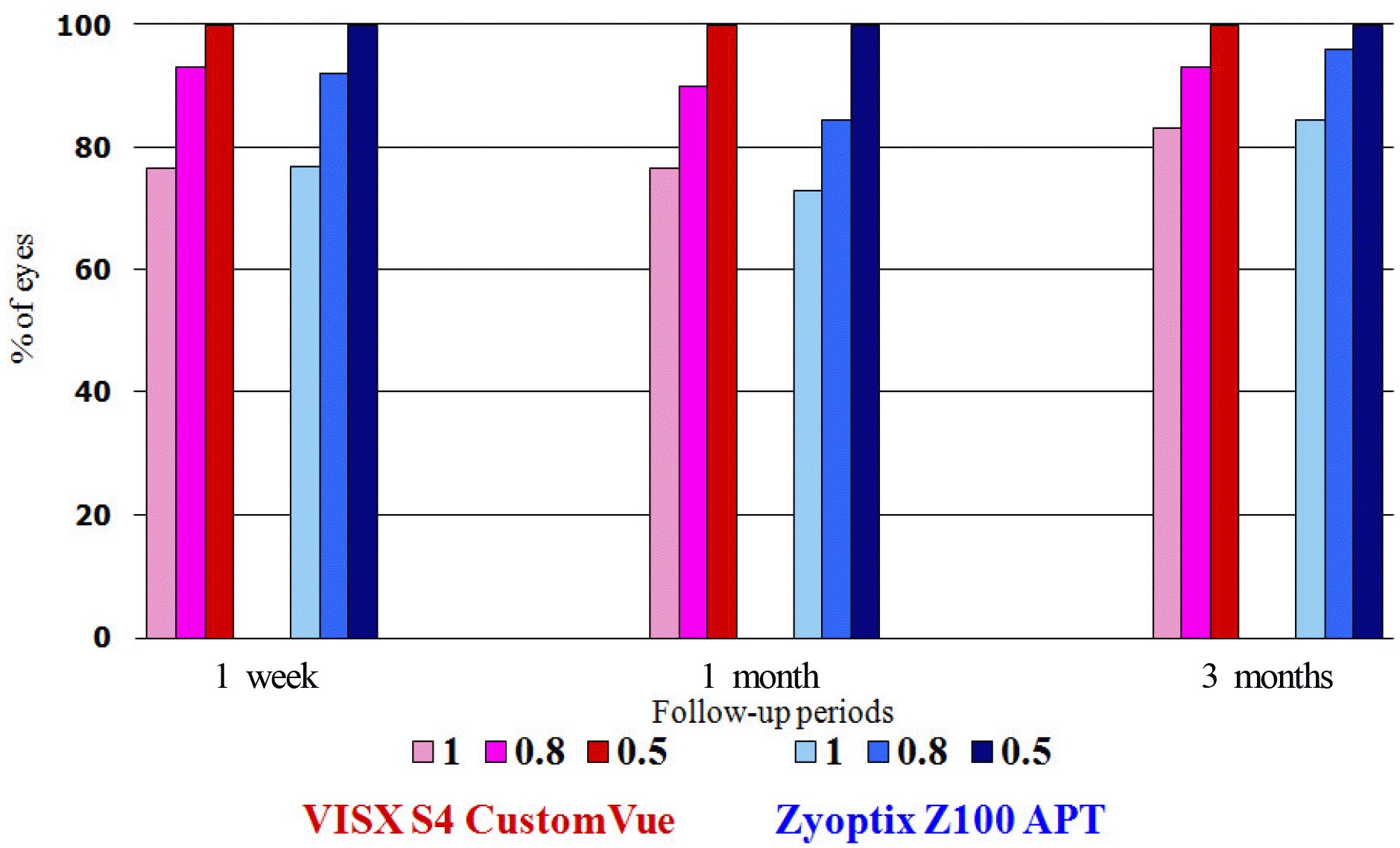 | Figure 1.Uncorrected visual acuity (UCVA) after LASIK. In VISX S4 CustomVue group, UCVA over 20/25 was presented in 93.3% at postoperative 1 week, 90% at postoperative 1 month and 93.3% at postoperative 3 months. In Zyoptix Z100 APT group, UCVA over 20/25 was presented in 92.3% at postoperative 1 week, 84.6% at postoperative 1 month and 96.2% at postoperative 3 months. |
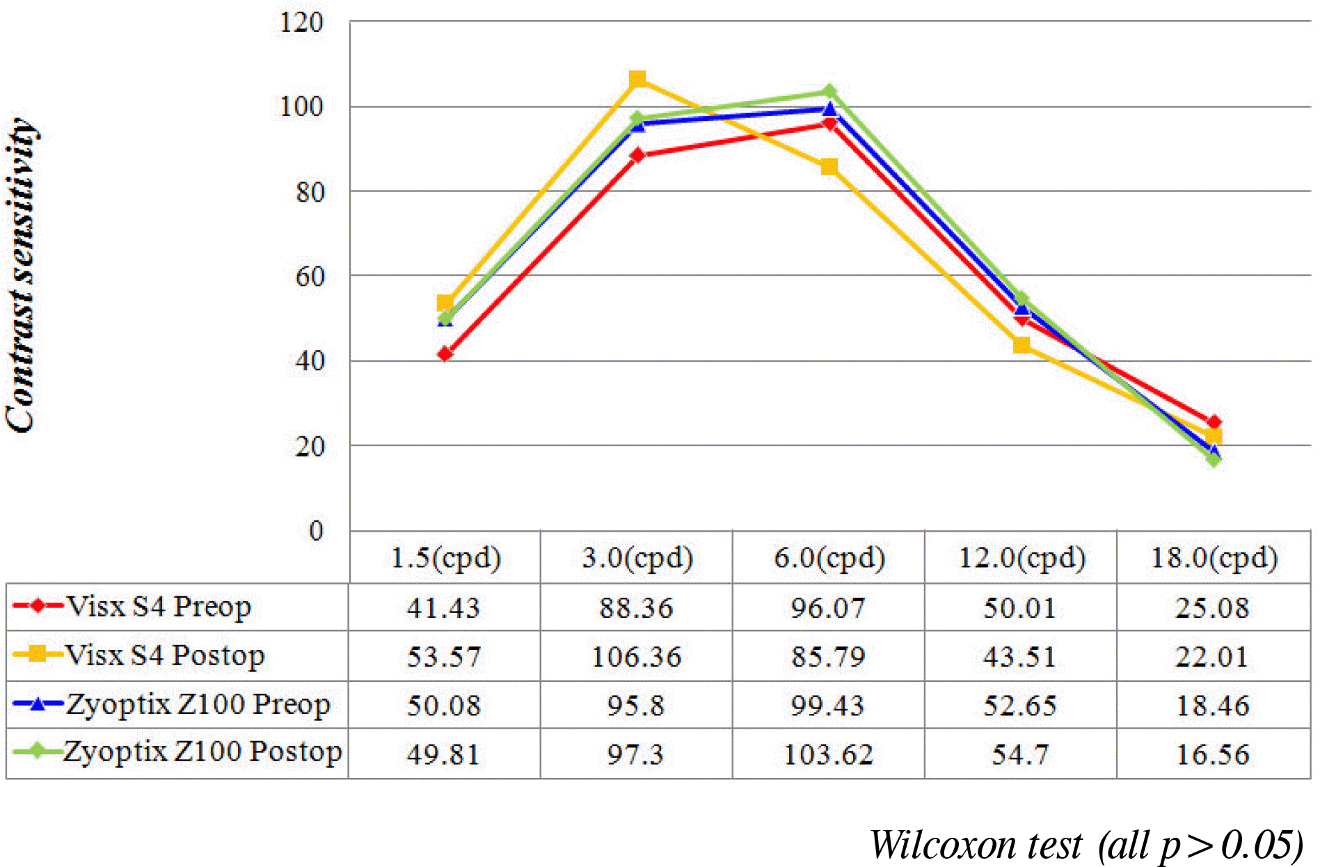 | Figure 3.Contrast sensitivity test result of CustomVue and APT group in photopic condition at preoperative and post-operative 3 month. In each group, there was no significant dif-fence in preoperative and postoperative contrast sensitivity values under photopic condition (Wilcoxon test, P>0.05). |
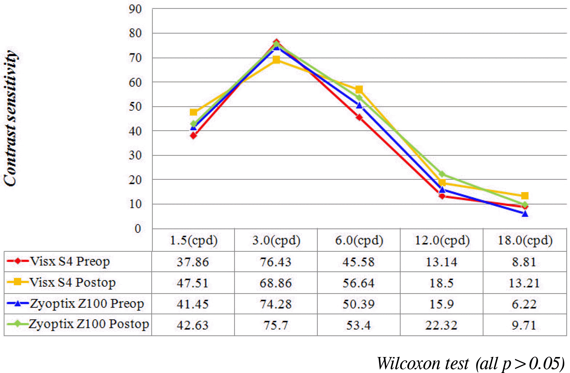 | Figure 4.Contrast sensitivity test result of CustomVue and APT group in mesopic condition at preoperative and post-operative 3 month. In each group, there was no significant dif-fence in preoperative and postoperative contrast sensitivity values under mesopic condition (Wilcoxon test, P>0.05). |
Table 1.
Patient demographics and characteristics
| |
Mean ± SD† |
P value* | |
|---|---|---|---|
| CustomVue | APT | ||
| Number of eyes | 30 | 26 | |
| Age (yrs) | 26.96 ± 5.37 | 26.80 ± 5.28 | 0.917 |
| M: F | 1:5 | 5:13 | |
| UCVA‡ | 0.11 ± 0.14 | 0.13 ± 0.12 | 0.351 |
| BCVA§ | 1.00 ± 0.00 | 1.00 ± 0.00 | |
Table 2.
Comparison of postoperative uncorrected visual acuity (UCVA), best corrected visual acuity (BCVA) between CustomVue group and APT group
| Results | Group |
Mean ± SD† |
|
|---|---|---|---|
| Post OP 1 mon | Post OP 3 mon | ||
| UCVA‡ (logMAR) | CustomVue | 0.05 ± 0.09* | 0.04 ± 0.06* |
| | APT | 0.04 ± 0.07* | 0.01 ± 0.04* |
| BCVA§ (logMAR) | CustomVue | 0.00 ± 0.00* | 0.00 ± 0.00* |
| | APT | 0.00 ± 0.00* | 0.00 ± 0.00* |
Table 3.
Changes in total high order aberration, spherical aberration, coma aberration in CustomVue group and APT group
| Results | Group |
Mean ± SD† |
||
|---|---|---|---|---|
| Pre OP | Post OP 1 mon | Post OP 3 mon | ||
| RMS HoA‡ | CustomVue | 0.49 ± 0.20 | 0.58 ± 0.39 | 0.59 ± 0.37 |
| | APT | 0.40 ± 0.13 | 0.55 ± 0.22 | 0.54 ± 0.19 |
| Spherical aberration | CustomVue | 0.17 ± 0.12 | 0.20 ± 0.14 | 0.19 ± 0.16 |
| | APT | 0.17 ± 0.19 | 0.35 ± 0.27* | 0.37 ± 0.23* |
| Vertical coma aberration | CustomVue | 0.19 ± 0.13 | 0.25 ± 0.33 | 0.27 ± 0.28 |
| | APT | 0.14 ± 0.11 | 0.17 ± 0.15 | 0.16 ± 0.19 |
| Horizontal coma aberration | CustomVue | 0.11 ± 0.11 | 0.18 ± 0.19 | 0.18 ± 0.21 |
| | APT | 0.12 ± 0.18 | 0.16 ± 0.16 | 0.15 ± 0.18 |
 XML Download