

 PDF
PDF ePub
ePub Citation
 Print
Print


Abstract
Purpose
To evaluate the long-term results of Descemet's stripping automated endothelial keratoplasty in Korea (DSAEK).
Methods
Seven patients with bullous keratopathy who underwent DSAEK and who were followed-up for more than 18 months were reviewed retrospectively. Best corrected visual acuity, refraction, corneal thickness, and endothelial cell count were examined before and after surgery.
Results
The mean follow-up period was 19.9 ± 2.9 months (18 to 24 months), and the mean age was 61.42 ± 10.13 years (46 to 76 years). Six patients (85.7%) showed successful results after surgery. Best corrected visual acuity (logMAR) was significantly improved from 1.62 (median) to 1.15 (median) (p = 0.027) at one month after surgery and was maintained until the final follow-up period. There were no statistical differences in spherical ametropia or astigmatism before or 18 months after the operation. Graft failure was observed in one case, in which penetrating keratoplasty was performed 12 months after DSAEK.
Go to : 
References
1. Melles GR, Eggink FA, Lander F, et al. A surgical technique for posterior lamellar keratoplasty. Cornea. 1998; 17:618–26.

2. Terry MA, Ousley PJ. Deep lamellar endothelial keratoplasty in the first United States patients: early clinical results. Cornea. 2001; 20:239–43.
3. Price FW Jr, Price MO. Descemet's stripping with endothelial keratoplasty in 50 eyes: a refractive neutral cornea transplant. J Refract Surg. 2005; 21:339–45.
4. Price FW Jr, Price MO. Descemet's stripping with endothelial keratoplasty in 200 eyes: early challenges and techniques to enhance donor adherence. J Cataract Refract Surg. 2006; 32:411–8.
5. Gorovoy MS. Descemet-stripping automated endothelial keratoplasty. Cornea. 2006; 25:886–9.
6. Bahar I, Kaiserman I, McAllum P, et al. Comparison of posterior lamellar keratoplasty techniques to penetrating keratoplasty. Ophthalmology. 2008; 115:1525–33.
7. Hyams M, Segev F, Yepes N, et al. Early postoperative complications of deep lamellar endothelial keratoplasty. Cornea. 2007; 26:650–3.
8. Yepes N, Segev F, Hyams M, et al. Five-millimeter-incision deep lamellar endothelial keratoplasty: one-year results. Cornea. 2007; 26:530–3.
9. Melles GR, Lander F, Beekhuis WH, et al. Posterior lamellarkeratoplasty for a case of pseudophakic bullous keratopathy. Am J Ophthalmol. 1999; 127:340–1.
10. Bahar I, Kaiserman I, Srinivasan S, et al. Posterior lamellar keratoplastyaccommodative–comparison of deep lamellar endothelial keratoplasty and Descemet stripping automated endothelial keratoplasty in the same patients: a patient's perspective. Br J Ophthalmol. 2009; 93:186–90.
11. Covert DJ, Koenig SB. New triple procedure: Descemet's stripping and automated endothelial keratoplasty combined with phacoemulsification and intraocular lens implantation. Ophthalmology. 2007; 114:1272–7.
12. Covert DJ, Koenig SB. Descemet stripping and automated endothelial keratoplasty (DSAEK) in eyes with failed penetrating keratoplasty. Cornea. 2007; 26:692–6.
13. Price PO, Price FW Jr. Descemet's stripping with endothelial keratoplasty: comparative outcomes with microkeratome-dissected and manually dissected donor tissue. Ophthalmology. 2006; 113:1936–42.
14. Hjortdal J, Ehlers N. Descemet's stripping automated endothelial keratoplasty and penetrating keratoplasty for Fuchs' endothelial dystrophy. Acta Ophthalmol. 2009; 87:310–4.
15. Koenig SB, Covert DJ, Dupps WJ Jr, Meisler DM. Visual acuity, refractive error, and endothelial cell density six months after Descemet stripping and automated endothelial keratoplasty (DSAEK). Cornea. 2007; 26:670–4.
16. Jeong HO, Lee HS, Na KS, Joo CK. Eight case of Descemet's stripping automated endothelial keratoplasty in eyes with bullous keratopathy. J Korean Ophthalmol Soc. 2009; 50:1115–9.
17. Fogla R, Padmanabhan P. Initial results of small incision deep lamellar endothelial keratoplasty (DLEK). Am J Ophthalmol. 2006; 141:346–51.
18. Koenig SB, Covert DJ, Dupps WJ Jr, et al. Visual acuity, refractive error, and endothelial cell density six months after Descemet stripping and automated endothelial keratoplasty (DSAEK). Cornea. 2007; 26:670–4.
19. Yoo SH, Kymionis GD, Deobhakta AA, et al. One-year results and anterior segment optical coherence tomography findings of descemet stripping automated endothelial keratoplasty combined with Phacoemulsification. Arch Ophthalmol. 2008; 126:1052–5.
20. Rose L, Briceno CA, Stark WJ, et al. Assessment of eye bank-prepared posterior lamellar corneal tissue for endothelial keratoplasty. Ophthalmology. 2008; 115:279–86.
21. Terry MA, Chen ES, Shamie N, et al. Endothelial cell loss after Descemet's stripping endothelial keratoplasty in a large prospective series. Ophthalmology. 2008; 115:488–96.
22. Terry MA, Shamie N, Chen ES, et al. Endothelial keratoplasty for Fuchs' dystrophy with cataract: complications and clinical results with the new triple procedure. Ophthalmology. 2009; 116:631–9.
23. Jun B, Kuo AN, Afshari NA, et al. Refractive change after descemet stripping automated endothelial keratoplasty surgery and its correlation with graft thickness and diameter. Cornea. 2009; 28:19–23.
24. Koenig SB, Covert DJ. Early results of small-incision Descemet's stripping and automated endothelial keratoplasty. Ophthalmology. 2007; 114:221–6.
25. Shih CY, Ritterband DC, Palmiero PM, et al. The use of postoperative slit-lamp optical coherence tomography to predict primary failure in descemet stripping automated endothelial keratoplasty. Am J Ophthalmol. 2009; 147:796–800.
Go to :
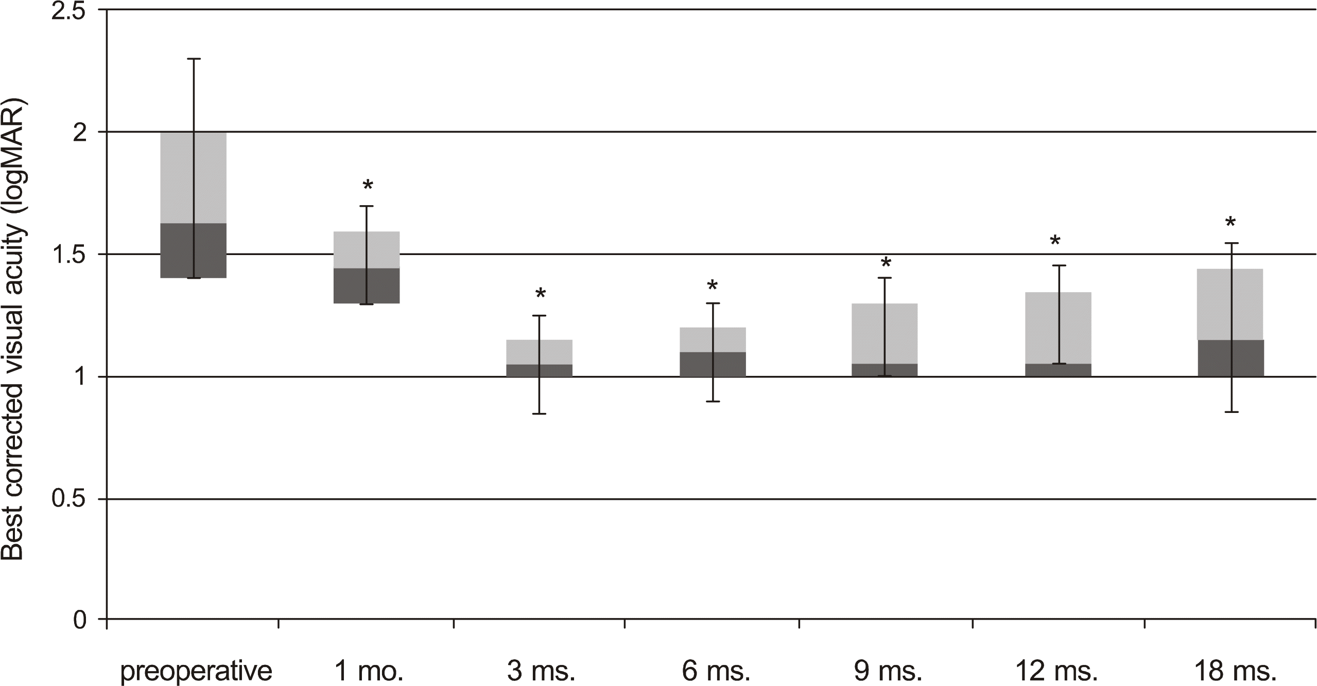 | Figure 1.Boxplot showing changes of best corrected visual acuity in the patients who underwent Descemet's stripping automated endothelial keratoplasty (* Wilcoxon signed rank test, p<0.05 between preoperative and postoperative results). |
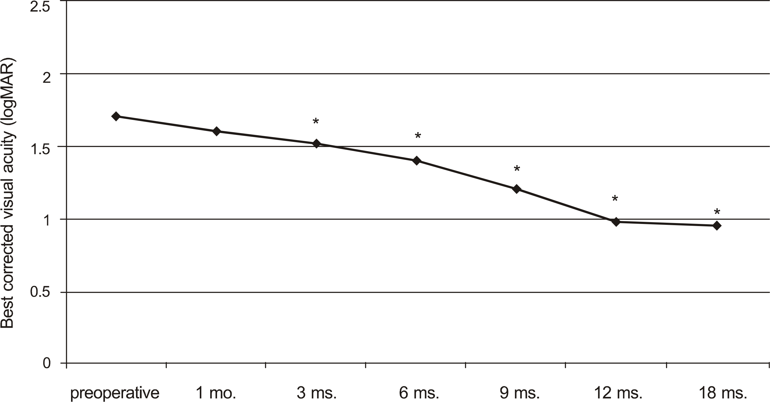 | Figure 2.Graphs showing changes in mean best corrected visual acuity over time in the patients who underwent penetrating keratoplasty (* Paired t-test, p<0.05 between pre-operative and postoperative results). |
 | Figure 3.(A) Preoperative slit lamp photograph showing edema and diffuse haziness in the cornea. (B) At 18 months after Descemet's stripping automated endothelial keratoplasty, the corneal graft remains clear. (C) Pentacam demonstrates interface smoothness and regular thickness of the cornea. (D) Specular microscope reveals viable endothelial cells. |
Table 1.
Demographics of patients who underwent Descemet's stripping automated endothelial keratoplasty
| Case | Age (yrs) | Sex | Preoperative diagnosis | Preoperative CCT* (μ m) | Preoperative BCVA† (logMAR) | Endothelial cell count (cells/mm2) | Combined procedure |
|---|---|---|---|---|---|---|---|
| 1 | 46 | M‡ | Bullous keratopathy | 924 | 1.4 | Error | |
| 2 | 55 | M | Bullous keratopathy | 1,014 | 1.4 | Error | |
| 3 | 56 | M | Bullous keratopathy, aphakia | 941 | 1.4 | 371 | Scleral fixation of IOLΠ |
| 4 | 61 | M | Bullous keratopathy, aphakia | 749 | 2.3 | Error | Scleral fixation of IOL |
| 5 | 70 | F§ | Bullous keratopathy, cataract | 723 | 1.7 | Error | Phaco# with IOL implantation |
| 6 | 76 | M | Bullous keratopathy, pseudophakia | 1,006 | 1.7 | 281 | |
| 7 | 66 | M | Bullous keratopathy, pseudophakia | 864 | 1.7 | 498 | |
Table 2.
Refractive ametropia, astigmatism, central corneal thickness, and endothelial cell density before and after Descemet's stripping automated endothelial keratoplasty
| | Before operation median (range) | Postoperative 12 months median (range) | Postoperative 18 months median (range) |
|---|---|---|---|
| Sphere (D) | −0.13 (1.5∼2.75) | +0.5 (0.25∼0.75) | +0.75 (0.25∼1.25) |
| Cylinder (D) | 0.0 (2.5∼1.5) | +1.25 (−1.25∼2.0) | +0.75 (1.25∼2.0) |
| Spherical equivalent (D) | −0.5 (1.5∼3.5) | +1.0 (0.0∼1.5) | +1.0 (0.25∼2.25) |
| Central corneal thickness (μ m) | 932.5 (723∼1014) | 601* (600∼716) | 651* (611∼701) |
| Endothelial cell density (cells/mm2) | Not applicable | 1359 (1258∼1587) | 1125 (998∼1262) |
Table 3.
Overall visual and surgical results at 18 months and at last follow-up after Descemet's stripping automated endothelial keratoplasty
| Case | Age (yrs) | Sex | Follow-up duration (mon) | Postoperative BCVA* (logMAR) at 18 mon after DSAEK# | Postoperative BCVA* (logMAR) at last follow-up | Postoperative CCT† (μ m) at 18 mon after DSAEK | Endothelial cell count (cells/mm2) at 18 mon after DSAEK | Complication and management |
|---|---|---|---|---|---|---|---|---|
| 1 | 46 | M‡ | 18 | 0.7 | 0.7 | 611 | 1,154 | |
| 2 | 55 | M | 18 | 0.7 | 0.7 | 646 | 1,262 | |
| 3 | 56 | M | 24 | 0.9 | 0.8 | 628 | 998 | Graft detachment → Air injection |
| 4 | 61 | M | 24 | 1.1 | 0.9 | 700 | 1,256 | |
| 5 | 70 | F§ | 18 | | | | | Graft failure → PKPΠ |
| 6 | 76 | M | 18 | 0.4 | 0.4 | 698 | 1,096 | |
| 7 | 66 | M | 20 | 0.7 | 0.7 | 656 | 1,011 | |
Table 4.
Refractive ametropia, astigmatism, central corneal thickness, and endothelial cell density after Descemet's stripping automated endothelial keratoplasty and penetrating keratoplasty
| |
Postoperative 12 mon |
Postoperative 18 mon |
||
|---|---|---|---|---|
| DSAEK* | PKP† | DSAEK | PKP | |
| | median (range) | mean ± SD | median (range) | mean ± SD‡ |
| Sphere (D) | +0.5 (0.25∼0.75) | −3.78 ± 1.64 | +0.75 (0.25∼1.25) | −3.98 ± 1.86 |
| Cylinder (D) | +1.25 (−1.25∼2.0) | −4.26 ± 2.33 | +0.75 (−1.25∼2.0) | −4.52 ± 1.82 |
| Central corneal thickness (μ m) | 601 (600∼716) | 598.33 ± 46.81 | 651 (611∼701) | 609.18 ± 27.86 |
| Endothelial cell density (cells/mm2) | 1,359 (1258∼1587) | 1,386.28 ± 192.92 | 1,125 (998∼1262) | 1,298.00 ± 251.76 |
 XML Download