

 PDF
PDF ePub
ePub Citation
 Print
Print


Abstract
Purpose
To evaluate the pattern VEP in adult amblyopic patients seen in consultation for ophthalmic evaluation as a past of physical examinations for conscription.
Methods
We retrospectively analyzed, 67 men, 20-year-old or older, who had pattern VEP done for the diagnosis of amblyopia from January 2004 to May 2009. P100 latency and P100 amplitude were analyzed.
Results
Thirteen patients were non-amblyopic, and 54 patients had amblyopia. Binocular amblyopia and monocular amblyopia were found in 23 and 31 patients, respectively. In the binocular amblyopic patients, four patients were hyperopic, seven patients were myopic, and 12 patients were astigmatic amblyopia. In the monocular amblyopic patients, 15 patients were anisometropic, 12 patients were strabismic, and four patients had organic amblyopia. The value of P100 latency and P100 amplitude were statistically significantly different between non-amblyopic and amblyopic eyes, with check size of 32×32. However, the types of amblyopia among the patients were not different at a statistically significant level. Visual acuity and P100 amplitude were significantly positively correlated.
References
1. von Noorden GK, Campos EC. Binocular vision and ocular motility. 6th ed.St. Louis: Mosby;2002. p. 246.
2. Wanger P, Nilson BY. Visual evoked response to pattern reversal stimulation in patients with amblyopia and/or defective binocular functions. Acta Ophthalmol. 1978; 56:617–27.
3. Chung W, Hong S, Lee JB, Han SH. Pattern visual evoked potential as a predictor of occlusion therapy for amblyopia. Korean J Ophthalmol. 2008; 22:251–4.

4. Lee SJ, Park S, Shin H. Pattern-VEP in child amblyopia. J Korean Ophthalmol Soc. 1995; 36:924–9.
5. Kriss A, Thompson D, Lloyd I, et al. Pattern VEP findings in young children treated for unilateral congenital cataract. Cottlier E, editor. Congenital cataract. Austin: Laudes;1994. p. 79–88.
6. Sokol S. Abnormal evoked potential latencies in amblyopia. Br J Ophthalmol. 1983; 67:310–4.
7. Hoe JW, Kim CW, Kim SM. VEP and pattern ERG in adult amblyopes. J Korean Ophthalmol Soc. 1990; 31:1481–8.
8. Wanger P, Persson HE. Visual evoked response to pattern-reversal stimulation in childhood amblyopia. Acta Ophthalmol. 1980; 58:697–706.
9. Sokol S, Fishman GA. Electrophysiologic testing in disorders of the retina, optic nerve, and visual pathway. In electrophysiologic testing. San Francisco: American academy of ophthalmology;1990. p. 123–5.
10. Park HK, Kim MM, Hahn DK. Evaluation of VEP in optic nerve diseases and amblyopia. J Korean Ophthalmol Soc. 1995; 36:1568–73.
11. Feinsod M, Hoyt WF, Wilson WB, Spire JP. Visually evoked response: use in neurologic evaluation of posttraumatic subjective visual complaint. Arch Ophthalmol. 1976; 94:237–40.
12. Kramer KK, La Piana FG, Appleton B. Ocular malingering and hysteria: diagnosis and management. Surv Ophthalmol. 1979; 24:89–96.
13. Saitoh E, Usami EA, Mizota A, Fujimoto N. Comparison of visual evoked potentials in patients with psychogenic visual disturbance and malingering. J Pediatr Ophthalmol Strabismus. 2001; 38:21–6.
Figure 1.
Correlation between visual acuity, and P100 amplitude and P100 latency by different check size. Visual acuity and P100 amplitude by check size 16×16 (A) and 32×32 (C) have positive correlation which is statistically significant (* p<0.05). Visual acuity and P100 latency by check size 16×16 (B) and 32×32 (D) have negative correlation which is statistically non-significant (p>0.05).
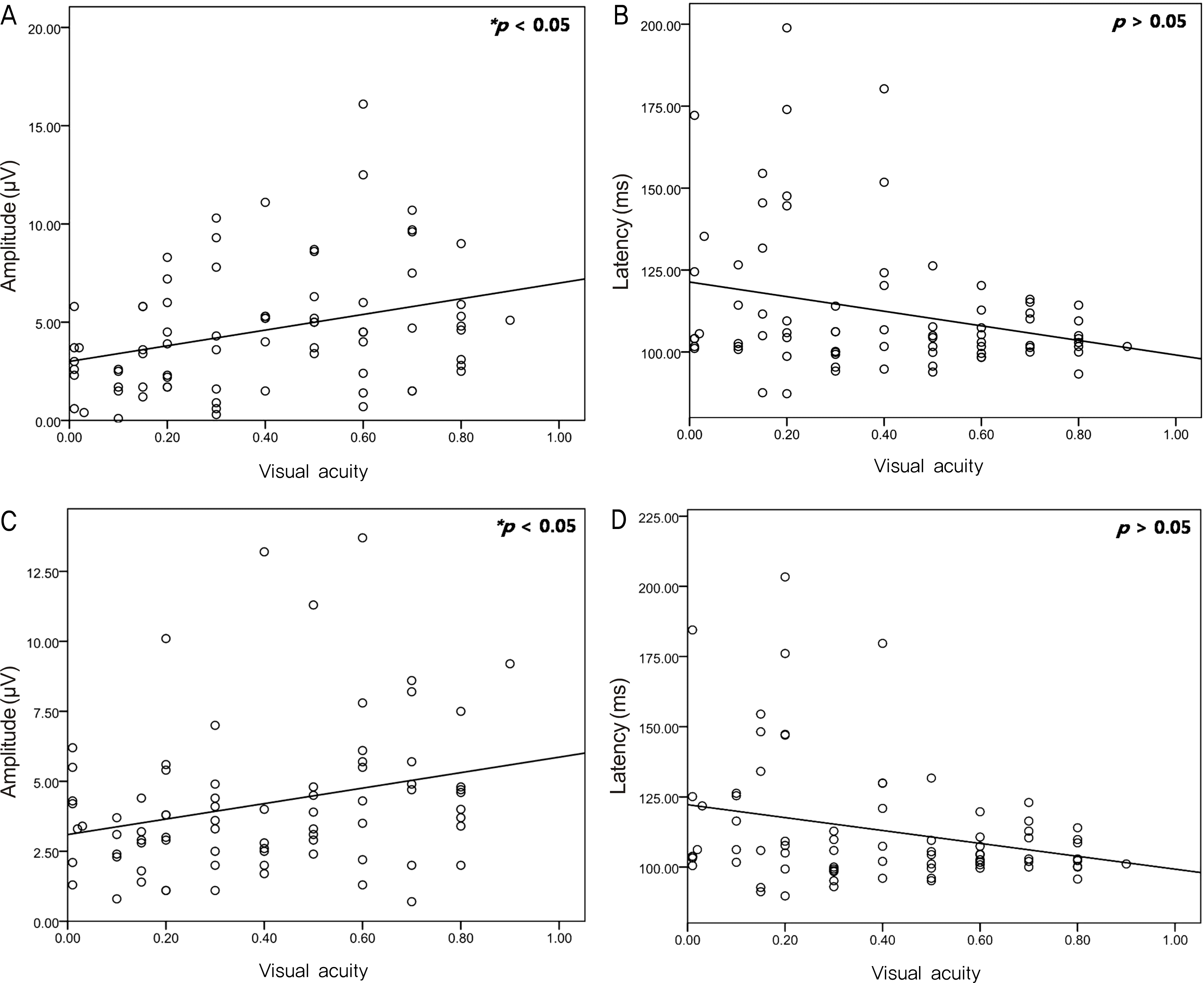
Table 1.
Causes of amblyopia and number of patients by diagnosis
| Unilateral | No. of pts. | Bilateral | No. of pts. |
|---|---|---|---|
| Anisometropic | 15 | Visual deprived | |
| Strabismic Visual deprived | 12 0 | Hypertropic Myopic | 4 7 |
| Organic Total | 4 31 | Astigmatic Total | 12 23 |
Table 2.
P100 amplitude and latency of pVEP in 26 normal control eyes
| Check size | Amplitude (μ V) | Latency (ms) |
|---|---|---|
| 16×16 | 5.29 ± 2.045 | 104.09 ± 5.586 |
| 32×32 | 5.38 ± 2.381 | 104.16 ± 5.234 |
Table 3.
P100 amplitude and latency between right and left eye in control group
Table 4.
P100 amplitude and latency of pVEP in 77 amblyopic eyes
| Check size | Amplitude (μ V) | Latency (ms) |
|---|---|---|
| 16×16 | 4.57 ± 3.166 | 112.56 ± 21.544 |
| 32×32 | 4.19 ± 2.628 | 113.14 ± 22.008 |
Table 5.
Interocular difference of P100 amplitude and latency between both eyes in normal control and monocular amblyopic group
| Check size | | Group | Difference of ampllitude (μ V) | Difference of Latency (ms) |
|---|---|---|---|---|
| 16×16 | Control | 1.42 ± 1.204 | 1.59 ± 1.145 | |
| Amblyopia | 2.68 ± 1.891 | 10.92 ± 21.586 | ||
| | P value | | 0.012* | 0.023* |
| 32×32 | Control | 1.72 ± 1.591 | 2.05 ± 2.486 | |
| Amblyopia | 2.97 ± 2.302 | 11.46 ± 23.008 | ||
| | P value | | 0.047* | 0.032* |
Table 6.
Inte rocular ampl litude difference ratio of control and monoc ular amblyopic group
| Check size | | Group | IADR |
|---|---|---|---|
| 16×16 | Control | 0.24 ± 0.227 | |
| Amblyopia | 0.42 ± 0.246 | ||
| | P value | | 0.034* |
| 32×32 | Control | 0.28 ± 0.219 | |
| Amblyopia | 0.43 ± 0.215 | ||
| | P value | | 0.047* |
Table 7.
P100 amplitude and P100 latency between types of amblyopia
Table 8.
The comparison of visual acuity with P100 amplitude and P100 latency
 XML Download