

 PDF
PDF ePub
ePub Citation
 Print
Print


Abstract
Purpose
To report a case of Candida parapsilosis keratitis with atypical presentation demonstrated by subepithelial white dot deposits without peripheral inflammatory reaction.
Case summary:
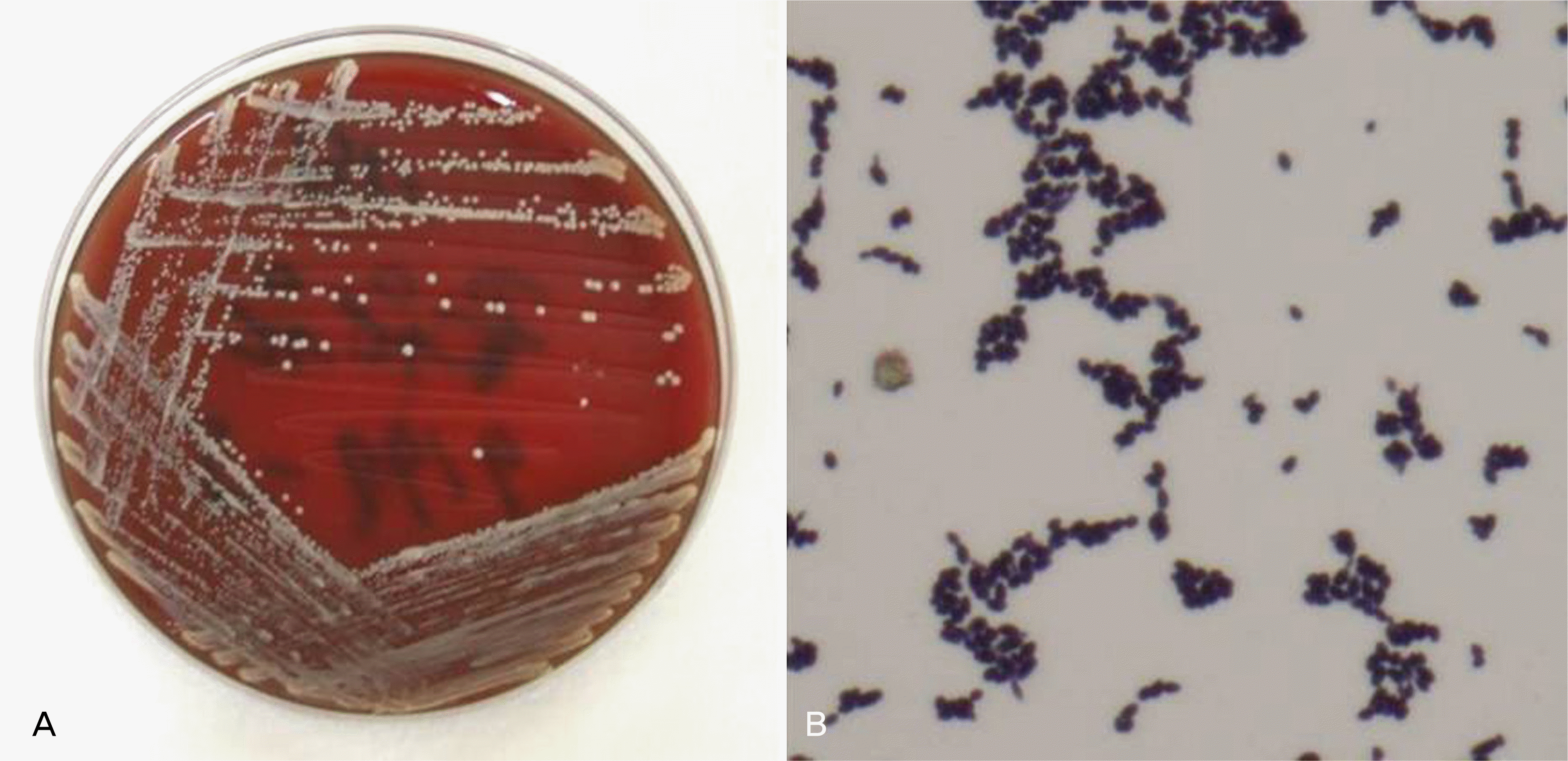
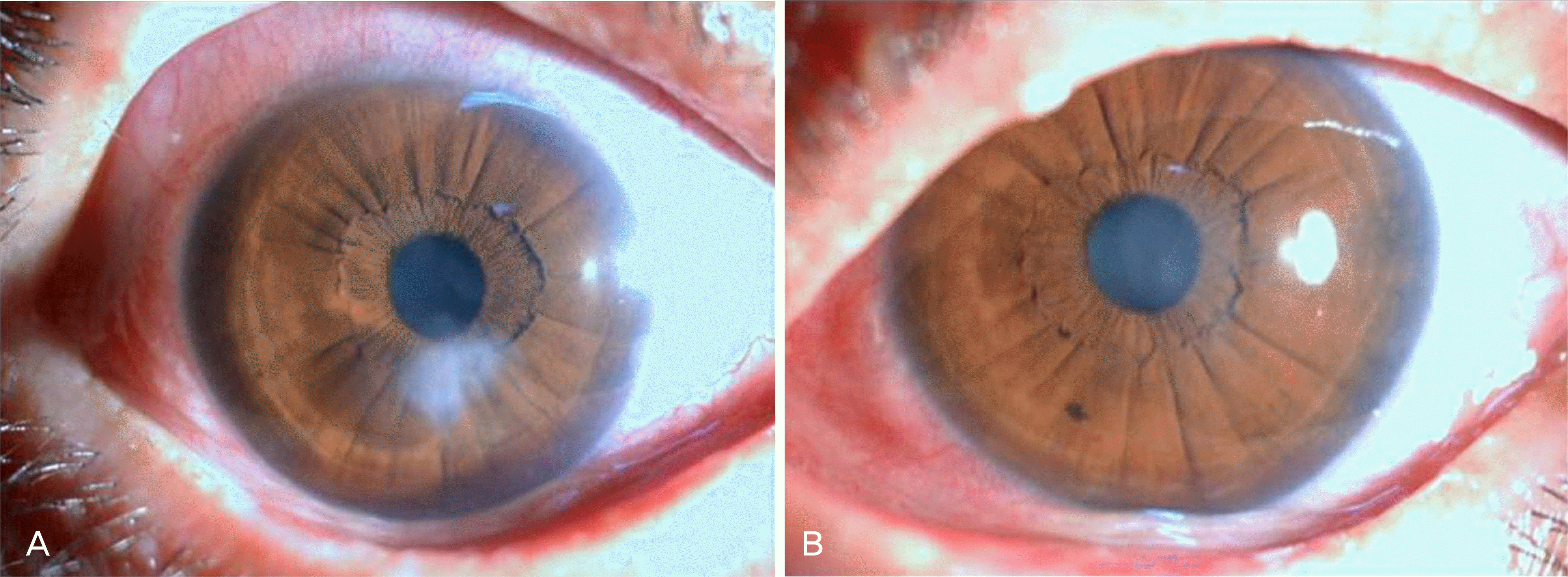
A 45-year-old woman with Stevens-Johnson syndrome had used topical corticosteroid and bandage contact lens due to recurrent epithelial defect and keratitis. Multiple subepithelial white dot deposits were revealed on the central corneal area without surrounding inflammation. The corneal lesion was improved after epithelial debridement with topical antibiotics and steroid eyedrops. A few months later, however, the corneal lesion recurred. Smear cytology was performed, and yeast-formed fungi and pseudohyphae were found. C. parapsilosis was identified in the culture study. Therefore, the topical steroid was withdrawn and 0.15% topical Amphotericin was applied. The corneal lesion improved and corneal opacity did not progress.
Conclusions
The case reported in this study is C. parapsilosis keratitis with multiple subepithelial white dot deposits without typical presentations of fungal kertitis. Although no typical infectious indication was evident, infection should be suspected in patients who show abnormal corneal lesion under immunosuppressive treatment.
References
1. Hahn YH, Hahn TW, Tchah HW, et al. Epidemiology of Infectious Keratitis (II): A Multicenter Study. J Korean Ophthalmol Soc. 2001; 42:247–65.
2. Sun RL, Jones DB, Wilhelmus KR. Clinical Characteristics and Outcome of Candida Keratitis. Am J Ophthalmol. 2007; 143:1043–5.

3. Bourcier T, Touzeau O, Thomas F, et al. Candida Parapsilosis aberrations. Cornea. 2003; 22:51–5.
4. Muallem MS, Alfonso EC, Romano AC, et al. Bilateral Candida Parapsilosis Interface Keratitis after In Situ Keratomileusis. J Cataract Refract Surg. 2003; 29:2002–5.
5. Weems JJ. Candida Parapsilosis: Epidemiology, Pathogenicity, Clinical Manifestations, and Antimicrobial Susceptibility. Clin Infect Dis. 1992; 14:756–66.
6. Solomon R, Biser SA, Donnenfeld ED, et al. Candida Parapsilosis Keratitis Following Treatment of Epithelial Ingrowth After Laser In Situ Keratomileusis. Eye Contact Lens. 2004; 30:85–6.
7. Fekrat S, Haller JA, Green WR, et al. Pseudophakic Candida aberrations Endophthalmitis with Consecutive Keratitis. Cornea. 1995; 14:212–6.
Figure 1.
Anterior segment photograph of both eyes at initial presentation. Multiple white dot deposits on left corneal surface without corneal edema and stromal infiltration (A). Multiple corneal epithelial defects on the right corneal surface (B).
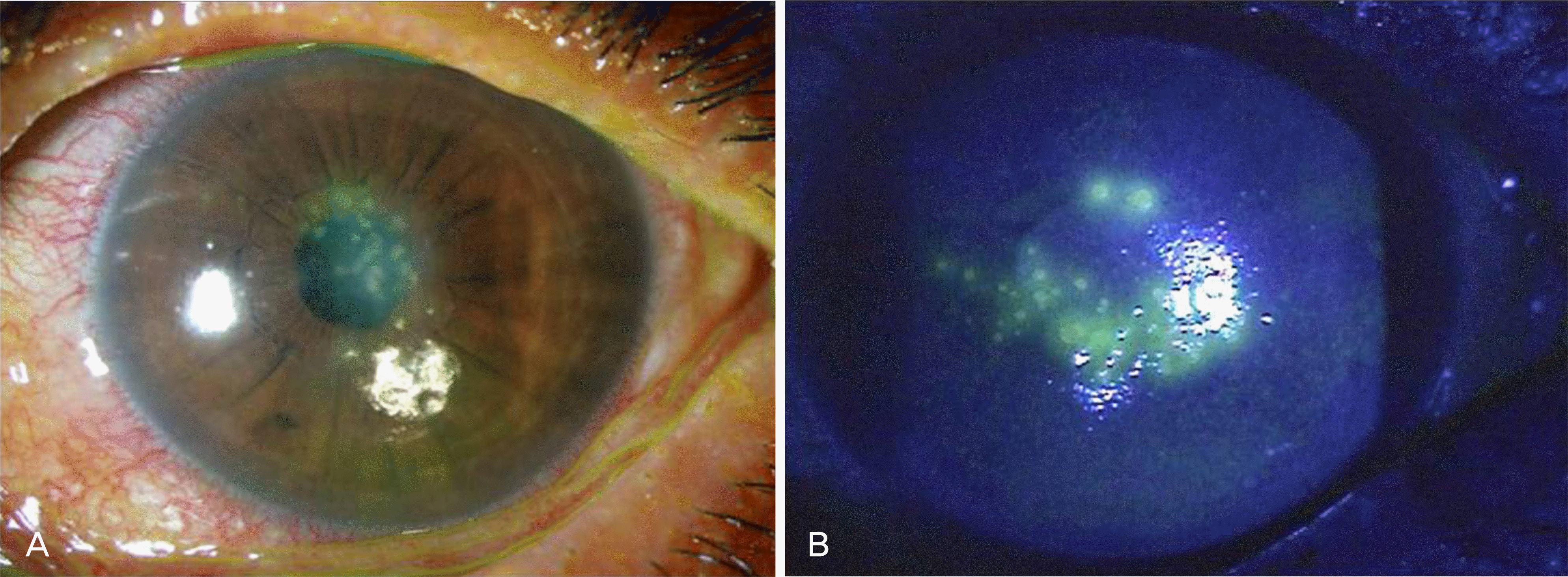
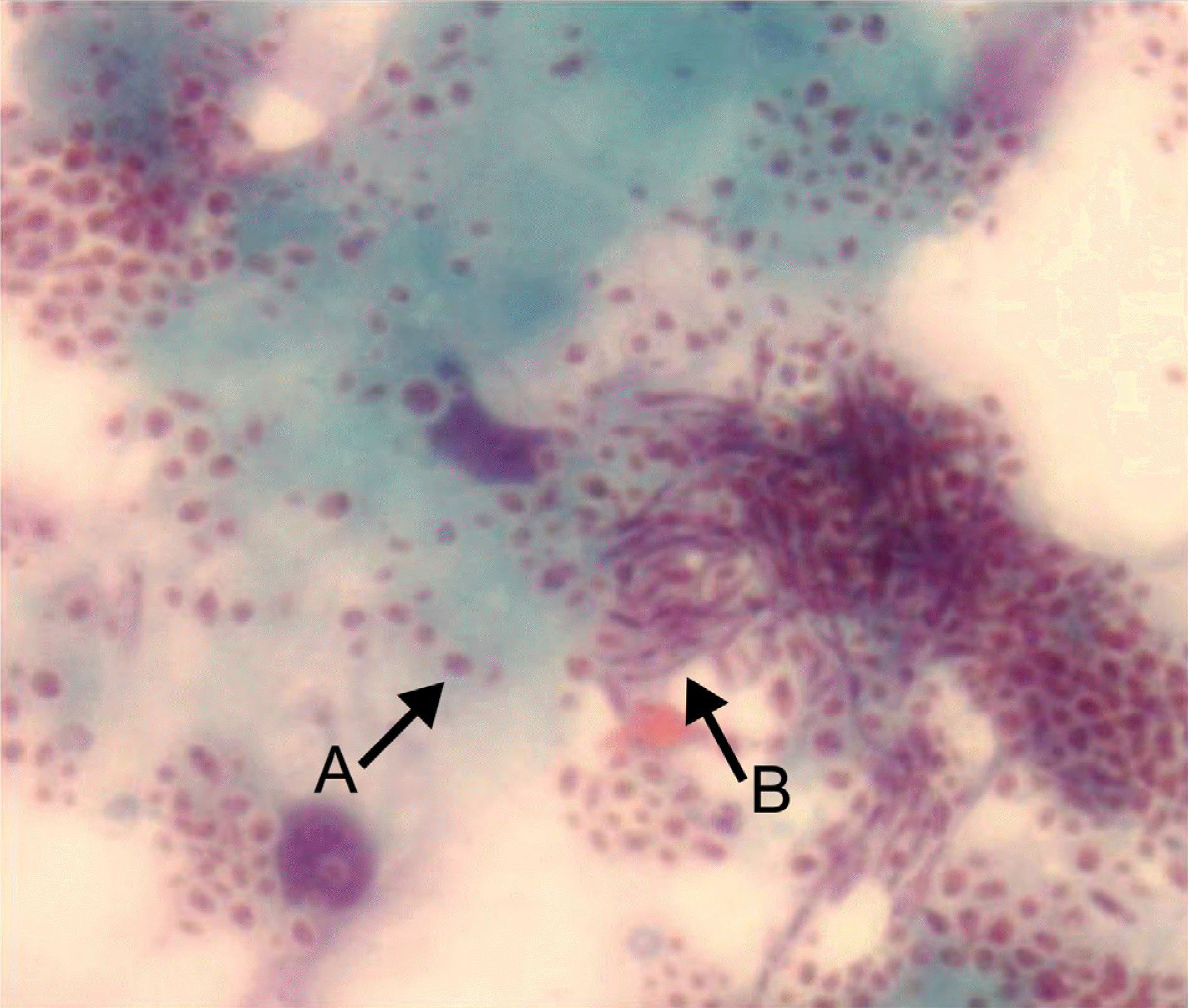
Figure 2.
Smear cytology with Papanicolaou stain (×800). Yeast-formed organisms (A) and pseudohyphae-like structures (B) are seen.
 XML Download