

 PDF
PDF ePub
ePub Citation
 Print
Print


Abstract
Purpose
To present a case of corneal endothelial damage caused by inflow of 5-FU after subconjunctival 5-FU injection in a patient undergoing trabeculectomy.
Case Summary
A 65-year-old female patient diagnosed with chronic angle-closure glaucoma underwent trabeculectomy using mitomycin C. Three weeks after the surgery, her intraocular pressure (IOP) elevated, and the follicles had disappeared by trabeculectomy. Subsequently, subconjunctival 5-FU injection was performed. Following the fourth injection, visual acuity abruptly decreased, and corneal edema was observed. Upon presumption of inflow of 5-FU into the anterior chamber, anterior chamber irrigation was performed within 40 minutes. On postoperative day 1, visual acuity decreased from 0.8 to counting fingers, and diffuse corneal edema and anterior capsular opacity of the lens were noted. Three months after the irrigation, visual acuity improved to 0.8. Corneal edema and capsular opacity also improved, however the density of the corneal endothelial count decreased.
Figures and Tables
Figure 1
Anterior segment findings of the patient, 2 months after inadvertent 5-FU inflow. (A) The upper part of the cornea is clear and the lens shows negligible anterior capsular opacities. (B) The lower segment of the cornea shows Descemet's folds.
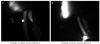
Figure 2
Specular microscopic findings according to timeline. (A) Preoperative specular microscopic findings. (B) Specular microscopic findings 2 months after inadvertent 5-FU inflow. Decreased cell density and obscured margins between cells were noted. (C) Specular microscopic findings 3 months after 5-FU inflow. Compared with preoperative findings, cell density was decreased, but margins between cells became clearer.
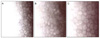
Table 1
Numeric data from specular microscopy according to timeline
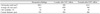
Note that corneal endothelial findings became worse 2 months after inadvertent 5-FU inflow compared with preoperative findings. Afterwards, corneal endothelial findings got improved at 3 months after inadvertent 5-FU inflow, but compared to the preoperative status, the corneal endothelial density was not restored.
References
1. Allingham RR, Damji KF, Freedman S, et al. Shields' Textbook of Glaucoma. 2005. 5th ed. Philadelphia: Lippincott Williams & Wilkins;577–579.
2. Lattanzio FA Jr, Sheppard JD Jr, Allen RC, et al. Do injections of 5-fluorouracil after trabeculectomy have toxic effects on the anterior segment? J Ocul Pharmacol Ther. 2005. 21:223–235.
3. Bhermi GS, Holak S, Murdoch IE. Inadvertent exposure of corneal endothelium to 5-fluorouracil. Br J Ophthalmol. 1999. 83:376–377.
4. Mazey BJ, Siegel MJ, Siegel LI, Dunn SP. Corneal endothelial toxic effect secondary to fluorouracil needle bleb revision. Arch Ophthalmol. 1994. 112:1411.
5. Lama PJ, Fechtner RD. Antifibrotics and wound healing in glaucoma surgery. Surv Ophthalmol. 2003. 48:314–346.
6. Nuyts RM, Pels E, Greve EL. The effects of 5-fluorouracil and mitomycin C on the corneal endothelium. Curr Eye Res. 1992. 11:565–570.
7. Mannis MJ, Sweet EH, Lewis RA, et al. The effect of fluorouracil on the corneal endothelium. Arch Ophthalmol. 1988. 106:816–817.
8. Krachmer JH, Mannis MJ, Holland EJ. Cornea. 2005. Vol. 1:2nd ed. Elsevier;1285–1294.
9. Shaarawy TM, Sherwood MB, Hitchings RA, Crowston JG. Glaucoma. Vol. 2 Surgical Management. 2009. Saunders Elsevier;223–229.
 XML Download