

 PDF
PDF ePub
ePub Citation
 Print
Print


Abstract
Purpose
To investigate levels of severity in dry eye syndrome according to Delphi panel classification.
Methods
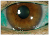
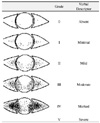
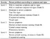
Three hundred and twenty-two eyes of 166 patients with dry eye syndrome that did not have coexisting lid margin disease or altered tear distribution and clearance were categorized into 1 of 4 levels of severity, according to the symptoms and signs in the Delphi panel classification. A symptom score was proposed depending on the frequency and impact on the quality of life. Degree of corneal staining using Lissamin green was categorized using the Oxford Scheme. In addition, measurement of the tear film break-up time (BUT) and Schirmer test were performed.
Results
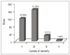
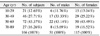
According to the levels of severity in the Delphi panel classification, 120 eyes (37.26%) were categorized as level1, 166 eyes (51.55%) as level 2, 30 eyes (9.31%) as level 3, and 6 eyes (1.86%) as level 4. Negative correlations were observed between the levels of severity, BUT, and measurements of the Schirmer test (p<0.001).
Figures and Tables
References
1. Schaumberg DA, Sullivan DA, Buring JE, Dana MR. Prevalence of dry eye syndrome among US women. Am J Ophthalmol. 2003. 136:318–326.
2. Lin PY, Tsai SY, Cheng CY, et al. Prevalence of dry eye among an elderly Chinese population in Taiwan: the Shihpai Eye Study. Ophthalmology. 2003. 110:1096–1101.
3. Brewitt H, Sistani F. Dry eye disease: the scale of the problem. Surv Ophthalmol. 2001. 45:S199–S202.
4. Behrens A, Doyle JJ, Stern L, et al. Dysfunctional tear syndrome: A Delphi approach to treatment recommendation. Cornea. 2006. 25:900–907.
5. Bron AJ, Evans VE, Smith JA. Grading of corneal and conjunctival staining in the context of other dry eye tests. Cornea. 2003. 22:640–650.
6. Kim WC, Kim HS, Kim MS. Current Trends in the Recognition and Treatment of Dry Eye: A Survey of Ophthalmologists. J Korean Ophthalmol Soc. 2007. 48:1614–1622.
7. Stern ME, Gao J, Siemasko KF, et al. The role of the lacrimal functional unit in the pathophysiology of dry eye. Exp Eye Res. 2004. 78:409–416.
8. Dana MR, Hamrah P. Role of immunity and inflammation in corneal and ocular surface disease associated with dry eye. Adv Exp Med Biol. 2002. 506:729–738.
9. Cho BJ, Lee JH, Shim OJ. The Relation Between Clinical Manifestations of Dry Eye Patients and Their BUTs. J Korean Ophthalmol Soc. 1992. 33:297–302.
10. Nichols KK, Nichols JJ, Mitchell GL. The lack of association between signs and symptoms in patients with dry eye disease. Cornea. 2004. 23:762–770.
11. Chia EM, Mitchell P, Rochtchina E, et al. Prevalence and associations of dry eye syndrome in an older population: the Blue Mountains Eye Study. Clin Experiment Ophthalmol. 2003. 31:229–232.
12. Viso E, Rodriguez-Ares MT, Gude F. Prevalence of and Associated Factors for Dry Eye in a Spanish Adult Population (The Salnes Eye Study). Ophthalmic Epidemiol. 2009. 16:15–21.
13. Lee JK, Ki CW, Roh KG. The Significance of the Tear Film Break-up Time (BUT) in the Diagnosis of the Dry Eye Syndrome. J Korean Ophthalmol Soc. 1985. 26:1311–1315.
14. Begley CG, Caffery B, Chalmers RL, et al. Dry Eye Investigation(DREI) Study Group. Use of the dry eye questionnaire to measure symptoms of ocular irritation in patients with aqueous tear deficient dry eye. Cornea. 2002. 21:664–670.
15. Hamano H, Hori M, Hamano T, et al. A new method for measuring tears. CLAO J. 1983. 9:281–289.
16. Asbell PA, Chiang B, Li K. Phenol-red thread test compared to Schirmer test in normal subjects. Ophthalmology. 1987. 94:128.
17. Nichols KK, Mitchell GL, Zadnik K. The repeatability of clinical measurements of dry eye. Cornea. 2004. 23:272–285.
 XML Download