

 PDF
PDF ePub
ePub Citation
 Print
Print


Abstract
Case summary:
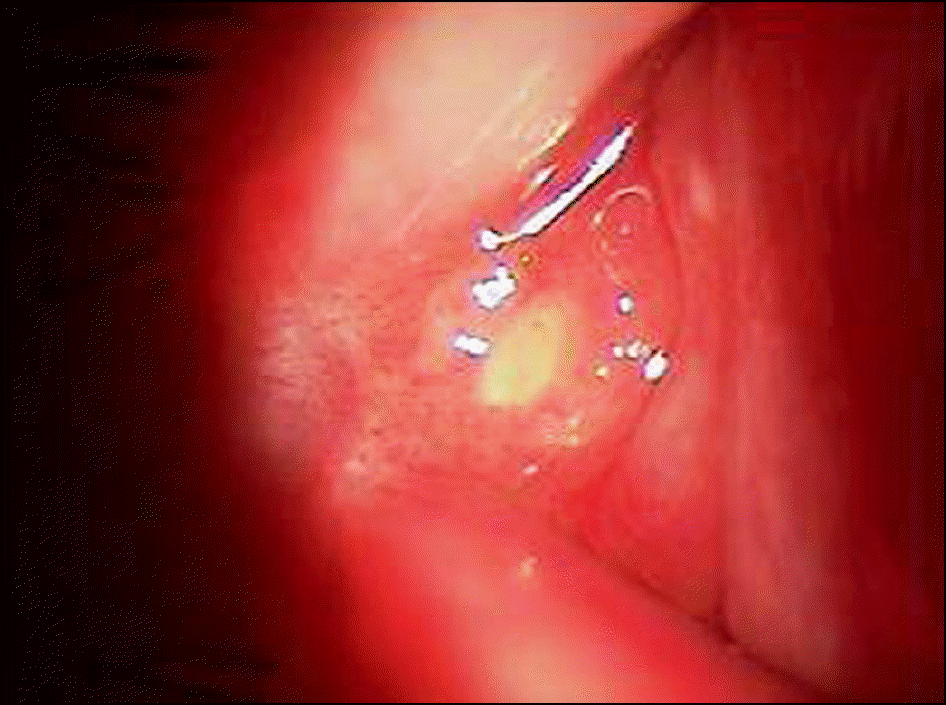
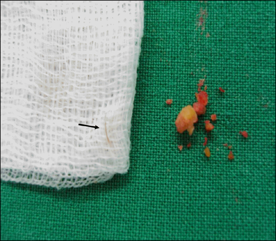
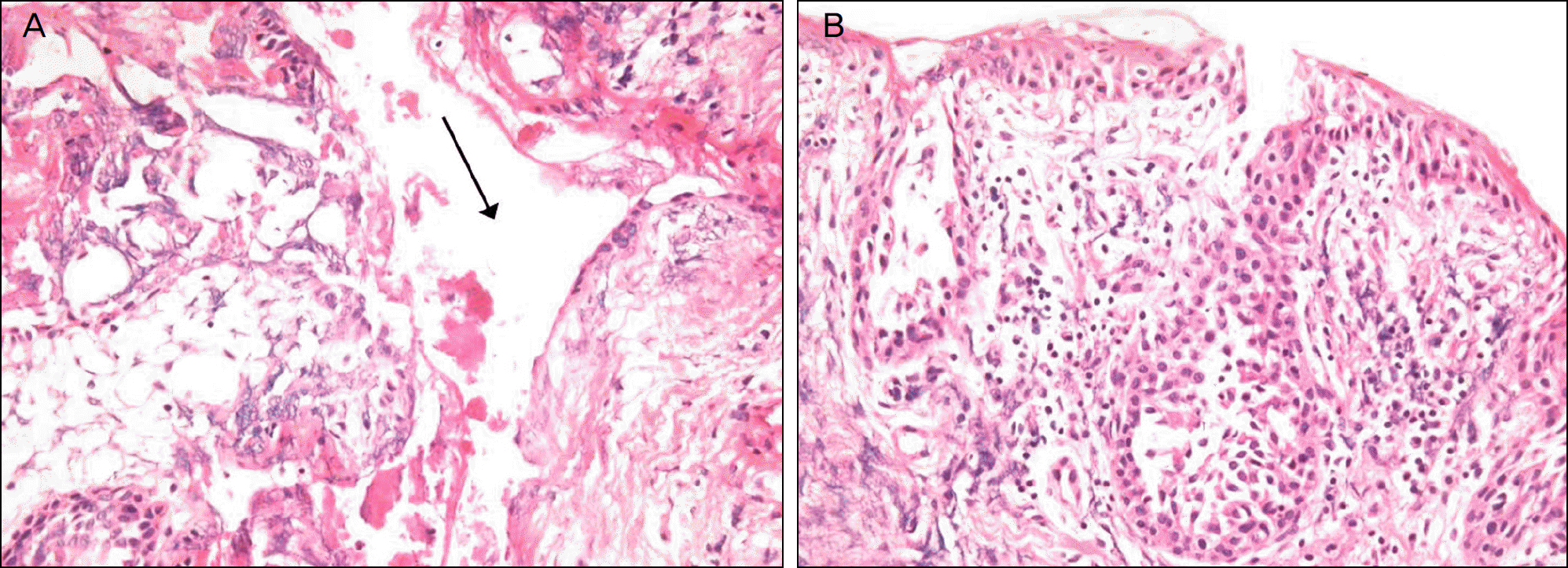
A 31-year-old woman presented with foreign body sensation, pain, and conjunctival injection in the lateral palpebral conjunctiva of her right eye over 6 months in duration. The physical examination revealed a small, firm nodule at the lateral canthal area. The excisional biopsy was performed, and the mass was a concretion that contained an eyelash in the center. During one-year follow-up, the patient showed no signs of recurrence or complication after excision.
Go to : 
References
1. Zafar A, Jordan DR, Brownstein S, Faraji H. Asymptomatic lacrimal ductule dacryolithiasis with embedded cilia. Ophthal Plast Reconstr Surg. 2004; 20:83–5.

2. Baratz KH, Bartley GB, Campbell RJ, Garrity JA. An eyelash nidus for dacryoliths of the lacrimal excretory and secretory systems. Am J Ophthalmol. 1991; 111:624–7.
3. Jay JL, Lee WR. Dacryolith formation around an eyelash retained in lacrimal sac. Br J Ophthalmol. 1976; 60:722–5.
4. Baker RH, Bartley GB. Lacrimal gland ductule stones. Ophthalmolgy. 1990; 97:531–4.
5. Duke-Elder S, editor. System of ophthalmology. Vol. XIII The ocular adenexa part 2: lacrimal, orbital and paraorbital diseases. St. Louis: CV Mosby Co.;1974. p. 672.
6. Mawn LA, Sanon A, Conlon MR, Nerad JA. Pseudomonas dacryoadenitis secondary to a lacrimal gland ductule stone. Ophthal Plast Reconstr Surg. 1997; 13:135–8.
7. Halborg J, prause JU, Toft PB, et al. Stones in the lacrimal gland: a rare condition. Acta Ophthalmol. 2009; 87:672–6.
8. Marthin JK, Lindegaard J, Prause JU, Heegaard S. Lesions of the lacrimal drainage system: a clinicopathological study of 643 biopsy specimens of the lacrimal drainage system in Denmark 1910–1999. Acta Ophthalmol Scand. 2005; 83:94–9.
9. Garfin SW. Etiology of dacryocystitis and epiphora. Arch ophthalmol. 1942; 27:167–88.
Go to :
 XML Download