Abstract
Case summary:
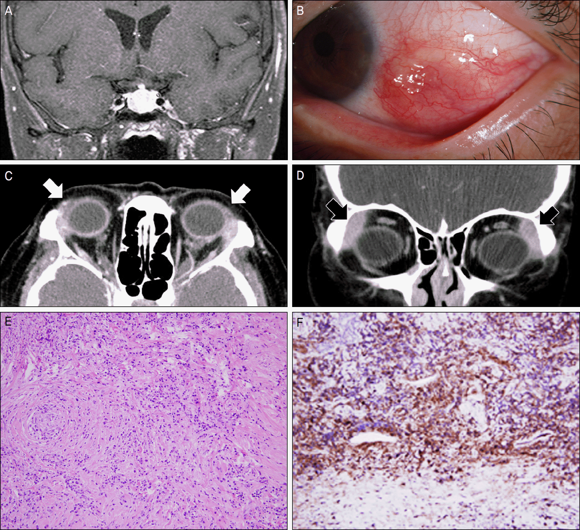
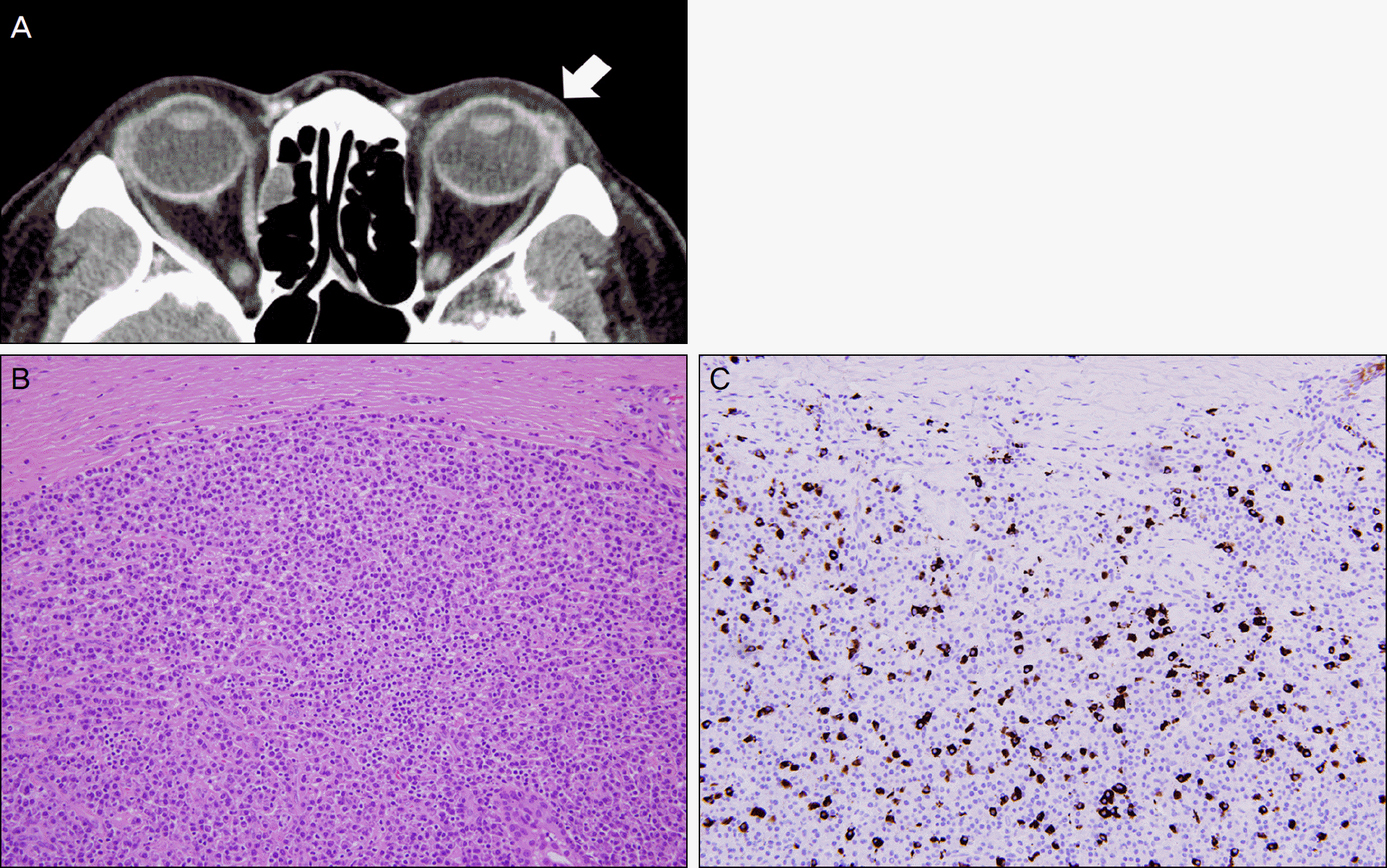
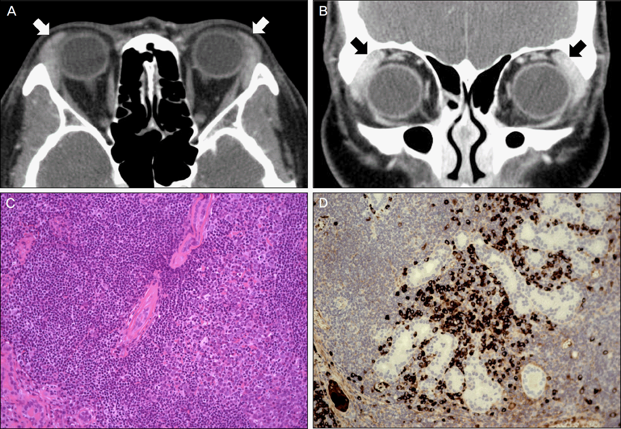
A 66-year-old woman presented with swelling of the bilateral upper eyelids with ocular pain that began 1 year before. Nodular episcleral injection of the left eye and other generalized symptoms, such as cough, decreased hearing ability, multiple nodular lesions of the bilateral lungs and right kidney, together suggested rheumatic disease. Orbital computed tomographic images revealed diffuse swelling of the bilateral lacrimal glands. After immunostaining a surgi-cally-biopsied specimen from the lacrimal gland for IgG4 expression, 15% of infiltrated lymphoplasmacytic cells were IgG4-positive. Similar findings were shown with biopsied specimens from the lung and kidney; therefore, the patient was diagnosed with Hyper-IgG4 syndrome. A 49-year-old woman complained of a mass in the left upper eyelid that began 4 years earlier. Orbital computed tomographic images showed a 5-mm-sized mass in the left upper eyelid. Ocular adnexal Hyper-IgG4 syndrome was confirmed by the immunostained biopsy from the left upper eyelid, showing infiltration of IgG4-positive lymphoplasmacytic cells. A 51-year-old woman presented with swelling of the bilateral lacrimal glands. Enlargement of the bilateral lacrimal glands were apparent in orbital computed tomographic images. After anti-IgG4 antibody staining of a biopsied specimen from the right lacrimal gland, dense infiltration of IgG4-positive lymphoplasmacytic cells was observed. The patient was also diagnosed with Hyper-IgG4 syndrome.
References
1. Kamisawa T, Funata N, Hayashi Y, et al. A new clinicopathological entity of IgG4-related autoimmune disease. J Gastroenterol. 2003; 38:982–4.

2. Yamamoto M, Takahashi H, Ohara M, et al. A new conceptualization for Mikulicz's disease as an IgG4-related plasmacytic disease. Mod Rheumatol. 2006; 16:335–40.

3. Sato Y, Ohshima K, Ichimura K, Imai K. Ocular adnexal IgG4- re-lated disease has uniform clinicopathology. Pathology International. 2008; 58:465–70.
4. Yamamoto M, Takahashi H, Sugai S, et al. Clinical and pathological characteristics of Mikulicz's disease (IgG4-related plasmacytic exocrinopathy). Autoimmun Rev. 2005; 4:195–200.

5. Naitoh I, Nakazawa T, Ohara H, et al. Endoscopic transpapillary intraductal ultrasonography and biopsy in the diagnosis of IgG4-related sclerosing cholangitis. J Gastroenterol. 2009; 44:1147–55.

6. Hamano H, Kawa S, Ochi Y, et al. Hydronephrosis associated with retroperitoneal fibrosis and sclerosing pancreatitis. Lancet. 2002; 359:1403–4.
7. Morgan WS, Castleman B. A clinicopathologic study of Mikulicz's disease. Am J Pathol. 1953; 29:471–503.
8. Tsubota K, Fujita H, Tsuzaka K, Takeuchi T. Mikulicz's disease and Sjogren syndrome. Invest Ophthalmol Vis Sci. 2000; 41:1666–73.
9. Hamano H, Kawa S, Horiuchi A, et al. High serum IgG4 concentrations in patients with sclerosing pancreatitis. N Engl J Med. 2001; 344:732–8.

10. Kamisawa T, Nakajima H, Egawa N, et al. IgG4-related sclerosing disease incorporating sclerosing pancreatitis, cholangitis, sialadenitis and retroperitoneal fibrosis with lymphadenopathy. Pancreatology. 2006; 6:132–7.

11. Kamisawa T, Okamoto A. IgG4-related sclerosing disease. World J Gastroenterol. 2008; 14:3948–55.

12. Neild GH, Rodriguez-Justo M, Wall C, Connolly JO. Hyper-IgG4 disease: report and characterisation of a new disease. BMC Medicine. 2006; 4:23.

13. Masaki Y, Dong L, Kurose N, et al. Proposal for a new clinical entity, Multiorgan lymphoproliferative syndrome: IgG4-positive analysis of 64 cases of IgG4-related disorders. Ann Rheum Dis. 2009; 68:1310–5.
Figure 1.
(A) Sellar MRI of the patient 1. Pituitary gland is enlarged, but there is no definite focal mass. (B) Temporal nodular injection is observed in the left eye. (C, D) Orbital CT revealed bilaterally enlarged lacrimal glands (arrows). (E) Biopsied specimens from lacrimal gland showed dense fibrosis, prominent lymphoplasmacytic infiltration, obliterative arteritis and periductal fibrosis (H&E, ×200). (F) About 15% of infiltrated lymphoplasmacytic cells are IgG4-positive (Anti-IgG4 Ab, ×200).

Figure 2.
(A) Orbital CT image of patient 2 shows 5-mm-sized nodular mass in left upper eyelid (arrow). (B) Biopsied specimen from nodular mass in left upper eyelid shows abundant lymphoplasmacytic infiltration, lymphoid follicles and interstitial fibrosis (H&E, ×200). (C) About 15∼20% of infiltrated lymphoplasmacytic cells are IgG4-positive (Anti-IgG4 Ab, ×200).

Figure 3.
(A, B) Orbital CT images of patient 3 shows bilaterally enlarged lacrimal glands (arrows). (C) Biopsied specimen from lacrimal gland shows abundant lymphoplasmacytic infiltration with interstitial fibrosis (H&E, ×200). (D) Dense infiltration of IgG4-positive lymphoplasmacytic cells is observed (Anti-IgG4 Ab, ×200).


 PDF
PDF ePub
ePub Citation
 Print
Print


 XML Download