

 PDF
PDF ePub
ePub Citation
 Print
Print


Abstract
Purpose
To experimentally investigate the effect for muscle weakness after superior rectus Z-myotomy and histological changes.
Methods
Superior rectus muscle fibers of rabbits (16 rabbits, 32 eyes) were cut transversely with scissors across 75% of the muscle in two different positions on opposite sides. In group 1 (16 eyes), myotomies were performed at 2 and 7 mm from the muscle insertion (5 mm gap) and in group 2, performed at 2 and 5 mm (3 mm gap). The change of mark, eyeball position, and muscle tension after myotomy and 4 weeks postoperatively was evaluated, the location of the mark was examined, and muscle tissue biopsy was performed.
Results
After Z-myotomy, the marks of the two groups moved significantly posteriorly from insertion within groups (p<0.05), with no significant differences between groups (p=0.469). Eyeball positions of the two groups moved significantly inferiorly (p<0.05); the amount of position change of group 1 was greater than group 2 (p<0.05). When the globe was pulled in opposite directions for muscle action, the degree of change decreased with significant difference within groups (p<0.05), but there were no significant differences between groups (p=0.32).
Go to : 
References
1. Helveston EM, Cofield DD. Indications for marginal myotomy and technique. Am J Ophthalmol. 1970; 70:574–8.

2. de Faber JT, von Noorden GK. Medial rectus muscle marginal myotomy for persistent esotropia. Am J Ophthalmol. 1991; 112:702–5.
3. Mellott ML, Scott WE, Ganser GL, Keech RV. Marginal myotomy of the minimally overacting inferior oblique muscle in asymmetric bilateral superior oblique palsies. J AAPOS. 2002; 6:216–20.
4. Almeida HC, Alvares MA. Split lengthening of the inferior oblique muscles. Graefe's Arch Clin Exp Ophthalmol. 1988; 226:181–2.
5. von Noorden GK. Binocular Vision and Ocular Motility: theory and management of strabismus. 6th ed.St. Louis: Mosby;2002. p. 101–7.
6. Helveston EM. Atlas of strabismus surgery. 3rd ed.St. Louis: CV Mosby;1985. p. 254–9.
7. Kumar K, Prasad HN, Monga S, Bhola R. Hang-back recession of inferior oblique muscle in V-pattern strabismus with inferior oblique overaction. J AAPOS. 2008; 12:401–4.
8. Lim KH, Yu YS, Chang BL. The effect of 2% methylhydrox-ypropylcellulose and hyaluronic acid on adjustable force in experimental adjustable strabismus surgery in rabbits. J Korean Ophthalmol Soc. 1997; 38:466–73.
Go to :
 | Figure 1.Procedures of superior rectus Z-myotomy. (A) Superior rectus muscle is exposed with muscle hooks after conjunctival dissection. (B) Marking with 5-0 black silk on the superior rectus muscle 9.0 mm from the muscle insertion site. (C) The hemostat is placed to grasp the muscle that will be cut. (D) The muscle is cut along the crushed area using small snips with scissors. (E)(F) Noticeable lengthening of the muscle is achieved. |
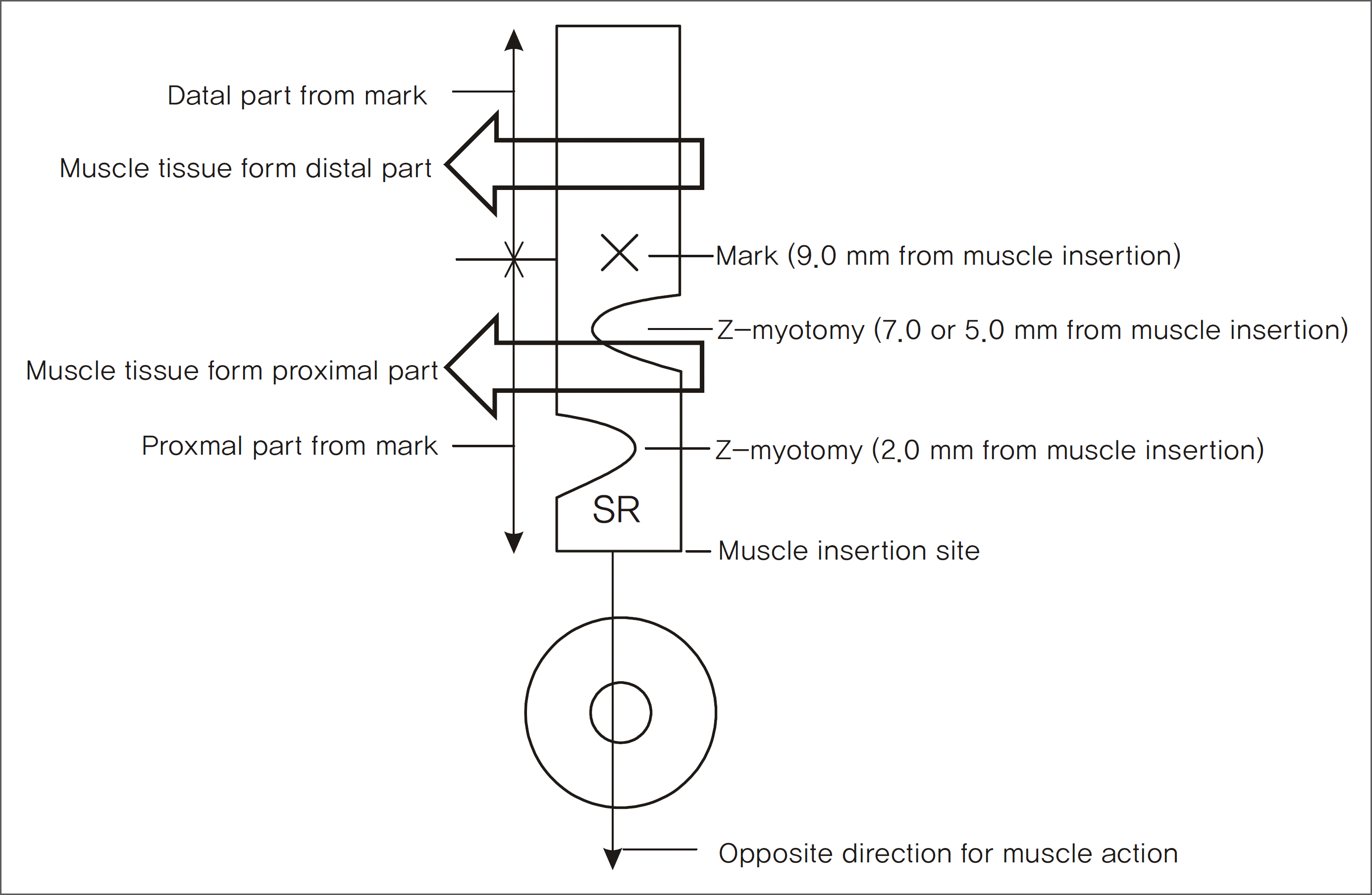 | Figure 2.This diagram depicts the position of the mark and Z-myotomy in the superior rectus muscle. |
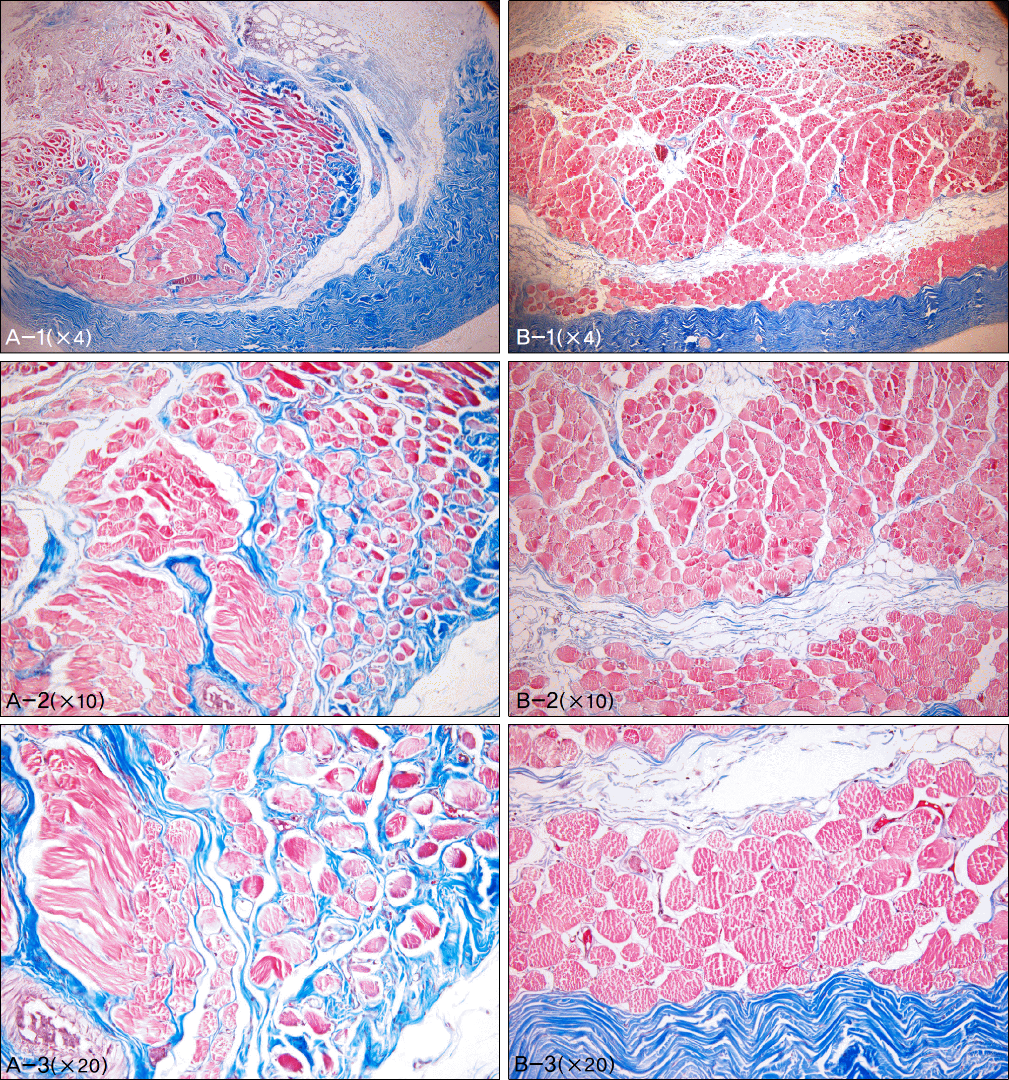 | Figure 4.Histologic findings of Z-myotomy site of a group 1 rabbit at 4 weeks after surgery (Masson's trichrome stain). (A-1, A-2, A-3) Proximal portion from the mark, injured muscle by myotomy. This muscle tissue showed vague margin between the orbital layer and global layer, collagen fiber proliferation among muscle fibers, and severely distorted EOM structure. (B-1, B-2, B-3) Distal portion from the mark, intact muscle. This intact muscle showed well defined orbital layer (small sized muscular fibers, surround global layer) and global layer (large sized muscular fibers, surrounded by orbital layer). |
Table 1.
The location of mark on superior rectus muscle before and after Z-myotomy (initial position of mark is on 9.0 mm from muscle insertion site)
| Before myotomy | Immediately after myotomy | 4 weeks after myotomy | * p-value Multivariate Tests | |
|---|---|---|---|---|
| Group 1 | 9.0 mm | 10.06 ± 1.59 mm | 9.50 ± 1.40 mm | <0.05 |
| Group 2 | 9.0 mm | 9.75 ± 1.23 mm | 10.53 ± 2.01 mm | <0.05 |
| Tests of Between-Groups: * p-value=0.469 | ||||
Table 2.
The change in the eyeball position before and after Z- myotomy
|
Eyeball position |
Amount of change | |||
|---|---|---|---|---|
| Before myotomy (A) | Immediately after myotomy (B) | * p-value (A-B) | ||
| Group 1 | 3.84 ± 0.53 mm | 1.53 ± 0.38 mm | <0.05 | 2.31 ± 0.44 mm |
| Group 2† | 3.71 ± 0.54 mm | 1.93 ± 0.65 mm | <0.05 | 1.78 ± 0.65 mm |
| † P value | 0.564 | 0.073 | <0.05 | |
Table 3.
The change in the eyeball position after traction with 50 g tension to opposite direction of muscular action
|
Eyeball position |
Amount of change | |||
|---|---|---|---|---|
| Before myotomy (A) | Immediately after myotomy (B) | * p-value (A-B) | ||
| Group 1 | 4.03 ± 1.05 mm | 1.93 ± 0.91 mm | <0.05 | 2.09 ± 0.75 |
| Group 2 | 4.12 ± 0.90 mm | 2.28 ± 0.79 mm | <0.05 | 1.84 ± 0.65 |
| † P value | 0.838 | 0.224 | 0.32 | |
 XML Download