

 PDF
PDF ePub
ePub Citation
 Print
Print


Abstract
Purpose
To evaluate the effectiveness of Mitomycin C used as a combined therapy along with limbal-conjunctival autograft for primary pterygium.
Methods
Thirty eyes of 29 patients received Mitomycin C (0.02% MMC 3 minutes) with limbal-conjunctival autograft, and 30 eyes of 28 patients received limbal-conjunctival autograft alone. Recurrence and complications were evaluated in the patients at 2 weeks, 1, 3, 6 and 12 months postoperatively.
Results
Mean follow-up periods were 13.4±2.1 and 13.9±2.9 months, respectively. Between the two groups, recurrence in the conjunctiva or the cornea was not observed during the follow-up period. In the Mitomycin C use group, complications included a granuloma at the donor site (1 eye, 3.3%), wound dehiscence (2 eyes, 6.7%), and subgraft hemorrhage (2 eyes, 6.7%). In comparison, in the group treated with limbal-conjunctival autograft alone, complications included granuloma at the donor site (1 eye, 3.3%), pseudopterygium at the donor site (1 eye, 3.3%), wound dehiscence (3 eyes, 10%), and subgraft hemorrhage (2 eyes, 6.7%).
References
1. Dushku N, John MK, Schultz GS, Reid TW. Pterygia pathogenesis: corneal invasion by matrix metalloproteinase expressing altered limbal epithelial basal cells. Arch Ophthalmol. 2001; 119:695–706.
2. Pinkerton OD. Immunologic basis for the pathogenesis of pterygium. Am J Ophthalmol. 1984; 98:225–8.

3. Lee SH, Jeong HJ. Immune reactions in pterygium. J Korean Ophthalmol Soc. 1987; 28:933–7.
4. Baharassa F, Datta R. Postoperative beta irradiation treatment of pterygium. Int J Radiat Oncol Biol Phys. 1983; 9:679–84.
5. Kunitomo N, Mori S. Studies on the pterygium. Report IV. A treatment of the pterygium by mitomycin C instillation. Nippon Ganka Gakkai Zasshi. 1963; 67:601–7.
6. Hayasaka S, Noda S, Yamamoto Y, Setogawa T. Postoperative instillation of low dose mitomycin C in the treatment of primary pterygium. Am J Ophthalmol. 1988; 106:580–3.
7. Frucht-Pery J, Siganos CS, Ilsar M. Intraoperative application of topical mitomycin C for pterygium surgery. Ophthalmology. 1996; 103:674–7.
8. Rubinfeld RS, Pfister RR, Stein RM, et al. Serious complications of topical mitomycin C after pterygium surgery. Ophthalmology. 1992; 99:1647–54.
9. Hirst LW. The treatment of pterygium. Surv Ophthalmol. 2003; 48:145–80.
10. Kenyon KR, Wagoner MD, Hettinger ME. Conjunctival autograft transplantation for advanced and recurrent pterygium. Ophthalmology. 1985; 92:1461–70.
11. Gris O, Güell JL, del Campo Z, et al. Limbal-conjunctival autograft transplantation for the treatment of recurrent pterygium. Ophthalmology. 2000; 107:270–3.
12. Shimazaki J, Yang HY, Tsubota K. Limbal autograft transplantation for recurrent and advanced pterygia. Ophthalmic Surg Lasers. 1996; 27:917–23.
13. Al Fayez MF. Limbal versus conjunctival autograft transplantation for advanced and recurrent pterygium. Ophthalmology. 2002; 109:1752–5.
14. Park JM, Ahn HB, Park WC. The effect of combined amniotic membrane and limbal transplantation for recurrent pterygium or pseudopterygium. J Korean Ophthalmol Soc. 2003; 44:1504–11.
15. Kilic A, Gurler B. The efficiency of limbal conjunctival autografting in pterygium surgery. Eur J Ophthalmol. 2006; 16:365–70.
16. Tan DT, Chee SP, Dear KB, et al. Effect of pterygium morphology on pterygium recurrence in a controlled trial comparing conjunctival autografting with bare sclera excision. Arch Ophthalmol. 1997; 115:1235–40.
17. Prabhasawat P, Barton K, Burkett G, Tseng SC. Comparison of conjunctival autografts, amniotic membrane grafts, and primary closure for pterygium excision. Ophthalmology. 1997; 104:974–85.
18. Verweij J, Pinedo HM. Mitomycin C: mechanism of action, usefulness and limitations. Anticancer Drugs. 1990; 1:5–13.
19. Kenyon KR, Tseng SC. Limbal autograft transplantation for ocular surface disorders. Ophthalmology. 1989; 96:709–23.
20. Tseng SC. Concept and application of limbal stem cells. Eye. 1989; 3:141–57.
21. Wong VA, Law FC. Use of mitomycin C with conjunctival autograft in pterygium surgery in Asian-Canadians. Ophthalmology. 1999; 106:1512–5.
22. Mutlu FM, Sobaci G, Tatar T, Yildirim E. A comparative study of recurrent pterygium surgery: limbal conjunctival autograft transplantation versus mitomycin C with conjunctival flap. Ophthalmology. 1999; 106:817–21.
23. Frucht-Pery J, Raiskup F, Ilsar M, et al. Conjunctival autografting combined with low-dose mitomycin C for prevention of primary pterygium recurrence. Am J Ophthalmol. 2006; 141:1044–50.
24. Kim HH, Mun HJ, Park YJ, et al. Conjunctivolimbal autograft using a fibrin adhesive in pterygium surgery. J Korean Ophthalmol Soc. 2008; 22:147–54.
25. Barraquer JI. Etiology, pathogenesis, and treatment of the pterygium. Trans New Orleans Acad Ophthalmol. 1980; 167–78.
26. Solomon A, Pires RT, Tseng SC. Amniotic membrane transplantation after extensive removal of primary and recurrent pterygia. Ophthalmology. 2001; 108:449–60.
27. Hirst LW. Prospective study of primary pterygium surgery using pterygium extended removal followed by extended conjunctival transplantation. Ophthalmology. 2008; 115:1663–72.
28. Bahar I, Kaiserman I, Weisbrod M, et al. Extensive versus limited pterygium excision with conjunctival autograft: outcomes and recurrence rates. Curr Eye Res. 2008; 33:435–40.
Figure 1.
Dashed line is extensive excision profiles of a pterygium. The extensive excision was designed to resect the maximum surface of pterygium, while preserving the conjunctiva.
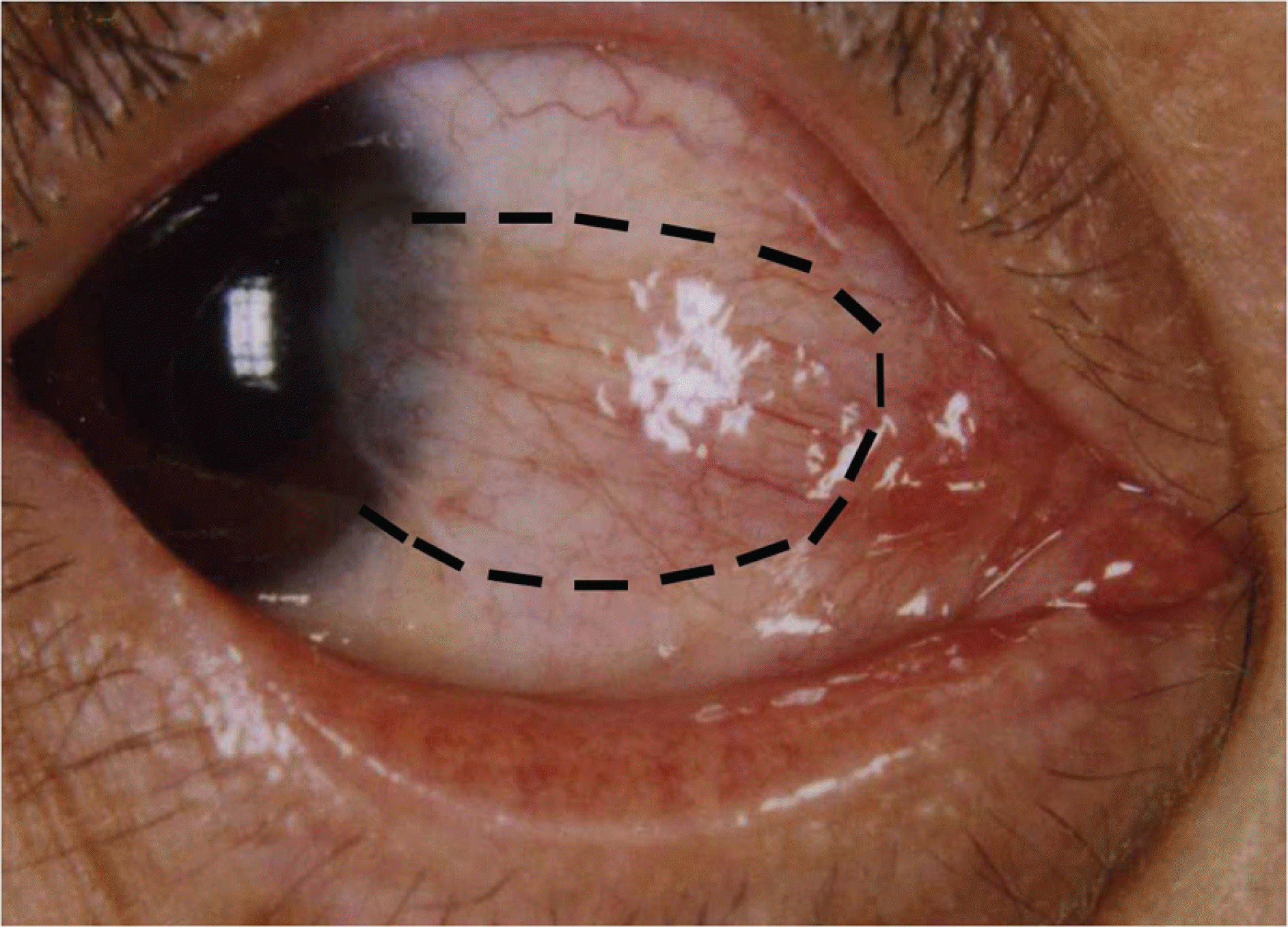
Figure 2.
The photograph of the anterior segment on day 1 after pterygium excision and superior limbal conjunctival autograft transplantation with temporary amniotic membrane patch. Under the temporary amniotic membrane transplantation, notice the relatively low inflammatory appearance of the eye, well-positioned graft and reestablishment of the normal limbal architecture.

Figure 3.
Left column (A, B: Limbal-conjunctival autograft without 0.02% MMC, C, D: Limbal-conjunctival autograft with 0.02% MMC) Images of preoperative fleshy grade 3 nasal primary pterygium. Right column (E, F: Limbal-conjunctival autograft without 0.02% MMC, G, H: Limbal-conjunctival autograft with 0.02% MMC) Images of the LCAT of the same patients 6 months after the surgery. Well-positioned graft and reestablishment of the normal limbal architecture are observed with no signs of recurrences in all subjects. Notice: The lack of scar on the donor and recipient conjunctiva due to the use of bioadhesives, and the cosmetic advantage due to appropriate size of conjuctival flap after extensive pterygium excision are apparent.

Figure 4.
Postoperative complications. (A) Granuloma at donor site. (B) Wound dehiscence (arrowhead). (C) Excessive fibrovascular tissue proliferation at donor site (similar to pseudopterygium). (D) Subgraft hemorrhage.

Table 1.
Previous reports of conjunctival autografts combined MMC
| Author (s) | Pterygium type | Number of Eyes | Type of surgery (MMC%, minutes) | Recurrence Rate (%) | Mean follow-up (months) | Donor Site of CAT* |
|---|---|---|---|---|---|---|
| Wong et al32 | Primary | 98 | CAT | 18% | 12 | Superior |
| | | 76 | CAT&MMC† | 9% | 12 | |
| | | | (0.025%, 1 min) | | | |
| Mutlu et al33 | Recurrent | 41 | LCAT‡ | 14.6% | 16±1.9 | Superotemporal |
| | | 40 | CAT&MMC | 12.5% | 15.5±1.5 | Superior |
| | | | (0.02%, 3 min) | | | |
| Frucht-Pery | Primary | 30 | CAT&MMC | 0% | 21.3±2.4 | Superior |
| et al34 | | | (0.02%, 1 min) | | | |
| | | 30 | MMC (0.02%, 3 min) | 6.6% | 31.5±3.4 | |
| | | 30 | CAT | 13.3% | 29.3±2.5 | Superior |
| | | 30 | Bare sclera | 46.6% | 36.2±2.8 | |
| This study | Primary | 30 | LCAT&MMC | 0% | 13.4±2.1 | Superior |
| | | | (0.02%, 3 min) | | | |
| | | | + TAMP§ | | | |
| | | 30 | LCAT+TAMP | 0% | 13.9±2.9 | Superior |
Table 2.
Demographic data of patients in the two study groups
| | LCAT*+MMC† group‡ | LCAT group | p |
|---|---|---|---|
| No. of eyes (patients) | 30 (29) | 30 (28) | |
| Age (years) | 56.9±7.8 | 59.6±8.5 | 0.217§ |
| Range | (44–71) | (45–72) | |
| Gender | | | 0.559Π |
| Male | 9 (32.0%) | 7 (30.8%) | |
| Female | 21 (68.0%) | 23 (69.2%) | |
| Follow-up (months) | 13.4±2.1 | 13.9±2.9 | 0.487§ |
| Grade# | | | 0.194Π |
| T1 / T2 / T3 | 0 / 11 / 19 | 0 / 17 / 13 | |
 XML Download