Abstract
Purpose
To analyze the clinical features of orbital wall fracture with diplopia between the surgical treatment group and the conservative treatment group.
Methods
The study comprised of 109 eyes of 109 patients with orbital wall fracture and diplopia. The patients were divided into two groups: the surgical treatment group (59 cases) and the conservative treatment group (50 cases). The groups were analyzed retrospectively according to age, gender, cause, CT, the period and severity of diplopia, and enophthalmos with time.
Results
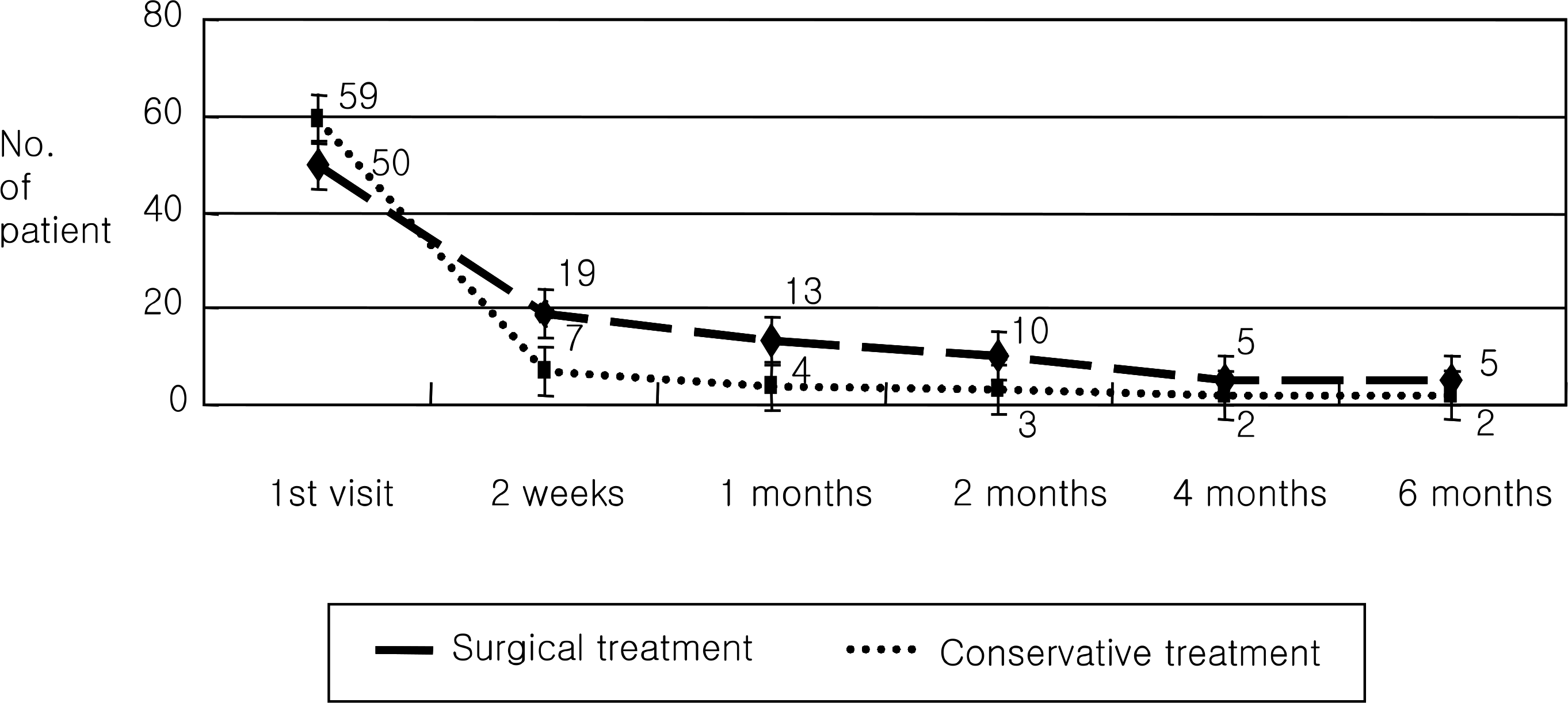
In the conservative treatment group, 38 cases (64.4%) had medial wall fracture, and the average fracture size was 26% of the inferior wall and 33% of the medial wall. In addition, at the first visit, the patients showed diplopia within 45.5 degrees, and diplopia disappeared completely within 17 days on average (57 cases, 96.6%). In the group that underwent the reconstruction of orbital wall fracture, 27 cases (54.0%) had inferior wall fracture, and the average fracture size was 41% of the inferior wall and 35% of the medial wall. Additionally, in the first visit, the patients showed diplopia within 20.3 degrees. The muscle incarceration occurred in 12 cases (24%). In the surgical treatment group, diplopia disappeared completely within 30 days on average (45 cases, 90.0%).
Conclusion
In the group of conservative treatment, they showed diplopia within 45.5 degrees at the first visit. Diplopia disappeared completely within 17 days on average (57 cases, 96.6%). In the group of surgical treatment, they showed diplopia within 20.3 degrees at the first visit. Diplopia disappeared completely within 30 days on average (45 cases, 90.0%).
Go to : 
References
1. Hosal BM, Beatty RL. Diplopia and enopthalmos after surgical repair of blowout fracture. Orbit. 2002; 21:27–33.
2. Putterman AM, Stevens T, Urist MJ. Nonsurgical management of blowout fractures of the orbital floor. Am J Ophthalmol. 1974; 77:232–9.

3. Gilbard SM, Mafee MF, Lagouros PA, Langer BG. Orbital blowout fractures. The prognostic significance of computed tomography. Ophthalmology. 1985; 92:1523–8.

4. Pearl RM. Treatment of enopthalmos. Clin Plast Surg. 1992; 19:99–111.
5. Iliff NT. The opthalmic implications of the correction of late enophthalmos following severe midfacial trauma. Trans Am Ophthalmol Soc. 1991; 89:477–548.
6. Hwang JH, Kwag MS. Residual diplopia and enophthalmos after reconstruction of rbital wall fractures. J Korean Ophthalmol Soc. 2003; 44:1959–65.
7. Kim HY, Kim YI, Won IK. Clinical analysis of blowout fracture with ocualr motion limitation: comparison of surgical and conservative treatment. J Korean Ophthalmol Soc. 1999; 40:632–8.
8. Kim SK, Chang HK. The clinical study of treatment of blowout fracture. J Korean Ophthalmol Soc. 1995; 36:1629–35.
9. Kroll M, Wolper J. Orbital blowout fracture. Am J Ophthalmol. 1967; 64:1169–72.
10. Yang PJ, Chi NC, Choi GJ. Comparison of sugical outcome between early and delayed repair of orbital wall fracture. J Korean Ophthalmol Soc. 2004; 44:1278–84.
11. Haug RH, Van Sickels JE, Jenkins WS. Demographics and treatment options for orbital roof fractures. Oral Surg Oral Med Oral Pathol Oral Radiol Endod. 2002; 93:238–46.

12. Smith B, Regan WF Jr. Blowout fracture of the orbit. Mechanism and correction of internal orbital fracture. Am J Ophthalmol. 1957; 44:739–9.
13. Putterman AM. Management of blow out fracture of the orbital floor: The conservative approach. Surv Ophthalmol. 1991; 35:292–8.
14. Millman AL, Della Rocca RC, Spector S, et al. Steroid and orbital blowout fractures, A new systematic concept in medical management and surgical decision-making. Adv Ophthalmic Plast Reconstr Surg. 1987; 6:291–300.
15. Koorneef L. Current concepts on the management of orbital blowout fracture. Ann Plast Surg. 1982; 9:195–200.
16. Everhard-Halm YS, Koornneef L, Zonneveld FW. Conservative therapy frequently indicated in blowout fractures of the orbit. Ned Tijdschr Geneeskd. 1991; 135:1226–8.
Go to : 
 | Figure 1.Cause of diplopia on the computed tomography of orbital wall fracture. (A) Soft tissue swelling, (B) Extraocular muscle swelling, (C) Soft tissue incarceration, (D) Extraocular muscle incarceration. |
Table 1.
Sex and age distribution of orbital wall fracture
| | | Surgical treatment | Conservative treatment | |
|---|---|---|---|---|
| Sex | Male | 44 (88%) | 41 (69.5%) | p=0.022* |
| (No.) | Female | 6 (12%) | 18 (30.5%) | |
| Age | mean | 28.8 | 36.1 | p=0.022* |
| (years) | ≤20 | 15 (30%) | 9 (15.3) | |
| | 21–40 | 26 (52%) | 26 (44.1) | |
| | ≥41 | 9 (18%) | 24 (40.7%) | |
Table 2.
The characteristics of orbital wall fracture
| | | Surgical treatment | Conservative treatment | |
|---|---|---|---|---|
| Cause of fracture | Violence | 18 (36%) | 21 (35.6%) | p=0.059* |
| (No) | Traffic accident | 5 (10%) | 18 (30.5%) | |
| | Fall down | 12 (24%) | 12 (20.3%) | |
| | Projectile object | 8 (16%) | 3 (5.1%) | |
| | Sports, Working | 6 (12%) | 5 (8.5%) | |
| Fracture site | Inferior wall | 27 (54%) | 6 (10.2%) | p<0.001* |
| (No) | Medial wall | 13 (26%) | 38 (64.4%) | |
| | Inf & med wall | 10 (20%) | 15 (25.4%) | |
| Size of fracture | Inferior wall | 41.5±15.0 | 26.2±9.6 | p<0.01† |
| (%) | Medial wall | 35.9±15.5 | 34.0±9.4 | p=0.51† |
Table 3.
The characteristics of diplopia
| | | Surgical treatment | Conservative treatment | |
|---|---|---|---|---|
| Degree of diplopia on 1 st visiit | 20.3±17.4 | 45.5±9.0 | p<0.001† | |
| Cause of diplopia (No)‡ | Soft tissue swelling | 0 (0%) | 37 (62.7%) | p<0.001* |
| Muscle swelling | 10 (20%) | 10 (16.9%) | ||
| Soft tissue incarceration | 28 (56%) | 9 (15.3%) | ||
| Muscle incarceration | 12 (24%) | 0 (0%) | ||
| Muscle paralysis | 0 (0%) | 3 (5.1%) | ||
| Loss of diplopia (days) | | 30 | 17 | p=0.009† |
Table 4.
The clinical feature of diplopia according to cause
| Cause of diplopia | Patient (No†) | Degree of diplopia on 1st visit(degree) | Residual diplopia on (No/degree) | Loss of diplopia (days) | |
|---|---|---|---|---|---|
| Soft tissue swelling | 37 | 45.68 | 0 (0.00%) | 0 | 16.24 |
| Muscle swelling | 20 | 35.25 | 2 (10.0%) | 35.00 | 16.03 |
| Soft tissue incarceration | 37 | 28.51 | 3 (8.1%) | 38.33 | 34.58 |
| Muscle incarceration | 12 | 9.58 | 2 (16.7%) | 22.50 | 39.62 |
| Muscle paralysis | 3 | 45.00 | 0 (0.0%) | 0 | 8.05 |
| Total | 109 | | p=0.17* | | |


 PDF
PDF ePub
ePub Citation
 Print
Print


 XML Download