Abstract
Purpose
To report a case of functional bilateral hemianopia which was not associated with any organic causes.
Case summary
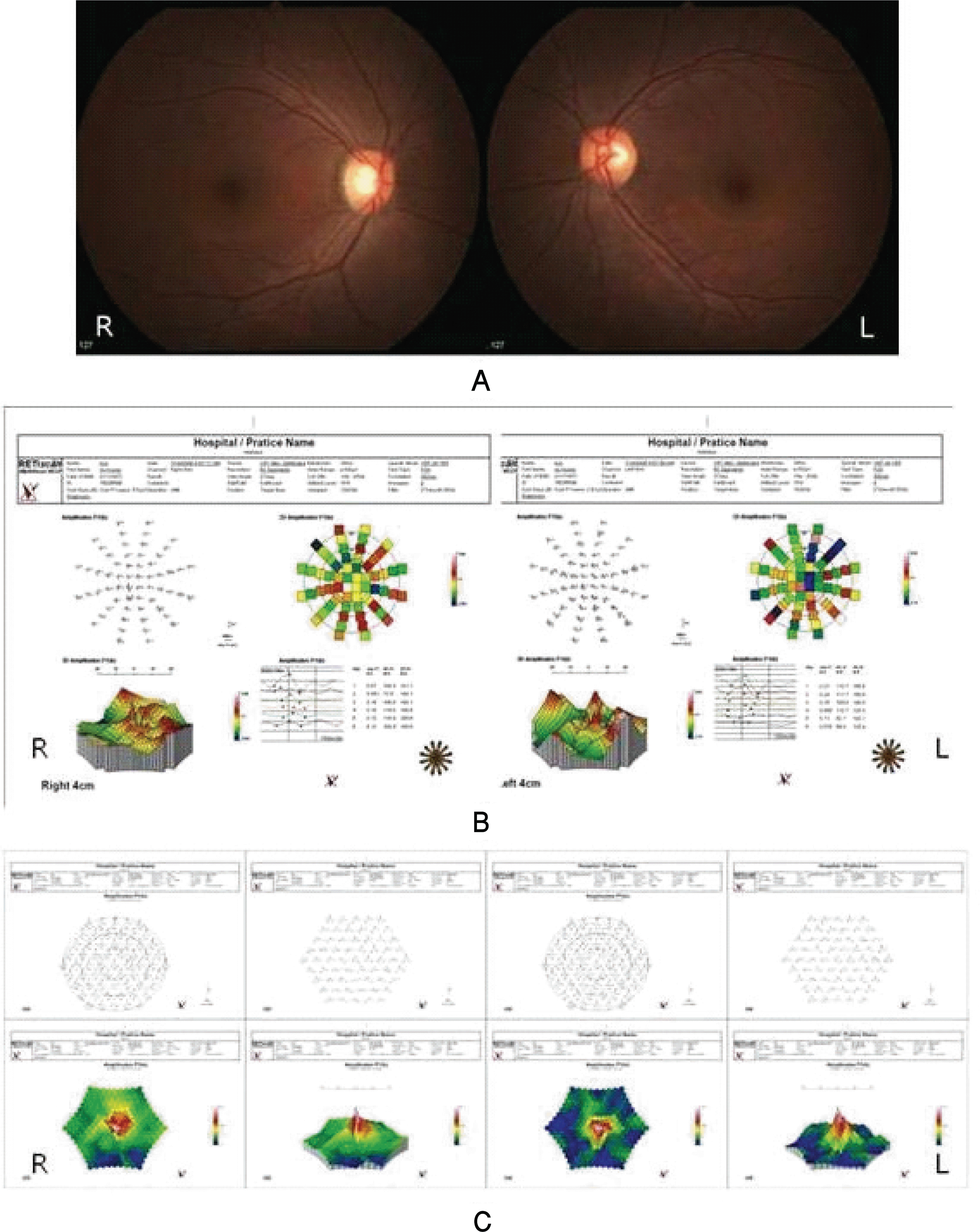
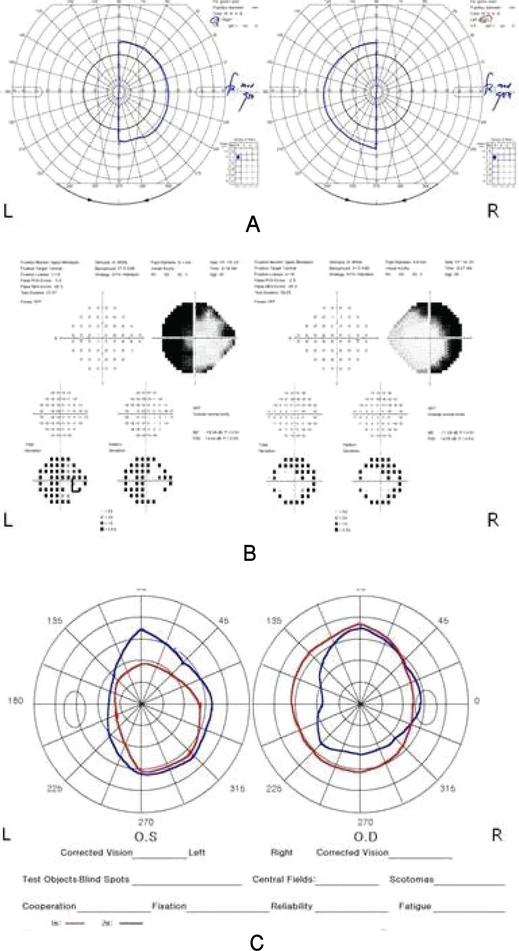
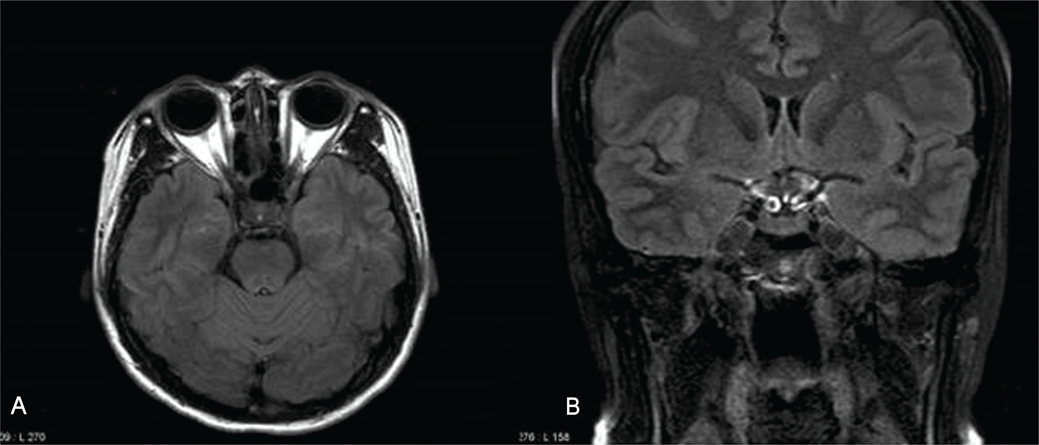
A 35-year-old female patient presented with bilateral disturbance of visual acuity and visual field, which had begun 8 months prior. Goldmann perimetry showed bitemporal hemianopsia respecting the vertical meridian. Pupillary response was normal, and the anterior segment, fundus, and optic nerve were also normal bilaterally. However, the tangent screen test and Humphrey visual field test showed a widening of hemianopia not respecting the vertical meridian, and the crossing of isopters at 1 m and 2 m with the tangent screen test. In addition, multifocal electroretinogram and multifocal visual evoked potential did not reveal any abnormal findings corresponding to the bitemporal hemianopia. Brain magnetic resonance imaging showed no abnormal findings in the orbit and brain.
Go to : 
References
1. Palmowski AM, Fischer A, Ruprecht KW. Multifocal examination techniques in malingering: case report of a patient with monocular vertical hemianopia. Graefes Arch Clin Exp Ophthalmol. 2003; 241:70–1.

3. Bakker SL, Hasan D, Bijvoet HW. Compression of the visual pathway by anterior cerebral artery aneurysm. Acta Neurol Scand. 1999; 99:204–7.

4. Hilton GF, Hoyt WF. An arteriosclerotic chiasmal syndrome. Bitemporal hemianopia associated with fusiform dilatation of the anterior cerebral arteries. JAMA. 1966; 196:1018–20.

5. Shikishima K, Kitahara K, Mizobuchi T, Yoshida M. Interpretation of visual field defects respecting the vertical meridian and not related to distinct chiasmal or postchiasmal lesions. J Clin Neurosci. 2006; 13:923–8.

6. Smith TJ, Baker RS. Perimetric findings in functional disorders using automated techniques. Ophthalmology. 1987; 94:1562–6.

7. Ohkubo H. Visual field in hysteria-reliability of visual field by Goldmann perimetry. Doc Ophthalmol. 1989; 71:61–7.

8. Thompson JC, Kosmorsky GS, Ellis BD. Field of dreamers and dreamed-up fields: functional and fake perimetry. Ophthalmology. 1996; 103:117–25.
9. Kim SY, Lee DH, Park SH. An analysis of visual fields in patients with posttraumatic functional visual loss. J Korean Ophthalmol Soc. 2004; 45:469–79.
10. Keltner JL, Johnson CA, Spurr JO, Beck RW. Baseline visual field profile of optic neuritis. The experience of the optic neuritis treatment trial. Optic Neuritis Study Group. Arch Ophthalmol. 1993; 1112:31–4.
13. Johnson LN, Rabinowitz YS, Hepler RS. Hemianopia respecting the vertical meridian and with foveal sparing from retinal degeneration. Neurology. 1989; 39:872–3.

14. Miele DL, Odel JG, Behrens MM, et al. Functional bitemporal quadrantopia and the multifocal visual evoked potential. J Neuroophthalmol. 2000; 20:159–62.

15. Massicotte EC, Semela L, Hedges TR 3rd. Multifocal visual evoked potential in nonorganic visual field loss. Arch Ophthalmol. 2005; 123:364–7.

16. Goldberg I, Graham SL, Klistorner AI. Multifocal objective perimetry in the detection of glaucomatous field loss. Am J Ophthalmol. 2002; 133:29–39.
Go to : 
 | Figure 1.(A) Fundus photography showed normal optic disc and fundus in both eyes. (B) Multifocal visual evoked potential revealed that there was neither interocular nor nasal-temporal difference in the response in both eyes. (C) Multifocal electroretinogram did not show any abnormal naso–temporal differences in amplitude or latency in both eyes (R: right, L: left). |
 | Figure 2.(A) Goldmann visual field test showed bitemporal hemianopia respecting the vertical meridian. (B) Humphrey visual field test showed bilateral temporal visual field defect not respecting the vertical meridian. (C) Tangent screen test did not show the bitemporal hemianopia detected in Goldmann visual field test. The visual field at 1 m (red line) and 2 m (blue line) crossed in the right eye and the visual field at 2 m (blue line) is smaller than that at 1 m (red line) in the left eye, suggesting the functional visual loss (R: right, L: left). |


 PDF
PDF ePub
ePub Citation
 Print
Print


 XML Download