Abstract
Case summary
The medical records of 9 patients with complications related to cosmetic contact lenses were retrospectively reviewed. All 9 patients had corneal erosion, and 2 patients had corneal epithelial defects. Corneal neovascularization more than 2 mm from the limbus was observed in 3 patients and one of the patients received a permanent impairment of visual acuity. Seven patients were not educated on the management of contact lenses and 2 patients had previous experience using contact lenses. None of the patients acquired any information or proper instructions regarding their cosmetic contact lenses. During the follow-up examination, 5 out of 6 patients had symptom relief after: 1) discontinuance of using the cosmetic contact lenses or 2) proper education of contact lens use with topical antibiotics and artificial tears.
Conclusions
Many cosmetic contact lens users have insufficient information on usage of contact lenses. Providing proper education to cosmetic contact lens users is very important. Cosmetic contact lens users should have ophthalmic checkups on a regular basis. In addition, illegal production and sales of cosmetic contact lenses must be strictly regulated to prevent complications caused by inferior products.
References
1. Lee DK, Choi SK, Song KY. Clinical survey of corneal complications associated with contact lens wear. J Korean Ophthalmol Soc. 1994; 35:895–901.
2. Ruben M, Guillon M. Contact lens practice. 1st ed.London, New York: Chapman & Hall;1994. p. 667–720.
3. Steinemann TL, Pinninti U, Szczotka LB, et al. Ocular complications associated with the use of cosmetic contact lenses from unlicensed vendors. Eye Contact Lens. 2003; 29:196–200.

4. Insler MS, Hendricks C, George DM. Visual field constriction caused by colored contact lenses. Arch Ophthalmol. 1988; 106:1680–2.

5. Josephson JE, Caffery BE. Visual field loss with colored hydrogel lenses. Am J Optom Physiol Opt. 1987; 64:38–40.

6. Lutzi FG, Chou BR, Egan DJ. Tinted hydrogel lenses perma-nency of tint. Am J Optom Physiol Opt. 1985; 62:329–3.

7. Schanzer MC, Mehta RS, Arnold TP, et al. Irregular astigmatism induced by annular tinted contact lenses. CLAO J. 1989; 15:207–11.
8. Holden BA, Mertz GW. Critical oxygen levels to avoid corneal edema for daily and extended wear contact lenses. Invest Ophthalmol Vis Sci. 1984; 25:1161–7.
9. Colin J, Aitali F, Malet F, et al. Bilateral infectious keratitis in a patient wearing cosmetic soft contact lenses. J Fr Ophtalmol. 2006; 29:665–7.
10. Lee JS, Hahn TW, Choi SH, et al. Acanthamoeba keratitis related to cosmetic contact lenses. Clin Experiment Ophthalmol. 2007; 35:775–7.

11. Teenan DW, Beck L. Contact lens-associated chemical burn. Cont Lens Anterior Eye. 2001; 24:175–6.

12. Schein OD, Glynn RJ, Poggio EC, et al. The relative risk of ulcerative keratitis among users of daily-wear and extended-wear soft contact lenses. A case-control study. Microbial Keratitis Study Group. N Engl J Med. 1989; 321:773–8.
13. Bennett ES, Weissman BA. Clinical contact lens practice. Philadelphia: Lippincott;1991. p. 1–22.
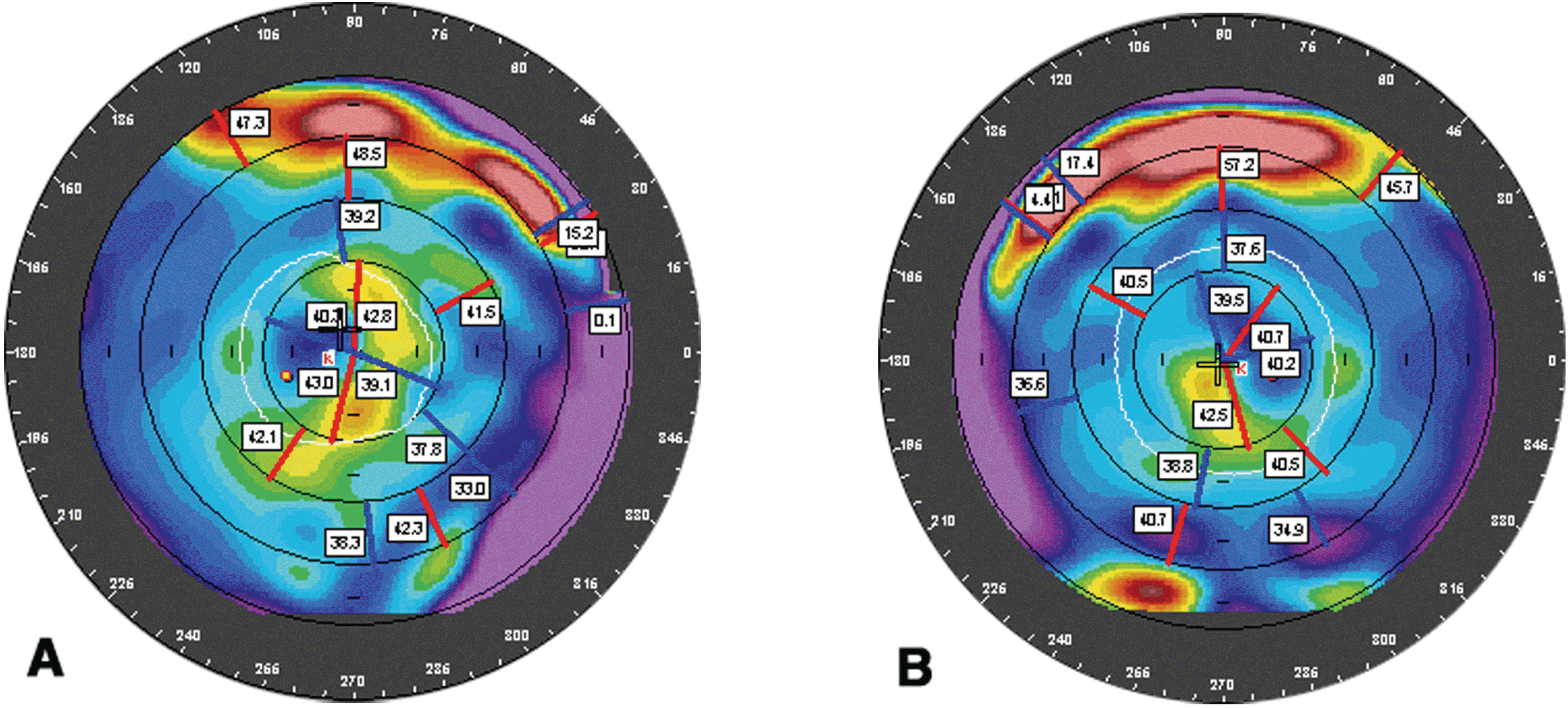
Figure 2.
Tangential power maps of corneal topography. Note the increased tangential power of the superior cornea (A=right eye; B=left eye).

Figure 3.
(A, B) New vessels from the limbus to center the of the cornea and subepithelial opacity of whole cornea (A=right eye; B=left eye), (C, D) Diffuse punctuate epithelial erosion (C=right eye; B=left eye).

Figure 4.
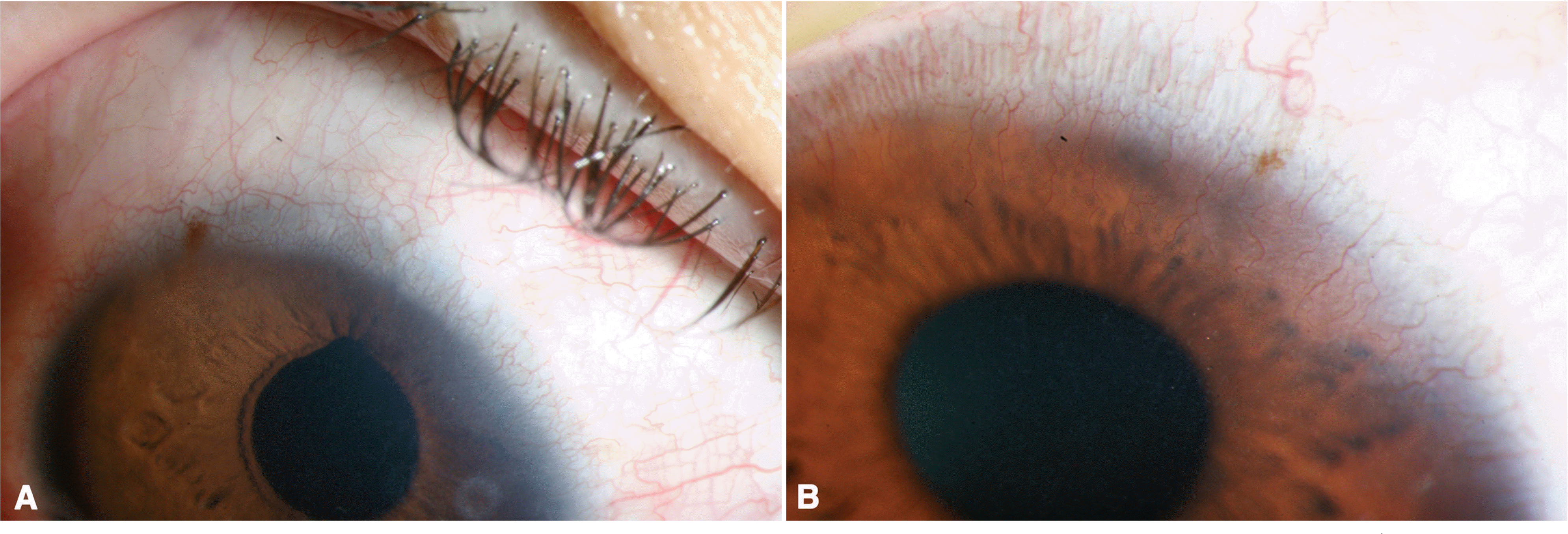
Ingrowth of new vessels about 2∼3 mm from the superior limbus to center of cornea (A=right eye; B=left eye).

Figure 5.
Ingrowth of new vessels about 4∼6 mm (A) and 2∼3 mm (B) from the superior limbus to the center of the cornea (A=right eye; B=left eye)


 PDF
PDF ePub
ePub Citation
 Print
Print


 XML Download